Vol. 34, No. 03, pp. 647 – 657, July – September, 2017 dx.doi.org/10.1590/0104-6632.20170343s20150649
* To whom correspondence should be addressed This is an extended version of the work presented at the
CHARACTERIZATION AND EVALUATION OF
SILVER-NANOPARTICLE-INCORPORATED IN
COMPOSITE GRAPHITE AIMING AT THEIR
APPLICATION IN BIOSENSORS
V. M. Santos
1, R. S. A. Ribeiro
1, A. J. T. Bosco
2,
E. M. Alhadeff
2and N. I. Bojorge
1*1Universidade Federal Fluminense. Departamento de Engenharia Química e Petróleo Rua Passo da Pátria, 156, Campus da Praia Vermelha, São Domingos, 24210-310 Niterói, RJ, Brasil.
*E-mail: [email protected]
E-mail: [email protected]; [email protected]
2Universidade Federal do Rio de Janeiro. Centro de Tecnologia. Escola de Química. Departamento de Engenharia Bioquímica Avenida Athos da Silveira Ramos, 149, Cidade Universitária, 21941-909 Rio de Janeiro, RJ, Brasil.
E-mail: [email protected]; [email protected]
(Submitted: October 14, 2015; Revised: April 26, 2016; Accepted: May 24, 2016)
Abstract - Biosensors based on nanomaterial composites have been investigated for their potential to function as high sensitivity signal response devices. In the present study, we report the fabrication of silver nanoparticles (AgNPs) on a graphite epoxy composite electrode (GEC) and mixed with the polyaniline (a conductive emeraldine salt form polymer) composite electrode (AgNPs/PANI/GEC), in order to compare the performance of the generated electrochemical response signals. Cyclic voltammetry tests were conducted to compare the quality and intensity of signals from the
different prepared electrodes. Tests for the AgNPs/PANI/GEC electrodes were made with and without the enzymes
alcohol oxidase and horseradish peroxidase immobilized on the composite surface. The prepared AgNPs/PANI/GEC nanocomposite was evaluated by thermal analysis. Scanning electron microscopy images and EDX were obtained for characterization of the electrode surface morphology. Square wave voltammetry techniques were then employed for ethanol analysis with the AOX/HRP/AgNPs/PANI/GEC biosensor achieving good results in a range of 0.37M to 0.65 M.
Keywords: Biosensor, Alcohol oxidase, Horseradish peroxidase, Polyaniline, Silver nanoparticles, Voltammetry.
INTRODUCTION
The growth and development of the field of
nanotechnology have ushered in the advent of new nanomaterials and their application in the development of novel, high sensitivity analytical devices (Wang et al., 2009). Biosensor-based nanomaterials, which represent
the integration of material science, molecular engineering, chemistry and biotechnology, can markedly improve the
sensitivity and specificity of biomolecular detection.
Detection devices based on nanomaterial sensors hold the capability of detecting or manipulating atoms and molecules. The biosensor-based nanomaterials composite have great potential in applications such as biomolecule
of Chemical
recognition, pathogenic diagnosis, and environmental monitoring (Zhang, Guo and Cui, 2009; Narayanan, 2012;
Santana et al., 2014; Alhadeff and Bojorge, 2011).
Incorporation of nanoparticles has greatly influenced the field of electrochemical biosensors due to their ability to strongly influence the mechanical properties, such as stiffness and elasticity, of the sensor material.
The inclusion of nanoparticles provides a combination of properties such as high surface area, good electrical conductivity, and chemical stability (Jianrong et al., 2004; Solanki et al., 2011; Berti and Turner, 2011), Furthermore, nanoparticles provide biocompatible environments for enzyme immobilization.
One example of a nanoparticle that is easily and cheaply prepared is the silver nanoparticle. The most frequently used method for preparing silver nanoparticles as stable colloidal dispersions in water or organic solvents is a chemical reduction (Shahverdi et al., 2007). The syntheses of nanoparticles by chemical reduction methods is generally performed in the presence of stabilizers to avoid unwanted agglomeration of the colloidal suspensions of nanoparticles (Jeong and Park, 2014; Abou El-Nour et al., 2010).
The characterization of these composites involves thermal analysis in order to verify thermic stability and conductivity. Wei and Hsueh (1989) studied the thermal characteristics of chemically synthesized polyaniline with various dopants by TGA/DSC showing that the HCl-doped polyaniline exhibits three major weight losses at around 100, 200, and 500°C, which were assigned to the removal of H2O and HCl, and decomposition of the polymer, respectively. Thermal aging of the HCl-doped polyaniline performed at 100, 150, and 200°C for various periods of time results in a decrease in conductivity. After the thermal treatments, the polymer can be re-doped with HCl to partially recover the conductivity. However, both the conductivity and the doping level cannot be restored to the level of the original materials owing probably to changes in morphology, crosslinking, or other chemical reactions (Wei and Hsueh, 1989). Recently, Rasool and Majid (2014) reported the synthesis of a PANI composite with cobalt monoethanolamine complex via in situ oxidative polymerization by ammonium persulfate and its positive
effect on thermostability of the polyaniline.
The objective of this work investigates the effect of the
incorporation of silver nanoparticles in ethanol biosensors, using as biological recognition elements the immobilized alcohol oxidase and horseradish peroxidase enzymes.
Several different composite electrode types were analyzed in order to compare the effect that incorporation of silver
nanoparticles had on the electrochemical response signals. Three composites were prepared, all of them with recently prepared silver nanoparticles: (a) a silver nanoparticles-graphite epoxy composite alone; (b) silver nanoparticles
incorporated in a graphite-epoxy composite (AOX/HRP/ GEC/AgNPs/GEC); and (c) a similar silver nanoparticles-graphite nanocomposite but also containing polyaniline (AOX/HRP/GEC/AgNPs/PANI/GEC).
MATERIALS AND METHODS
Materials
The reagents used for this research were: polyaniline (emeraldine salt), sodium borohydride, powder, 98%, albumin, glutaraldehyde, and enzyme alcohol oxidase EC 1.1.3.13 (Pichia pastoris) purchased from Sigma-Aldrich; enzyme horseradish peroxidase EC 1.11.1.7 from Toyobo Brazil; sodium citrate, 4-aminoantipyrine, hydrogen peroxide, phenol and ethanol 95% BP and sodium
phosphate buffer pH 7 were acquired from Vetec; silver
nitrate (Synth); epoxy resin DER 332 and graphite powder (FP, 99.5% pure) were purchased from Fluka.
Syntheses of silver nanoparticles
Silver nanoparticles (AgNPs) were synthesized by the method of the reaction between 0.001M silver nitrate solution and an ice-cold solution of 0.0020 M sodium borohydride (NaBH4) as per the literature (Mavani and Shah, 2013).
Construction of composite electrodes
Without extracting the Ag nanoparticles, 150 mg of powdered graphite was directly dissolved in the obtained Ag nanoparticles solution and left to dry in an oven at 100°C for 12 hours. The resulting Ag nanoparticles/
graphite mixture, as conductive fillers, was dispersed in
epoxy and the mixture was stirred to obtain a uniform composite matrix of 40 wt% epoxy matrix and 60 wt%
conductive fillers and placed in a Teflon tube fitted with a
19 Gauge metal wire. Figure 1 depicts the construction of
the composite modified microdisk electrode. Subsequently,
the electrode was rinsed with distilled water several times
and further dried in air before use. The final obtained
electrode is denoted as AgNPs/GEC.
Figure 1. A) Schematic diagram of the process for preparing the AgNPs/GEC electrode; B) Schematic illustration of the bi-enzyme biosensor with reaction steps and electron-transfer pathway from ethanol via AOX and enzymatically generated H2O2 via HRP and polymer-bound to the AgNPs composite surface.
Bi- enzymatic Immobilization Methodology
The components used to bind the enzyme to the composite surface were 2.5% (v/v) glutaraldehyde, albumin 1 % (w/v) and an enzymatic solution composed of AOX (286 U) and HRP (2640 U). From the proportions suggested by a statistical analysis, 10 mL of immobilization solution were deposited on the electrode surface, which remained stored at 4 ºC for 12 hours.
Characterization
UV–visible spectroscopic analysis of synthesized silver nanoparticles
The reduction of silver ions was confi rmed by qualitative
testing of supernatant by UV–visible spectrophotometer. One ml samples of supernatant were withdrawn freshly made and 24, 48, and 120 hours after preparation and absorbance was measured by scanning the absorbance between 300-700 nm.
Thermal characterization of the composite constituents.
The thermal stability of the composite and its constituents was investigated by thermal gravimetric
analysis experiments in order to verify the infl uence of
AgNPs in the composite. The thermogravimetric analyzer (TGA) was a PerkinElmer, model Pyris, and used a scanning
rate of 10ºC/min from 23ºC to 800ºC, with nitrogen fl owing at 30 mL/min). Diff erential scanning calorimetry (DSC) was performed with an exploratory diff erential calorimeter
(PerkinElmer, Pyris Diamond DSC model) working with
nitrogen at 20 mL/min fl ow rate (scanning rate of 10ºC/
min from 20ºC to 350ºC).
TGA of the mixture composite (PANI + enriched AgNPs graphite) was performed as a preliminary analysis to determine the temperature range to be used in DSC. DSC analysis was performed by varying the temperature
of the diff erent samples (sample 1: PANI; sample 2:
A
graphite; sample 3: suspension of colloidal AgNPs; sample 4: AgNPs enriched with graphite and sample 5: composite prepared from 38% enriched with graphite and AgNPs and 62% of PANI).
Electrode Surface Morphological Characterization
A JEOL JSM-6460LV model scanning electron microscope (SEM) operating at 20 kV acceleration voltage was employed to analyze the morphology of the electrode surface. The electrode was placed in the sample port using an adhesive double-sided carbon to improve the electrical conductivity in order to analyze the electrode with and without enzyme immobilization.
Energy dispersive X-ray Analysis of silver nanoparticles composites
For electron dispersive X-ray spectroscopy (EDX) analysis, composite samples were prepared to the same
composition specified before and washed three times with
deionized water and subjected to EDX measurements on an EDX-720, Energy dispersive X-ray spectrometer, Shimadzu. Quantitative elemental analysis of samples was
performed by collecting EDX on 5 different regions and
averaging the obtained values.
Electrochemical Analysis
The parameters used during the tests with square wave voltammetry were as follows: 10 ml of electrolyte solution, frequency 25 Hz, scan rate of 40 mV/s, potential between 0.2 and 0.75 V. Measurements were made with
a potentiostat (Autolab, PGSTAT12 model), Ag/AgCl reference electrode (ALS model RE-012 167 1B), and counter electrode platinum wire.
RESULTS AND DISCUSSION
AgNPs Colloidal Dispersion Characterization
The absorption spectrum of freshly prepared Ag
nanocolloid solutions after five days of synthesis is
shown in Figure 2. All spectra exhibit an absorption maximum at approximately 400 nm, consistent with the results reported in the literature (Mavani and Shah, 2013;
Rashid et al., 2013; Rajkumar, 2016), confirming that their
synthesis was successful since the typical absorption band is in the range of 350 to 450 nm region. Such bands are unique physical properties of these nanoparticles. When
an external electromagnetic field, such as light, is applied
to a metal, the conduction electrons move in tandem to provide a distributed load disturbance known as plasmon, located close to the metal surface (Rashid et al., 2013). The absorbance of freshly prepared Ag nanocolloid suspension using 2 mL AgNO3 gave an absorbance of 0.666 at a wavelength of 398 nm. However, after 1 103.7oC week of the synthesis, the absorbance spectrum of the Ag nanocolloids showed a red shift (to around 400 nm) with the decrease in the peak absorbance value. The absorbance maximum was found to be 0.530. The absorption peak became broader when the spectrum was recorded after 1 week of preparation. This indicated that the Ag nanocolloids aggregated with time.
Adsorption of borohydride plays a key role in stabilizing growing silver nanoparticles by providing a particle surface charge (Solomon et al., 2007) and it must
be in sufficient amounts to stabilize the particles as the
reaction proceeds. However, later in the reaction, too much sodium borohydride increases the overall ionic strength and aggregation will occur, respectively.
Thermal Characterization
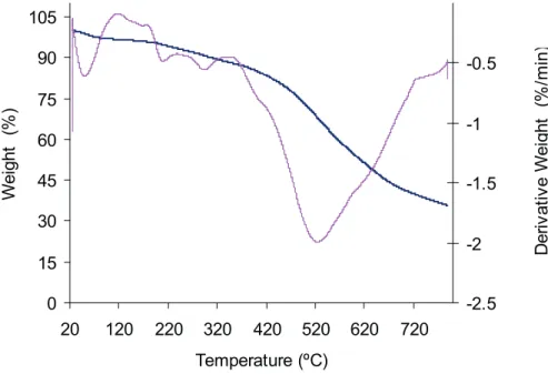
In this work, the thermal behavior of samples of graphite, graphite enriched with AgNPs, PANI and PANI/ graphite enriched with the AgNPS were analyzed by DSC according to the chemical nature and composition of matter. Figure 3 showed the TGA curves of percentage weight loss of the composite PANI/graphite enriched with AgNPs as a function of increasing temperature.
Three significant mass loss events can be observed
(Figure 3): 50.17°C (3.0%), between 200°C and 293.5°C
(7.8%), and a greater at 524.59°C (52.1%). The first loss
observed at low temperature (50.17°C) may be due to water evaporation from PANI, a highly hygroscopic polymer. The second and third recorded weight losses should be related to the decomposition of the polymer backbone to yield a residue of approximately 25%, which corresponds to the content of enriched with graphite AgNPs used in the composite preparation.
For the commercial PANI sample, two events were
observed: the first one endothermic (191°C) and the second
exothermic (292.2°C). The glass transition temperature (change of the amorphous state to the crystalline state)
verified in this study was 165.3°C, an intermediate
temperature between the values observed by Ding et al., (1999) around 70°C and 250°C for powdered PANI. Abdelkader et al. (2012) studied the thermal stability
of different states of PANI and observed distinct glass
transition temperatures: 74.1°C for the emeraldine basic form, 103.7oC for the emeraldine salt form and 126.9oC
0 15 30 45 60 75 90 105
20 120 220 320 420 520 620 720
Temperature (ºC)
W
ei
ght
(
%
)
-2.5 -2 -1.5 -1 -0.5
D
er
iv
at
iv
e W
ei
ght
(
%
/m
in)
Figure 3. AgNPs–PANI-graphite composite TGA curves (% weight loss - blue line). Derivative weight loss – rose line.
for the hydrochloric acid doped polyaniline. Chauhan et al. (2011), studying the thermal behavior of emeraldine
base PANI, reported two significant different events: one
in the range of 25°C to 60°C, credited to the evaporation of adsorbed water, and a second between 90°C to 115°C due possibly to cross-linking reactions between nearby quinoidal rings in the structure of the polymer.
Belaabed et al. (2010) found that the PANI-doped BSA
is thermally stable up to 600 ºC working in a nitrogen flow
(20 mL/min) and a heating rate of 5 ºC/min, reporting that
only 40% of weight loss was observed. Three significant events of weight loss were verified: the first in the range
PANI-doped BSA/epoxy resin up to 360 ºC, when a sharp weight loss happened with degradation of the composites.
In the DSC profile obtained for the composite PANI/
graphite enriched with AgNPs, two thermal events appeared, one endothermic at 195.9°C and another exothermic at
292.4°C. The first event may be due to evaporation of
the associated water and the second promoted by cross and recrystallization reactions promoted by the rising temperature. Comparing the DSC curves obtained for the
PANI and the PANI/Graphite+AgNPs samples the change in enthalpy (DH) and the areas under the peaks showed an increase of the exothermic event magnitude from 282.1 mJ (-75.5 J/g) to 1172.8 mJ (-252.5 J/g) which occurred at the same temperature, around 292°C. Table 1 shows the comparative values of temperature and amount of heat
generated in the thermal events for the different samples
in DSC studies.
Table 1. Temperature values and heat generated by the thermal events
Sample Tg (oC) DC
p (J/g.
oC) T 1 (
oC) DH
1 (J/g) Area1 (mJ) T2 (
oC) DH
2 (J/g) Area2 (mJ)
PANI 165.3 0.378 191.0 48.9 173.5 292.2 -79.5 -282.1
PANI/Graphite+AgNPs ND ND 195.9 73.3 340.5 292.4 -252.5 -1172.8
Colloidal AgNPs - - 106.3 143.8 2027.2 - -
-The DSC analysis for the graphite sample supplemented
with AgNPs showed five endothermic events that were
observed between 25°C and 350°C, possibly due to removal of adsorbed water by evaporation and the volatilization of some chemical residues formed during the AgNPs colloidal preparation). Khan et al. (2011) showed that in heating and TGA and DTA measurements, carried out in a nitrogen atmosphere at 10°C/min, for powder silver nanoparticles, only 14.58% of mass loss were observed at 200ºC. An intense exothermic peak between 200°C and 300°C was attributed to crystallization and thermal decomposition of silver nanoparticles.
The AgNPs colloidal sample, in the temperature range investigated by DSC, showed a single endothermic event at temperature 106.3°C, probably due to evaporation of water. Mani et al. (2013) observed a mass loss of about 28% of AgNPs by TGA working with a temperature range up to 800°C and a DSC endothermic event at 69°C, studying a phytochemical method to promote the reduction of Ag+ to Ago (nanoparticles) with diameter between 20 and 30 nm, that generated a thermally stable compound at low temperatures. Khan et al. (2011) reported a degradation of about 14.6% at temperatures above 300°C, the occurrence of exothermic peak between 200°C and 300°C credited to desorption of water and organic volatiles, with a possible crystallization of the nanoparticles, and an increase of the average diameter (17.5 nm).
The results obtained by TGA for the AgNPs/graphite/ PANI are in line with those reported previously (Mani et al., 2013; Majeed Khan, et. al., 2011) and the DSC analysis for AgNPs dispersion performed in this work, which showed a single endothermic peak at 106.3°C (water evaporation).
Scanning Electron Microscopy (SEM) and Energy dispersive X-ray (EDX) Studies
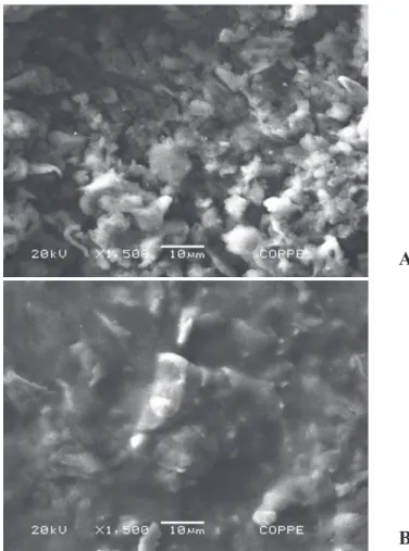
The surface analysis by scanning electron microscopy revealed that the electrodes had a degree of surface roughness (Figure 4(A)). These ridges promote an increase
in electrode contact surface, contributing to the increased number of active sites on the surface thereof, which, in turn, contribute to an increase in the area available for enzyme immobilization.
The surface modified electrode, shown in Figure 4(B),
is similar to the results obtained previously (Silva et al., 2011), where there is a large reduction in the number of grooves on the electrode surface, indicating they are
completely filled during the glutaraldehyde crosslinking
with the AOX and HRP.
The EDX spectrometer analyses confirmed the presence
of an elemental silver signal of the silver nanoparticles (Figure 5). The vertical axis displays the number of x-ray counts whilst the horizontal axis displays energy in KeV.
Identification lines for the major emission energies for
silver (Ag) are displayed and these correspond with peaks
in the spectrum, thus giving confidence that silver has been correctly identified.
The EDX of the AgNPs/GEC composite sample (65% graphite/AgNPs + 35% Epoxy) showed around 24 wt % of silver as indicated in the table inset of Fig.5(A) and the AgNPs/PANI/GEC composite sample (40% graphite/ AgNPs + 25% PANI + 35% Epoxy) showed only 2 wt % of silver (Fig. 5(B)).
Electrochemical characterization
Bosco et al. (2015), in order to determine the
electrochemical effect of inclusion AgNPs in a composite
bare electrode, compared the performance of a PANI/ GEC electrode on standard solutions containing 10 mM K4Fe(CN)6. The authors reported that the redox couple of Fe(CN)63- / Fe(CN)64- appears quite reversible to both electrodes, without and with AgNPs. The peak currents
obtained are well-defined with Ipa/Ipc=1.28 for a PANI/
GEC bare electrode and 1.07 for the AgNPs/PANI/GEC bare electrode. Peak current enhancement accompanied by a peak potential shift suggests that electrocatalytic activity
Figure 4. SEM images of the electrode surface (1500X magnifi cation). A) The composite AgNPs-graphite-polyaniline without, and; B) With
the immo bilized AOX and HRP.
Figure 5. EDX Spectra of composite samples: (A) AgNPs/GEC and (B) AgNPs/PANI/GEC
A
B
A
when compared with the other electrode.
The eff ect of varying the scan rates on the cyclic
voltammograms of the Fe(CN)63-/Fe(CN) 6
4- couple using
AgNPs/GEC and AgNPs/PANI/GEC as the working electrodes in 10 mM K4Fe(CN)6 supporting electrolyte was studied over 10 – 100 mV/s in the potential range of +1.5 to -1.5V. It was observed that currents increase with the scan rate due to heterogeneous kinetics and a good area of potential was found at a low scan rate when compared with the GEC simple electrode (Figure 6). The oxidation and
reduction peak currents were measured for both composite electrodes, AgNPs/GEC, and AgNPs/PANI/GEC, working with the electrolytic Fe(CN)63-/Fe(CN)
6
4- solution.
Based on a plot of square root of scan rate versus Ipc for
the reduction current of the first cycle, straight lines were
obtained as shown in the inset of Figure 6A and Figure 6B,
satisfying the equation y = 0.348x + 0.0081 (R2 = 0.9655) for the AgNPs/PANI/GEC electrode and y = 0.3732x - 0.2694(R² = 0.9583) for the AgNPs/GEC.
Figure 6. Comparative cyclic voltammetry of AgNPs/GEC (A) and AgNPs/PANI/GEC (B) in 10 mM K3 [Fe(CN)6] at diff erent scan rates
from 10 to 150 mV/s versus Ag/AgCl. Inset: Calibration plots for square root of scan rate vs. Ipa and Ipc.
With the increase of scan rate, the anodic and cathodic
peak potentials of both composites modifi ed with AgNPs
showed a small shift and the redox peak currents increased linearly (Figure 6 insets), indicating a surface-controlled electrode process. According to the kinetic theory of the electrode reaction, the slope of Ip vs. square root of scan
rate should be 0.5 for a pure diff usion controlled process
and 1 for a pure adsorption controlled process. Hence, the
observed slopes further confi rmed that the electrochemical behavior of AgNPs at the modifi ed electrode is quasi-reversible and a diff usion-controlled electrochemical
process (Li et al., 2005).
To achieve rapid response and a wide linear range
of ethanol detection at the modifi ed electrode surface, voltammetric techniques can be more eff ective to probe
the ethanol electrocatalytic oxidation reaction. The performances of the disposable biosensors were tested by applying them to the determination of the ethanol concentration. In this study, the concentration of ethanol was determined by using the standard addition method
due to the elimination of matrix eff ects. Upon addition of
ethanol to 0.5mM pH 7.0 PBS, the cyclic voltammogram of AgNPs/GEC for the direct electron transfer of the
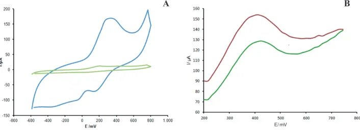
AOX/HRP complex changed dramatically, with an increase of reduction peak current and a decrease of oxidation peak current (Figure 7A), while the change of the cyclic voltammogram of bare or AgNPs/PANI/ GEC was negligible (not shown), displaying an obvious electrocatalytic behavior of the AOX/HRP complex for the reduction of ethanol.
As previously mentioned, the enzymatic complex of AOX/HRP was immobilized with glutaraldehyde on AgNPs incorporated in graphite composite electrodes (AOX/HRP/AgNPs/GEC and AOX/HRP/AgNPs/PANI/ GEC). Graphite in the composites acts as a bridge to provide an electrical contact or pathway for electron transfer between the immobilized enzymes and the surface of the AgNPs/graphite nanocomposite electrode. Thus, it was expected that direct electron communication between the bi-enzymatic complex and the conducting
substrate was easily bridged by the AgNPs, and verifi ed
Figure 7. (A) Cyclic voltammograms obtained with the AOX/HRP/AgNPs/GEC biosensor: Green curve in the absence; blue curve in the presence of ethanol (0.5mM) in 0.1 M phosphate buffer pH 7.0. Scan rate 50 mV/s. (B) Square wave voltammograms obtained with the AOX/ HRP/AgNPs/PANI/GEC biosensor for two different ethanol concentrations: 0.37M (Green line) and 0.65 M (Red line).
300mV, demonstrating that ethanol could be catalytically reduced within the optimal potential range for biosensors
between −600 and 800mV (versus Ag/AgCl). Figure 7B
shows the square wave voltammograms obtained with the
AOX/HRP/AgNPs/PANI/GEC biosensor for two different
ethanol concentrations: 0.37M and 0.65 M. The square
wave profiles generated for ethanol standard solutions
prepared in the range of 0.37M to 0.65 M were determined. This gave a calibration curve with a linear range of current intensity (Ic, mA) with the ethanol concentration (Eth, g/L)
according to: Ic = 0.004[Eth] + 0.1186, with a correlation
coefficient (R2) of 0.9833. The bi-enzymatic immobilized
electrode prepared with the AgNPs/PANI/GEC composite generated good response signals when the square voltammetry technique was applied as an electrochemical analytical method.
From the information presented in this section, it can be seen that the positive contribution in sensitivity was strongly dependent upon the AgNPs content in the
bulk composite and their effect in improving the current
sensitivity. An electrochemical biosensor is sensitive to changes in current through the device, which means any strategy for amplifying the current density variation will improve the sensing mechanism. The amount and activity of the immobilized biological elements in the electrochemical detection surface will determine the global sensitivity of biosensors and the ability to regenerate. The high surface area of AgNPs in relation to their volume and their biocompatibility, with a large number of interaction sites, causes AgNPs to be potential candidates for amplifying the area of the detection surface and maintain the bioactivity of detection, thus increasing the amount and activity of biological recognition elements (Pacioni et. al 2015; ElKaoutit et. al., 2012; Guo et al., 2015). In addition, the excellent stability can improve biocompatible
biological recognition and relatively high-density AgNPs allow for the application as a current enhancer.
CONCLUSIONS
In this study through diverse characterization
techniques the effect of incorporation of AgNPs into different graphite composites was observed. The thermal
analysis results obtained for the AgNPs/PANI/GEC composite agreed with those reported in the literature, with the initial loss of weight at a temperature of 50°C (3%). By cyclic voltammetry, an improvement of the electrochemical response signals was observed, with a reversible performance. For the bi-enzymatic immobilized AgNPs–graphite/epoxy composite electrode, cyclic voltammetry worked well as an electrochemical method applied for both validation and as an analytical procedure. The AgNPs–polyaniline/graphite/epoxy composite used to prepare the working electrode generated the best response signal for the cycle voltammetry tests, showing to be a good
methodology to define the best electrochemical parameters
and conditions analysis. On the other hand, for ethanol analytical performance, square wave voltammetry was
more sensitive, which was necessary to obtain amplified
response signals. The EDX studies for the AgNPs/PANI/ GEC sample detected only 2 wt% of silver nanoparticles in the global composition, and for the AgNPs/GEC sample around 24 wt% incorporated into the composite.
The difference in AgNPs weight percentages added to the
prepared composites, without and with PANI, is apparently
reflected in the quality and intensity of the response signal.
This behavior was observed for the ethanol analytical assays in which it was necessary to employ square wave voltammetry to obtain a more sensitive performance and amplify the response signal.
ACKNOWLEDGMENTS
The authors thank AGIR-UFF for a PIBITIC scholarship
and CENPES for financial support and Toyobo of Brazil
for the donation of horseradish peroxidase enzyme.
REFERENCES
Abdelkader, R., Amine, H. and Mohammed, B., Thermally stable forms of pure polyaniline catalyzed by an acid-exchanged
montmorillonite clay called maghnite-H+ as an effective
catalyst. International Journal of Polymer Science, 2012, pp. 1-7, (2012).
Abou El-Nour, K.M.M., Eftaiha, A., Al-Warthan, A. and Ammar, R. A., Synthesis and applications of silver nanoparticles. Arabian Journal of Chemistry, 3 (3), pp. 135-140 (2010). doi:http://dx.doi.org/10.1016/j.arabjc.2010.04.008.
Alhadeff, E. and Bojorge, N., Graphite-Composites Alternatives
for Electrochemical Biosensor. In: J. Cuppoletti, Metal, Ceramic and Polymeric Composites for Various, Rijeka, Croatia.: InTechWEB.ORG., pp. 597-620 (2011).
Belaabed, B., Lamouri, S., Naar, N., Bourson, P., Hamady, S. O. S., Polyaniline-doped benzene sulfonic acid/epoxy resin composites: structural, morphological, thermal and dielectric behaviors. Polymer Journal, 42, pp. 546–554 (2010). Berti, F. and Turner, A. P. (2011). New Micro- and Nanotechnologies
for Electrochemical Biosensor Development. Weinheim, Germany.: Verlag GmbH & Co. KGaA. Published Online: 19 MAR 2011.DOI: 10.1002/9783527635160.ch1.
Bosco, A. J., Alhadeff, E. M., Mihos, F. d., Yokoyama, L. M.,
Santos, V. and Bojorge Ramirez, N. I., Optimization of the method of preparing carbon paste electrode. Chemical Engineering Transactions, 25, pp. 2443-2448 (2015). Chauhan, N. P., Ameta, R., Ameta, R., and Ameta, S., Thermal
and conducting behavior of emeraldine base (EB) form of polyaniline (PANI). Indian Journal of Chemical Technology, 18, pp. 118-122 (2011).
Ding, L., Wang, X. and Gregory, R. V., Thermal properties of chemically synthesized polyaniline (EB) powder. Synthetic Metals, 104, pp. 73 - 78 (1999).
ElKaoutit. M., Kaoutit, M. E., Naggar, Naranjo-Rodrígueza I., H-H de Cisnerosa, J. L., Graphite grains studded with silver nanoparticles: Description and application in promoting direct biocatalysis between heme protein and the resulting carbon paste electrode, Colloids and Surfaces B: Biointerfaces 92, pp. 42– 49 (2012).
Guo L., Jackman J. A., Yang H-H, Chen P., Cho N-J., Kim
D-H., Strategies for enhancing the sensitivity of plasmonic nanosensors, Nano Today, 10 (2), pp. 213-239 (2015), ISSN 1748-0132.
Jeong, L. and Park, W. H., Preparation and Characterization
of Gelatin Nanofibers Containing Silver Nanoparticles.
International Journal of Molecular Sciences, 15(4), pp. 6857– 6879 (2014). doi:http://doi.org/10.3390/ijms15046857. Jiang, W., Nadeau, G., Zaghib, K., Kinoshita, K., Thermal
analysis of the oxidation of natural graphite - the effect of
particle size. Thermochimica Acta, 351, pp. 85-93 (2000)
Jianrong, C., Yuqing, M., Nongyue, H., Xiaohua, W. and
Sijiao, L., Nanotechnology and biosensors. Biotechnology Advances, 22(7), pp. 505-518 (2004).
Johansson, K., Jönsson-Pettersson, G., Gorton, L., Marko-Varga, G. and Csöregi, E., A reagentless amperometric biosensor for alcohol detection in column liquid chromatography based on co-immobilized peroxidase and alcohol oxidase in carbon paste. Journal of Biotechnology, 31(3), pp. 301-316 (1993). Khan, M. A. M., Kumar, S., Ahamed, M., Alrokayan, S. A.
and AlSalhi, M. S., Structural and thermal studies of silver nanoparticles and electrical transport study of their thin
films. Nanoscale Research Letters, 6, pp. 434 (2011).
doi:10.1186/1556-276X-6-434.
Li, P., Li, J., Wu, C., Wu, Q. and Li, J., Synergistic antibacterial
effects of β-lactam antibiotic combined with silver
nanoparticles. Nanotechnology, 16(9), pp. 1912-1917 (2005). Majeed Khan, M. A., Kumar, S., Ahamed, M., Alrokayan, S. A.
and AlSalhi, M. S., Structural and thermal studies of silver
nanoparticles and electrical transport study of their thin films.
Nanoscale Research Letters, 6 (1)(434), pp. 1-8 (2011). Mani, U.; Dhanasingh, S.; Arunachalam, R.; Paul, E., A simple and
green method for the synthesis of silver nanoparticles using Ricinus communis leaf extract. Progress in Nanotechnology and Nanomaterials, 2(1), pp. 21 -25 (2013).
Narayanan, R., Nanoparticles of Different Shapes for Biosensor
Applications. In: Functional Nanoparticles for Bioanalysis, Nanomedicine, and Bioelectronic Devices, vol. 1, pp. 281-292, ACS Symposium Series (2012)..
Pacioni, N.L., Borsarelli, C. D., Borsarelli, C.D., Rey, V., Synthetic Routes for the Preparation of Silver Nanoparticles. A Mechanistic Perspective, Springer International Publishing Switzerland, E.I. Alarcon et al. (eds.), Silver Nanoparticle Applications, Engineering Materials, pp. 13-46 (2015). DOI 10.1007/978-3-319-11262-6_2.
Rajkumar, K., Kanipandian, N.and Thirumurugan, R.Rajkumar, K. S.,. Toxicity assessment on haemotology, biochemical and histopathological alterations of silver nanoparticles-exposed
freshwater fish Labeo rohita. Applied Nanoscience, 6:19, pp.
1-11 (2016). DOI https://doi.org/10.1007/s13204-015-0417-7 Rashid, M. U., Bhuiyan, M. K. and Quayum, M. E., Synthesis of
Silver Nano Particles (Ag-NPs) and their uses for Quantitative Analysis of Vitamin C Tablets. Journal of Pharmaceutical Sciences, 12(1), pp. 29-33 (2013).
Rasool, R. and Majid, K., Synthesis, characterization, thermal and electrical properties of composite of polyaniline with cobaltmonoethanolamine complex. Bulletin of Materials Science, 37(5), pp. 1181–1190 (2014).
Santana, A. C., Southgate, E. F., Mendes, J. P., Dweck, J.,
Alhadeff, E. M. and Bojorge Ramirez, N. I., Characterization
of an HRP-AOX-polyaniline-graphite composite biosensor. Journal of Electrochemical Science and Engineering, 4(4), pp. 165-175 (2014).
Shahverdi, A. R., Fakhimi, A., Shahverdi, H. R. and Minaian, S.,
Synthesis and effect of silver nanoparticles on the antibacterial activity of different antibiotics against Staphylococcus
aureus and Echerichia coli. Nanomedicine: Nanotechnology, Biology and Medicine, 3, pp. 168-171 (2007).
D. and Furtado, R. F., Biossensor amperométrico para determinação de peróxido de hidrogênio em leite. Eclética Química. Eclética Química, 36(2), pp. 143-157 (2011). Solanki, P. R., Kaushik, A., Agrawal, V. and Malhotra, B.,
Nanostructured metal oxide-based biosensors. NPG Asia Materials, 3(1), pp. 17–24 (2011).
Solomon, S. D., Bahadory, M., Jeyarajasingam, A. V., Rutkowsky, S. A. and Boritz, C., Synthesis and Study of Silver Nanoparticles. Journal of Chemical Education, 84 (2), pp. 322 - 325 (2007).
Tsocheva, D., Zlatkov, T. and Termlemezyan, L., Thermoanalytical studies of polyaniline Emeraldine base. Journal Of Thermal Analysis And Calorimetry, 53, pp. 895-904 (1998).
Wang, G., Wang, W., Wu, J., Liu, H., Jiao, S. and Fang, B.,
Self-assembly of a silver nanoparticles modified electrode and its
electrocatalysis on neutral red. Microchimica Acta, 164(1), pp. 149-155 (2009).
Wei, Y. and Hsueh, K. F., Thermal analysis of chemically synthesized polyaniline and effects of thermal aging on
conductivity. Journal of Polymer Science Part A: Polymer Chemistry, 27(13), pp. 4351–4363 (1989).


![Figure 6. Comparative cyclic voltammetry of AgNPs/GEC (A) and AgNPs/PANI/GEC (B) in 10 mM K 3 [Fe(CN) 6 ] at diff erent scan rates from 10 to 150 mV/s versus Ag/AgCl](https://thumb-eu.123doks.com/thumbv2/123dok_br/16318297.719007/8.918.112.814.332.574/figure-comparative-cyclic-voltammetry-agnps-agnps-pani-versus.webp)