ABSTRACT
BACKGROUND AND OBJECTIVES: Submucosal dexa-methasone injection directly in the surgical area has been used in diferent dental procedures, but there are still few studies evalu-ating its eicacy as compared to oral route for impacted third molar surgeries. So, this study aimed to evaluate postoperative pain, edema and trismus after impacted third molar surgeries us-ing oral or submucosal local injection of dexamethasone. METHODS: his was a prospective, controlled, crossover and randomized study involving 36 patients with indication of lower third molar surgeries, who were randomly distributed in two groups: group A – submucosal local injection of dexamethasone (4mg/1mL) after local anesthesia, and group B – oral dexameth-asone tablet (4mg) one hour before procedure. Edema and tris-mus were clinically evaluated in the postoperative period and in the 1st, 2nd, 3rd and 7th postoperative days. Patients were oriented
to record pain intensity in the visual analog scale in periods zero (preoperative), 1h, 2h, 4h, 12h, 1 day, 2 days and 3 days and one week after surgery. Data were submitted to statistical analysis with signiicance level of 5%.
RESULTS: here were no signiicant diferences in surgical time with regard to operated sides (p=0.4). Edema and trismus values were not statistically diferent between observed groups (p>0.05). Mean pain values recorded in the visual analog scale were not statistically diferent between groups and patients have
Evaluation of effects of two dexamethasone formulations in impacted
third molar surgeries*
Avaliação dos efeitos de duas formulações de dexametasona em cirurgias de terceiros molares
inclusos
Marsal Moretto Noboa1, Juliana Cama Ramacciato2, Rubens Gonçalves Teixeira1, Carllini Barroso Vicentini3, Francisco Carlos
Groppo4, Rogério Heládio Lopes Motta2
*Received from São Leopoldo Mandic College, Campinas, SP, Brazil.
1. College São Leopoldo Mandic, Campinas, SP, Brazil.
2. College São Leopoldo Mandic, Area of Pharmacology, Anesthesiology and herapy, Campinas, SP, Brazil
3. College Tocantinense Institute Presidente Antonio Carlos, Department of Sugery, Porto Nacional, TO, Brazil
4. College of Dentistry of Piracicaba, FOP-UNICAMP, Area of Pharmacology, Anesthesiolo-gy and herapy, Piracicaba, SP, Brazil.
Submitted in February 18, 2014. Accepted for publication in June 13, 2014. Conlict of interests: none.
Correpondence to: Rogério Heládio Lopes Motta Rua José Rocha Junqueira, 13 13045-755 Campinas, SP, Brasil. E-mail: [email protected]
© Sociedade Brasileira para o Estudo da Dor
not reported major postoperative discomfort and had no need to prolong analgesic medication (p>0.05).
CONCLUSION: Both dexamethasone administration routes were efective to control pain, edema and trismus after lower third molar surgeries, presenting similar results.
Keywords: Dexamethasone, Edema, Oral surgery, Pain.
RESUMO
JUSTIFICATIVA E OBJETIVOS: A utilização da injeção submu-cosa de dexametasona diretamente na área cirúrgica tem sido real-izada em vários procedimentos odontológicos, mas ainda são escas-sos os estudos que avaliaram a sua eicácia em comparação com a via oral para exodontias de terceiros molares inclusos. Desta forma, o objetivo do presente estudo foi avaliar a dor, edema e trismo no pós-operatório de cirurgia de terceiros molares inclusos utilizando-se dexametasona, por via oral, ou por injeção local submucosa. MÉTODOS: Estudo prospectivo, controlado, cruzado e ran-domizado envolvendo 36 pacientes com indicação cirúrgica de terceiros molares inferiores que foram divididos em dois grupos: grupo A - injeção local submucosa de dexametasona (4mg/1mL) após a anestesia local e grupo B - 1 comprimido de dexametasona (4mg), por via oral, uma hora antes do procedimento. Edema e trismo foram avaliados clinicamente no pré-operatório, 1º, 2º, 3º e 7º dia de pós-operatório. Os pacientes foram orientados a reg-istrar a intensidade de dor na escala analógica visual nos períodos 0 (pré-operatório), 1h, 2h, 4h, 12h, 1 dia, 2 dias, 3 dias e uma semana após as cirurgias. Os dados obtidos foram submetidos a análise estatística com nível de signiicância de 5%.
RESULTADOS: Não revelaram diferenças signiicativas no tem-po cirúrgico em relação aos lados operados (p=0,4). Os valores de edema facial e trismo não demonstraram diferenças estatisti-camente signiicativas entre os grupos observados (p>0,05). Os valores médios de dor registrados na escala analógica visual não mostraram diferença estatística signiicativa entre os grupos e os pacientes não relataram grande desconforto pós-operatório e não necessitaram prolongar o fármaco analgésico (p>0,05).
CONCLUSÃO: As duas vias de administração da dexametasona mostraram-se eicazes no controle da dor, edema e trismo após cirurgias de terceiros molares inferiores, apresentando resultados semelhantes.
Descritores: Cirurgia bucal, Dexametasona, Dor, Edema.
INTRODUCTION
Impacted third molar extraction involves soft and bone tissue trauma and may result in considerable pain, edema and tris-mus. hese postoperative pathophysiological events are associ-ated to the release of inlammatory mediators resulting from arachidonic acid metabolism, which may bring discomfort to patients and afect their quality of life1-3.
Several professionals have emphasized the need for better con-trolling the postoperative inlammatory process of such sur-gical procedures, and diferent drugs have been proposed for such. Corticosteroids may inhibit the onset of inlammatory mediators synthesis and are considered potent drugs to control pain, edema and trismus4-7.
Dexamethasone has been widely used in dentistry in diferent doses and administration routes to decrease postoperative dis-comfort and when used for a short period presents less interfer-ence with chemotaxis for leucocytes8,9. Among administration
routes, submucosal injection has been reported by previous studies with signiicant efects on postoperative edema, but just a limited efect on trismus and pain10-13.
Notwithstanding several scientiic investigations on this sub-ject, there is still no consensus with regard to patients selection, corticosteriods dosage, time and administration route. Drug choices to control postoperative sequelae after oral surgery is normally supported by professional experience and personal preferences, and in this sense, crossover and randomized stud-ies are important to explain and guide the best therapeutic choices14.
his study aimed at comparing the eicacy of oral or submuco-sal dexamethasone for impacted third molar extraction.
METHODS
his is a prospective, crossover and randomized study where 36 volunteers of both genders, aged from 18 to 25 years and without systemic changes that could contraindicate the surgical procedure were selected. All volunteers had indication for bi-lateral extraction of lower third molars with similar impaction pattern, classiied as Class I or II and in position B, according to Pell and Gregory15 and vertical or mesio-angulated
impac-tion according to Winter16. In the initial visit, patients were
evaluated according to a clinical record where patients’ identi-ication medical and dental history, results of preoperative tests (blood count and coagulation time), date and time of surgery, surgery duration, operated side, number of anesthetic tubettes used and administration route of dexamethasone used during the session were recorded8,12,14,17,18.
Randomization and study groups
To accurately control patients and for better idelity of results a randomized clinical trial was proposed for the operated side and therapy, based on items 8-10 of the 2001 checklist of the Cochrane Collaboration (Oral Health Group, University of Manchester, UK), which determines the randomized method to generate sequence, to hide groups’ letterings and blindness
of involved parties19-21. Two pharmacological protocols were
proposed to control postoperative pain and edema: Group A – local submucosal injection of 1mL of 4 mg/mL dexamethasone (Decadron®, Aché Laboratórios Farmacêuticos S.A., Guarulhos,
SP, Brazil) immediately after locoregional anesthesia; Group B – 1 oral 4mg dexamethasone tablet (Decadron®, Aché
Labo-ratórios Farmacêuticos S.A., Guarulhos, SP, Brazil), one hour before the procedure.
Surgical procedures
Each volunteer was submitted to two surgical procedures per-formed by the same surgeon with proven experience and with 21-day interval between the irst and the second surgery22. his
crossover study has determined that in every surgical procedure the same patient should receive one of the proposed therapies for each operated side – submucosal injection of dexametha-sone (4mg/mL) in impacted third molar vestibular region or oral dexamethasone tablet (4mg).
For each surgical procedure patients were oriented to vigorous-ly mouthwash with 0.12% chlorhexidine solution (Proderma®,
Piracicaba, SP, Brazil) for one minute, were submitted to ex-traoral antisepsis with 2% chlorhexidine solution (Proderma®,
Piracicaba, SP, Brazil) and received postoperative recommenda-tions about diet, rest and oral hygiene23.
For teeth extraction, all surgical technique and asepsis prin-ciples were strictly followed. Inferior alveolar lingual and buc-cal nerves were blocked with 2% lidocaine with 1:100000 epinephrine (Alphacaine®, Rio de Janeiro, Brazil)24.
Immedi-ately after, group A has received subcutaneous dexamethasone in the side determined by randomization. Sulcular incision was performed with knife blade 15 (MedGoldman®, São José,
Brazil) and after detachment a mucoperiosteal lap was ob-tained. Osteoctomy and dental section were performed with rotary tool with abundant sterile saline irrigation. Nylon thread 3.0 (Polysuture®, São Sebastião do Paraíso, Brazil) was
used for suture.
After surgical procedure, all patients received a vial with 8 paracetamol tablets (750mg) (Tylenol®, Johnson & Johnson do
Brasil Indústria e Comércio de Produtos para Saúde Ltda., São José dos Campos, SP, Brazil) and were oriented to take one tablet every 6h for two days. If analgesia had to be prolonged, volunteers were oriented to record quantity and times of ad-ditional analgesics use.
Edema and trismus evaluation
Evaluation was carried out in moments: preoperative, 1st, 2nd,
3rd and 7th postoperative days by linear measurements. Edema
was measured between the tragus and the wing of the nose and between tragus and labial commissure, at the operated side, with silk thread 2.0. Trismus was evaluated by the in-terincisal distance, taken as from the incisal edge of upper and lower central incisive teeth at the operated side. Measures were taken with patients in maximum mouth opening, us-ing a gauged digital caliper rule (Pantec®, São Bernardo do
Campo, Brazil) in the preoperative, 1st, 2nd, 3rd and 7th
Pain evaluation
he visual analog scale (VAS) printed in 10 pages of a book-let with explanations about illing was used. Each page of this booklet represented one pain measurement moment (preopera-tive, immediate postopera(preopera-tive, 1, 2, 4, 12 hours; 1, 2, 3 and 7 postoperative days).
Patients were oriented to mark with a trace on the horizontal line to relect pain intensity, considering zero as no pain and 10 as maximum possible pain26. Volunteers were asked to
per-sonally return the booklet with illed VAS when they returned for the seventh postoperative day consultation. heir marks were then measured by gauged digital caliper rule (Pantec®,
São Bernardo do Campo, Brazil), considering the distance from zero to the trace recorded by patients in every measure-ment momeasure-ment27.
Statistical analysis
Descriptive statistical techniques were used through absolute and percentage distributions and inferential statistical meth-ods. Paired t, Levene, Shapiro-Wilks, ANOVA and Tukey tests were used with signiicance level of 5%, being that calcula-tions were obtained with the BioEstat 5.0 program (Fundação Mamirauá, Belém, PA).
This study was approved by the Human Beings Research Ethics Committee, Center of Dental Research and São Leo-poldo Mandic Dentistry School (Process 2009/0110), in compliance with Resolution 196/1996. All patients were in-formed about the objectives of the study and have accepted to participate by signing the Free and Informed Consent Term (FICT).
RESULTS
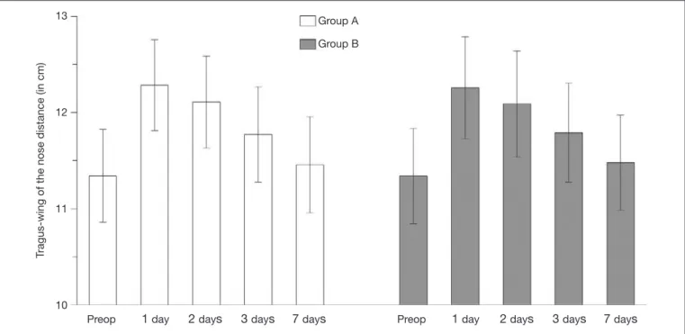
he analysis (paired t test) of surgical moments has shown no statistically signiicant diferences (p=0.7109) between the sur-gical time needed for surgeries of group B medicated by oral route, and of group A, by parenteral route. Levene test has shown that “tragus-wing of the nose” (p=0.9973), “tragus-com-missure” (p=0.1262), “mouth opening” (p=0.1210) and “pain” (p=0.0935) were homogeneous, being that Shapiro-Wilks test has shown normal distribution for the same measurements. So, measurements were submitted to ANOVA for repeated measures and to Tukey test. Figures 1 and 2 show edema evaluation results. With regard to tragus-wing of the nose measurement, data anal-ysis has shown no statistically signiicant diferences between groups in moments: preoperative (p=1.0), 1 day (p=0.8141), 2 days (p=0.8693), 3 days (p=0.8610) and 7 days (p=0.8508). However there have been statistically signiicant diferences be-tween each moment, both for group A (p<0.0001) and group B (p<0.0001).
Except for period “3 days” (p=0.0690), there have been no statistically signiicant diferences (p>0.05) between groups in remaining evaluated times. here have also been no statistically signiicant diferences between “preoperative” and “7 days” (p=0.3385); between “1 day” and “2 days” ((p=0.3146); and between “3 days” and “7 days” ((p=0.2120) for group B. Re-maining combinations of periods have shown signiicant dif-ferences (p<0.05) for this group. For group A, there have been no statistically signiicant diferences in “preoperative” and “7 days” (p=0.6032); and “1 day” and “2 days” (p=0.6002), being that remaining periods had signiicant diferences (p<0.05).
Figure 1. Tragus to wing of the nose distance (mean ± standard deviation) as a function of therapies used)
T
ragus-wing of the nose distance (in cm)
Group A
Group B 13
12
11
10
With regard to mouth opening, data analysis has shown no statistically signiicant diferences between groups in pre-operative (p=1.0), 1 day (p=0.6667), 2 days (p=0.6799), 3 days (p=0.6632) and 7 days (p=0.9539). However, there have been statistically signiicant diferences between each moment when considering just group A (p<0.0001) and just group B (p<0.0001).
With regard to pain evaluation, data analysis has shown no
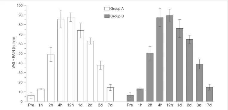
sta-tistically signiicant diferences between groups in any evalu-ated moment: preoperative (p=0.8981), 1 hour (p=0.8268), 2 hours (p=0.3254), 4 hours (p=0.2813), 12 hours (p=0.1978), 1 day (p=0.1185), 2 days (p=0.2180), 3 days (p=0.4030) and 7 days (p=0.7435). However, there have been statistically signii-cant diferences (p<0.0001) between the periods: preoperative, 1h, 2h, 2 days and 3 days both for group A and group B. How-ever, periods “4h” and “12h” and “1 day” and “7 days” were not statistically diferent for both groups (p>0.05).
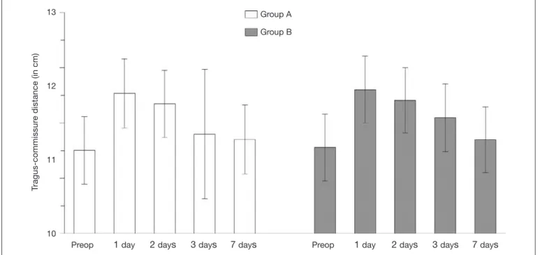
Figure 2. Tragus to labial commissure distance (mean ± standard deviation) as a function of therapies used
Figure 3. Mouth opening values (mean ± standard deviation) as a function of therapies used
T
ragus-commissur
e distance (in cm)
Group A
Group B 13
12
11
10
Preop 1 day 2 days 3 days 7 days Preop 1 day 2 days 3 days 7 days
Mouth-opening (in mm)
Group A
Group B 70
60
50
40
30
20
DISCUSSION
Impacted third molar extraction is in general associated to moderate to severe postoperative discomfort, even when the technique is subtly used, as shown in igure 4, which shows higher painful sensitivity in the periods of 4, 12 and 24h after the procedure1-3,10. here is a broad discussion about the best
drug to minimize postoperative discomfort in dentistry and among the best drugs of choice, dexamethasone is being evalu-ated by several scientiic studies due to its eicacy to control inlammatory complications as compared to its non-use5-7.
Meechan and Seymour28 have studied diferent complications
that appear after impacted third molar surgery and have con-cluded that the observation of such complications is important to comparatively evaluate the eicacy of several therapeutic measures. Other authors have shown that surgical procedure and immediate postoperative observations are a clinical model for the evaluation of the eicacy of diferent drugs14,18,29,30. In
the crossover model used in our study, the same patient was submitted to both proposed treatments (submucosal injection and oral route), one for each operated side during randomiza-tion. his model is an advantage for the prospective evaluation of clinical pharmacology, since patients assure the quality of their own control.
Some studies suggest the systemic use of corticosteroids for im-pacted third molar surgeries5-7,12,13. Markiewicz et al.4 in a
me-ta-analysis have concluded that corticosteroids administered in the preoperative period were of great value to decrease postop-erative inlammatory signs and symptoms. Speciically, patients receiving corticosteroids had signiicantly less postoperative edema, pain and trismus, both the early (after 1-3 days) and the late period (after 4-7 days). Notwithstanding such results,
there is still no consensus about the best administration route, dose and duration of treatment, in addition to diferences in methods used to evaluate clinical variables.
Oral dexamethasone administration involves later onset of ef-fect, which is inherent to its pharmacokinetics and requires pa-tients’ cooperation31. However, it is a convenient, safe and
low-cost route. Our study data showed that oral dexamethasone was efective to control pain and edema during the studied period, which is in line with other similar studies6,7,11,12.
Submucosal dexamethasone injection had signiicant efect on edema in two previous studies and both have reported signiicant decrease of edema in the immediate postoperative period as compared to controls1,13
. Our results have shown
that submucosal dexamethasone injection has signiicantly decreased edema in the irst postoperative days, in line with previous studes10,13. An interesting observation in this group
was the signiicant trismus decrease in the irst postopera-tive day, which is similar to group B (oral route), fact which might be result of the higher concentration of dexamethasone obtained immediately at injury site. hese results add more power to the concept that dexamethasone administered close to the surgical site is a valuable way to decrease edema and trismus5,6,12,32.
Our results, regarding Levene and Shapiro-Wilks tests, have not shown statistically signiicant diferences between groups with regard to postoperative pain, edema and trismus decrease after third molar extraction. his is in line with several authors who have observed the therapeutic eicacy of submucosal ad-ministration of corticosteroids in previous studies, shown that submucosal dexamethasone injection, as well as its oral admin-istration, may be a feasible alternative for more invasive dental procedures6,10,12,13
Figure 4. Means ± standard deviation obtained as from visual analog scale (VAS in mm) as a function of therapies used
V
AS – P
AIN (in mm)
Group A
Group B 100
90
80
70
60
50
40
30
20
10
0
CONCLUSION
Bolus administration of parenteral dexamethasone by sub-mucosal injection, and oral administration with tablets have shown similar efects to decrease pain, edema and trismus after impacted third molar extractions.
REFERENCES
1. Majid OW. Submucosal dexamethasone injection improves quality of life measures after third molar surgery: a comparative study. J Oral Maxillofac Surg. 2011;69(9):2289-97. 2. Herrera-Briones FJ, Prados Sánchez E, Reyes Botella C, Vallecillo Capilla M. Update on the use of corticosteroids in third molar surgery: systematic review of the literature. Oral Surg Oral Med Oral Pathol Oral Radiol. 2013;116(5):e342-51.
3. Mehra P, Reebye U, Nadershah M, Cottrell D. Eicacy of anti-inlammatory dru-gs in third molar surgery: a randomized clinical trial. Int J Oral Maxillofac Surg. 2013;42(7):835-42.
4. Markiewicz MR, Brady MF, Ding EL, Dodson TB. Corticosteroids reduce postopera-tive morbidity after third molar surgery: a systematic review and meta-analysis. J Oral Maxillofac Surg. 2008;66(9):1881-94.
5. Klongnoi B, Kaewpradub P, Boonsiriseth K, Wongsirichat N. Efect of single dose preoperative intramuscular dexamethasone injection on lower impacted third molar surgery. Int J Oral Maxillofac Surg. 2012;41(3):376-9.
6. Bhargava D, Sreekumar K, Deshpande A. Efects of intra-space injection of Twin mix versus intraoral-submucosal, intramuscular, intravenous and per-oral administration of dexamethasone on post-operative sequelae after mandibular impacted third mo-lar surgery: a preliminary clinical comparative study. Oral Maxillofac Surg. 2013;20 [Epub ahead of print].
7. Simone JL, Jorge WA, Horliana AC, Canaval TG, Tortamano IP. Comparative analy-sis of preemptive analgesic efect of dexamethasone and diclofenac following third molar surgery. Braz Oral Res. 2013;27(3):266-71.
8. Sotto-Maior BS, Senna PM, de Souza Picorelli Assis NM. Corticosteroids or cyclooxy-genase 2-selective inhibitor medication for the management of pain and swelling after third-molar surgery. J Craniofac Surg. 2011;22(2):758-62.
9. Li J, Wang X, Zhou C, Liu L, Wu Y, Wang D, Jiang H. Perioperative glucocorticos-teroid treatment delays early healing of a mandible wound by inhibiting osteogenic diferentiation. Injury. 2012;43(8):1284-9.
10. Grossi GB, Maiorana C, Garramone RA, Borgonovo A, Beretta M, Farronato D, et al. Efect of submucosal injection of dexamethasone on postoperative discomfort after third molar surgery: a prospective study. J Oral Maxillofac Surg. 2007;65(11):2218-26. 11. Apiliogullari S, Yildirim G, Ataoglu H. he supraperiosteal route of dexamethasone
should be considered for dental surgery. Oral Surg Oral Med Oral Pathol Oral Radiol Endod. 2010;109(2):165; author reply 165-6.
12. Antunes AA, Avelar RL, Martins Neto EC, Frota R, Dias E. Efect of two routes of administration of dexamethasone on pain, edema, and trismus in impacted lower third molar surgery. Oral Maxillofac Surg. 2011;15(4):217-23.
13. Warraich R, Faisal M, Rana M, Shaheen A, Gellrich NC, Rana M. Evaluation of postoperative discomfort following third molar surgery using submucosal dexametha-sone - a randomized observer blind prospective study. Oral Surg Oral Med Oral Pathol Oral Radiol. 2013;116(1):16-22.
14. de Sousa Santos JA, da Silva LC, de Santana Santos T, Menezes Júnior LR, de
Assun-ção Oliveira AC, Brandão JR. Comparative study of tramadol combined with dexa-methasone and diclofenac sodium in third-molar surgery. J Craniomaxillofac Surg. 2012;40(8):694-700.
15. Pell GJ, Gregory GT. Impacted mandibular third molars: classiication and modiied technique for removal. Dent Dig. 1933;39:e330-e8.
16. Winter GB. Principles of exodontia as applied to the impacted third molar. St. Louis: American Medical Books; 1926.
17. Graziani F, D’Aiuto F, Arduino PG, Tonelli M, Gabriele M. Perioperative dexametha-sone reduces post-surgical sequelae of wisdom tooth removal. A split-mouth randomi-zed double-masked clinical trial. Int J Oral Maxillofac Surg. 2006;35(3):241-6. 18. Laureano Filho JR, Maurette PE, Allais M, Cotinho M, Fernandes C. Clinical
compa-rative study of the efectiveness of two dosages of dexamethasone to control postope-rative swelling, trismus and pain after the surgical extraction of mandibular impacted third molars. Med Oral Patol Oral Cir Bucal. 2008;13(2):E129-32.
19. Moher D, Schulz KF, Altman D. CONSORT Statement: revised recommendations for improving the quality of reports of parallel-group randomized trials 2001. Explore (NY). 2005;1(1):40-5.
20. Siddiqi A, Morkel JA, Zafar S. Antibiotic prophylaxis in third molar surgery: A rando-mized double-blind placebo-controlled clinical trial using split-mouth technique. Int J Oral Maxillofac Surg. 2010;39(2):107-14.
21. Aznar-Arasa L, Harutunian K, Figueiredo R, Valmaseda-Castellón E, Gay-Escoda C. Efect of preoperative ibuprofen on pain and swelling after lower third molar removal: a randomized controlled trial. Int J Oral Maxillofac Surg. 2012;41(8):1005-9. 22. van Gool AV, Ten Bosch JJ, Boering G. Clinical consequences of complaints and
compli-cations after removal of the mandibular third molar. Int J Oral Surg. 1977;6(1):29-37. 23. Hedström L, Sjögren P. Efect estimates and methodological quality of randomized
controlled trials about prevention of alveolar osteitis following tooth extraction: a syste-matic review. Oral Surg Oral Med Oral Pathol Oral Radiol Endod.2007;103(1):8-15. 24. Gordon SM, Chuang BP, Wang XM, Hamza MA, Rowan JS, Brahim JS, et al.
he diferential efects of bupivacaine and lidocaine on prostaglandin E2 release, cyclooxygenase gene expression and pain in a clinical pain model. Anesth Analg. 2008;106(1):321-7.
25. Neupert EA 3rd, Lee JW, Philput CB, Gordon JR. Evaluation of dexamethasone for reduction of postsurgical sequelae of third molar removal. J Oral Maxillofac Surg. 1992;50(11):1177-83.
26. Kaczmarzyk T, Wichlinski J, Stypulkowska J, Zaleska M, Panas M, Woron J. Single--dose and multiSingle--dose clindamycin therapy fails to demonstrate eicacy in preventing infectious and inlammatory complications in third molar surgery. Int J Oral Maxillo-fac Surg. 2007;36(5):417-22.
27. Collins SL, Moore RA, McQuay HJ. he visual analogue pain intensity scale: what is moderate pain in millimetres? Pain. 1997;72(1-2):95-7.
28. Meechan JG, Seymour RA. he use of third molar surgery in clinical pharmacology. Br J Oral Maxillofac Surg. 1993;31(6):360-5.
29. Ong CK, Lirk P, Seymour RA, Jenkins BJ. he eicacy of preemptive analgesia for acute postoperative pain management: a meta-analysis. Anesth Analg. 2005;100(3):757-73. 30. Pektas ZO, Sener M, Bayram B, Eroglu T, Bozdogan N, Donmez A, et al. A
compa-rison of pre-emptive analgesic eicacy of dilunisal and lornoxicam for postoperative pain management: a prospective, randomized, single-blind, crossover study. Int J Oral Maxillofac Surg. 2007;36(2):123-7.
31. Dan AE, hygesen TH, Pinholt EM. Corticosteroid administration in oral and or-thognathic surgery: a systematic review of the literature and meta-analysis. J Oral Maxillofac Surg. 2010;68(9):2207-20.