Detection of Respiratory Viruses in
Nasopharyngeal Swab and Adenoid Tissue from
Children Submitted to Adenoidectomy: Pre- and
Postoperative Analysis
Osvaldo Vinícius Biill Primo
1Edmir Américo Lourenço
1Saulo Duarte Passos
21Department of Otorhinolaryngology, Faculdade de Medicina de Jundiaí, Jundiaí, São Paulo, Brazil
2Department of Pediatrics, Faculdade de Medicina de Jundiaí, Jundiaí, São Paulo, Brazil
Int Arch Otorhinolaryngol 2014;18:150–154.
Address for correspondence Osvaldo Vinícius Biill Primo, MSc, ENT, Department of Otorhinolaryngology, Faculdade de Medicina de Jundiaí-SP, Rua Francisco Telles, 250–Vila Arens, Jundiaí, São Paulo
13202-550, Brazil (e-mail: [email protected]).
Introduction
The Waldeyer lymphatic ring is formed by a conglomerate of lymphoid structures comprising the pharyngeal tonsils (ad-enoids) and palatine tonsils, beyond the lingual and peritubal tonsils. It is strategically located at the entrance of the
respiratory and digestive systems and is exposed to a wide variety of antigens present in air and food, which are contin-uously presented to the body.1–3
Hypertrophy of pharyngeal and palatine tonsils is one of the most common disorders found by otolaryngologists and is a major cause of upper airway obstruction, accounting
Keywords
►
viruses
►
adenoids
►
adenoidectomy
►
child
►
polymerase chain
reaction
Abstract
Introduction
The presence of respiratory viruses in lymphoid tissues of the
nasophar-ynx and oropharnasophar-ynx and its impact on recurrent infections and hypertrophy of these
tissues are not yet fully understood.
Objective
To identify and determine the prevalence of major respiratory viruses in
nasopharyngeal secretions and adenoid tissue pre- and postoperatively of children
undergoing adenoidectomy.
Methods
A prospective observational study was conducted in 36 patients under
12 years of age with upper airway lymphoid hypertrophy who were undergoing
adenoidectomy, in which various respiratory viruses were investigated using
real-time polymerase chain reaction in adenoid tissue and nasopharyngeal secretions
collected preoperatively and 30 days postoperatively.
Results
At least 1 viral agent was isolated in any of the samples collected in 58.3% of
children and 25.9% of total samples. Respiratory viruses were identi
fi
ed in 33.8% of
preoperative nasopharyngeal specimens and in 19.8% of postoperative secretion. Of the
21 patients with positive results for any respiratory virus, 6 (28.6%) had more than 1
virus. Considering all 36 respiratory viruses found, the main agent isolated was
rhinovirus (27.8%), followed by bocavirus (22.2%).
Conclusion
The virus found more frequently in all samples was rhinovirus. After
removal of adenoid tissue, there was a decrease in the prevalence of the virus contained
in nasopharyngeal secretion 30 days after surgery.
received
September 10, 2013 accepted
December 2, 2013
DOI http://dx.doi.org/ 10.1055/s-0034-1368135. ISSN 1809-9777.
Copyright © 2014 by Thieme Publicações Ltda, Rio de Janeiro, Brazil
for 75% of respiratory disorders in childhood, which may vary from primary snoring to severe cases of obstructive sleep apnea with cardiovascular commitment.4,5
The pathogenesis of inflammatory/infectious disease of tonsils and adenoids probably involves their anatomical location and its antigen-processing function. There is no certainty about what determines the onset of chronic infec-tion. Viral infection with bacterial infection may be one of the triggering mechanisms of chronic infection, but the effects of the environment, personal factors, and diet, among others, may also be involved. Several studies and theories have been published based primarily on allergic mechanisms or viral and/or bacterial infectious processes.6–9
Although it is a very common health problem in daily medical practice, the presence of respiratory viruses in lymphoid tissues of the nasopharynx and oropharynx and their impact on recur-rent infections and hypertrophy of these tissues are not yet fully understood and are being studied by numerous researches. Many authors have identified the presence of multiple viral agents in these tissues, latent in asymptomatic patients.6,10–15
Most of these studies in the literature are performed among symptomatic and hospitalized children with acute disease, which hampers a better understanding of this pathophysiology.
Aim
This study aims to identify and determine the prevalence of major respiratory viruses in nasopharyngeal secretions col-lected pre- and postoperatively and adenoid tissue of children undergoing adenoidectomy.
Methods
A prospective observational study was performed in 36 pa-tients under 12 years of age with upper airway lymphoid hypertrophy undergoing adenoidectomy or adenotonsillec-tomy, assisted by the otorhinolaryngology clinic of a university hospital. The study was approved by the institutional research ethics committee (protocol number 363/2011). All parents or guardians signed a consent form after being informed of the objectives, procedures, and responsibilities of the research, as well as received answers to any questions regarding the study. The study included children under 12 years of age with clinical and/or radiologic criteria for adenoidectomy or ad-enotonsillectomy (recurrent tonsillitis and/or hypertrophy of lymphoid tissues leading to upper airway obstruction). Chil-dren were excluded from the study if they had fever or any type of acute respiratory tract infection at the time of collec-tion of biological samples, if they had craniofacial malforma-tions, if they were immunocompromised, or if their parents did not agree with their participation in the research project. Several respiratory viruses (influenza A and B; parainfluenza 1, 2, 3, and 4; rhinovirus; respiratory syncytial virus; human bocavirus; coronaviruses; and metapneumovirus) were investi-gated by quantitative real-time polymerase chain reaction (q-PCR) in adenoid tissue removed surgically and nasal swab specimens collected preoperatively and at 1 month postopera-tive follow-up visit.
Nasal secretion was collected with the aid of a sterile metal rod nasal swab, following the rules of asepsis. The nasal cavity was previously humidified with 1 mL of sterile sodium chloride 0.9% solution. The swab was introduced directly into the nasal cavity without contact with the patient’s skin or other region. After collecting, this swab was homogenized in microtubes containing sterile Ringer lactate solution. The adenoid tissue was obtained by cold adenoidectomy under general anesthesia by curettage of adenoid tissue. Small fragments of adenoid tissue were stored in microtubes containing virus transport media, which is a solution for transportation and storage of viruses in tissues.
After collection, the samples were transported in liquid nitrogen in microtubes to the Research Laboratory of Virology and Molecular Biology, where they were frozen at 80°C, and subsequently viral genetic material in the secretion and ade-noid tissue was extracted according to a standardized protocol by the laboratory. After extraction, q-PCR was performed of respiratory viruses using the handset Applied Biosystems 7500 Real-Time PCR Systems (Life Technologies™, California, USA) following the manufacturer’s specifications and protocols. The FTD-Respiratory-21 kit from Fast-Track Diagnostics™ (Junglin-ster, Luxemburg) was used to identify respiratory viruses.
The database was created using Microsoft Excel program (Microsoft, Redmond, Washington, United States). The data were analyzed using absolute (n) and relative (%) frequencies, average, and standard deviation, depending on the variable studied. The averages of continuous outcome variables were compared by the Student t test. The association of the qualitative measures between groups was performed using the chi-square test or Fisher exact test to determine statistical significance. The soft-ware used for analysis was SPSS (Statistical Package for Social Sciences, IBM™, Armonk, New York, USA) version 13 and the assumed level of significance was 5% (p<0.05).
Results
The study evaluated 36 children between 3 and 12 years of age undergoing adenoidectomy or adenotonsillectomy from April 2012 to January 2013, with an average age of 7.3 years and a median of 7 years. Of these 36 children, 25 (69.4%) were girls and 11 (30.6%) were boys. A total of 108 pre- and postoperative nasopharyngeal specimens and adenoid tissue samples were removed surgically from these 36 patients (75 samples from girls and 33 from boys). At least 1 viral agent was isolated in any of the samples collected in 58.3% of 36 children (n¼21) and in 25.9% of 108 samples (n¼28), in 36.4% (n¼12) of 33 samples of boys and 21.3% (n¼16) samples from 75 girls. Of the respiratory viruses found in 36 samples, 75% (n¼27) were isolated in children under 7 years of age.
in patients under 6 years of age, and 2 of these patients had coinfections in 2 different samples.
Respiratory viruses were found in 33.3% of preoperative nasopharyngeal specimens (n¼12), with a single virus isolated in 83.3% (n¼10) and coinfection by 2 viral agents in 16.7% of samples (n¼2), totaling 14 respiratory viruses. In adenoid tissue samples, respiratory viruses were found in 30.6% (n¼11): single viral infection in 72.8% (n¼8) and coinfection in 27.2% (n¼3) of these positive samples (total-ing 14 viruses). Of postoperative nasopharyngeal secretions, the positivity was less—in only 13.9% of samples (n¼5) was a respiratory virus isolated: coinfection in 60% (n¼3) and single virus infection in 40% (n¼2;►Table 1).
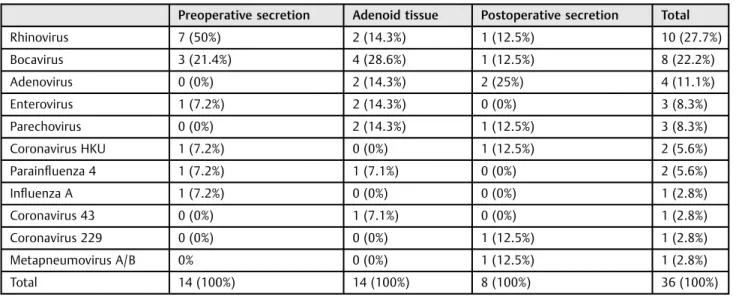
From the 14 respiratory viruses identified in preoperative nasopharyngeal secretions, 50% were rhinovirus (n¼7), present in 19% of 36 samples tested. In adenoid tissue, respiratory viruses were identified in 14, and the most frequent virus found was bocavirus, present in 11% of samples (n¼4), corresponding to 28.6% of the virus isolated. In nasopharyngeal secretions collected after surgery, only 8 respiratory viruses were identified, of which 25% (n¼2) were adenoviruses, present in 6% of samples.
Considering all 36 respiratory viruses isolated in 108 samples evaluated, the main agent isolated was rhinovirus (27.8%), followed by bocavirus (22.2%). Other viruses isolated with a lower frequency were adenovirus (11.1%),
enterovi-ruses (8.3%), parechovirus (8.3%), coronavirus HKU (5.6%), parainfluenza virus 4 (5.6%), influenza A (2.8%), coronavirus 43 (2.8%), coronavirus 229 (2.8%), and metapneumovirus A/B (2.8%). The respiratory syncytial virus was not isolated in the samples (►Table 2).
Discussion
In general, 58.4% of the patients (n¼21) and 25.9% of samples (n¼28) were positive for at least 1 respiratory virus. Studies in the literature show different prevalences, because some studies were virus-specific and others researched the prevalence of multiple respiratory viruses at the same time. Herberhold et al investigated several respiratory viruses in adenoid tissue from 30 children. The authors found a high positivity: at least 1 respiratory virus was identified in 97% of 52 samples.16Sato et al also searched respiratory viruses in adenoid tissue and found at least 1 respiratory virus in all samples.17In the study of Proenca-Modena et al, this positiv-ity was 97.5%, and the highest rate of viral detection was found in the adenoid tissue (85.7%), followed by nasal secre-tions (78.5%), tonsils (68.6%), and peripheral blood (1%).8
Considering all viruses found in this study, rhinovirus was isolated most frequently, followed by bocavirus. In the study of Herberhold et al, rhinovirus was also the most common respiratory virus, identified in 67% of samples, followed by
Table 1 Respiratory viruses (n¼36) found in 28 samples from 21 patients
Patient Sex Age Respiratory viruses
Preoperative secretion Adenoid tissue Postoperative secretion
1 Male 11 Rhinovirus – –
2 Female 5 – ParechovirusþBocavirus ParechovirusþCoronavirus 229
3 Male 5 Rhinovirus – RhinovirusþAdenovirus
4 Male 8 – Enterovirus Adenovirus
5 Male 4 Bocavirus – –
6 Male 5 Rhinovirus Adenovirus –
7 Male 9 Rhinovirus – –
8 Female 10 – – Coronavirus HKU
9 Female 6 RhinovirusþInfluenza A – –
10 Female 6 Enterovirus – –
11 Female 10 – Enterovirus –
12 Female 4 RhinovirusþBocavirus Parechovirus Metapneumovirus A/BþBocavirus
13 Female 3 – Coronavirus 43 –
14 Male 5 – Rhinovirus –
15 Female 9 Coronavirus HKU – –
16 Male 7 Parainfluenza 4 – –
17 Female 4 Rhinovirus – –
18 Female 5 – RhinovirusþBocavirus –
19 Female 9 – Adenovirus –
20 Male 5 – Bocavirus –
bocavirus in 53%, similar to our study but with different prevalences.16 In another study, adenovirus was the most common viral agent, isolated in 47.1% of patients, followed by enterovirus (40.5%), rhinovirus (38%), and bocavirus (29.8%).8 In the study from Sato et al, adenovirus was the most common viral agent, present in 80% of samples.17Alkhalaf et al ana-lyzed 106 palatine and pharyngeal tonsils of 57 patients undergoing routine tonsillectomy or adenoidectomy. Em-ploying the technique of real-time polymerase chain reaction, the authors identified 84 samples (72.4%) positive for adenovirus.18
In this study, 28.6% (n¼6) of patients with positive results presented coinfection with 2 viral agents, and 2 patients had viral coinfection in 2 samples, totaling 8 samples. Bocavirus was isolated in 75% (n¼6) of the coinfections. Rhinovirus was identified in all coinfections of preoperative nasopha-ryngeal secretions (n¼2), and bocavirus was isolated in all coinfections in adenoid tissue (n¼3). In coinfections of preoperative nasopharyngeal secretions, there was no pre-dominant viral agent.
Herberhold et al found multiple respiratory viruses in 83% of samples evaluated.16In the study of Proenca-Modena et al, the rate of coinfection was 69.5%. The authors evaluated 121 children who underwent adenotonsillectomy and found high rates of viral detection in adenotonsillar tissues. The authors were researching respiratory viruses on palatine tonsils, adenoid tissue, nasal secretions, and peripheral blood but did not evaluate the postoperative nasopharyn-geal secretions.8
Review of the literature found no studies comparing the prevalence of the virus in nasopharyngeal secretions pre- and postoperatively. Some studies revealed that the adenoid tissue may be a reservoir of viruses. Comparing the preva-lence of virus pre- and postoperatively, we aimed to analyze a possible influence of adenoid tissue in the maintenance of these viruses in secretions. After the analysis of preoperative nasopharyngeal samples, this study found that 33.3% of patients (n¼12) were positive for at least 1 respiratory virus.
Of the 14 respiratory viruses isolated, 50% were rhinovirus. After surgery, the positivity of respiratory viruses in naso-pharyngeal secretions of these patients decreased: in only 13.9% of patients (n¼5) was at least 1 viral agent isolated in the sample. In the postoperative secretions, 8 respiratory viruses were detected, and adenoviruses were identified in 2 patients. These results suggest a decrease in the prevalence of the virus after surgery (p¼0.0522). However, the number of patients in the study is relatively small, and a greater number of samples are required to confirm or reject this hypothesis. In addition to the small sample size, a seasonal effect may contribute to the discrepancies found between our results and other studies in the literature. We know that some respiratory viruses are found in certain periods of the year; however, as we have seen, apparently the virus may persist in the adenoid tissue and nasopharyngeal secretions even after the symptomatic phase of the disease. The climatic conditions of each region, air quality, and population characteristics may also contribute to a greater or lesser circulation of respiratory viruses in a given region and time of year.
The present study revealed that some patients with hy-pertrophy of pharyngeal tonsil exhibit respiratory viruses detected on nasopharyngeal and adenoid tissue, even if asymptomatic. These data suggest that the persistence or latency of respiratory viruses on nasopharyngeal and adenoid tissue after an acute infectious process may be related to the pathogenesis of lymphoid hypertrophy of the upper airways and that these tissues may function as a reservoir of virus, with possible influence on its transmission in the community. After adenoidectomy, a decrease in respiratory viruses pres-ent in nasopharyngeal secretions was observed. From this finding, we can infer a lower chance of occurrence of infec-tions as well as secondary superinfecinfec-tions and virus trans-mission to contacts after surgery. Considering the age group studied, in which there is a greater interpersonal contact, this decrease in the circulation of respiratory viruses becomes important for public health, with positive impact on the quality of life of these children.
Table 2 Frequency of viruses in the samples (n¼36)
Preoperative secretion Adenoid tissue Postoperative secretion Total
Rhinovirus 7 (50%) 2 (14.3%) 1 (12.5%) 10 (27.7%)
Bocavirus 3 (21.4%) 4 (28.6%) 1 (12.5%) 8 (22.2%)
Adenovirus 0 (0%) 2 (14.3%) 2 (25%) 4 (11.1%)
Enterovirus 1 (7.2%) 2 (14.3%) 0 (0%) 3 (8.3%)
Parechovirus 0 (0%) 2 (14.3%) 1 (12.5%) 3 (8.3%)
Coronavirus HKU 1 (7.2%) 0 (0%) 1 (12.5%) 2 (5.6%)
Parainfluenza 4 1 (7.2%) 1 (7.1%) 0 (0%) 2 (5.6%) Influenza A 1 (7.2%) 0 (0%) 0 (0%) 1 (2.8%) Coronavirus 43 0 (0%) 1 (7.1%) 0 (0%) 1 (2.8%)
Coronavirus 229 0 (0%) 0 (0%) 1 (12.5%) 1 (2.8%)
Metapneumovirus A/B 0% 0 (0%) 1 (12.5%) 1 (2.8%)
Conclusion
The rhinovirus was the most frequently found virus in all samples, followed by bocavirus. After removal of adenoid tissue, there was a decrease in the prevalence of viruses in nasopharyngeal secretions.
References
1 Dias EP, Rocha ML, Carvalho MOO, Amorim LMF. Detecção do vírus Epstein-Barr em tonsilites recorrentes. Braz J Otorhinolaryngol (Engl Ed) 2009;75(1):30–34
2 Dell’Aringa AR, Juares AJC, deMelo C, et al. Análise histopatológica de produtos de adenotonsilectomia de janeiro de 2001 a maio de 2003. Braz J Otorhinolaryngol 2005;71(1):18–22
3 MogoantăCA, IoniţăE, Pirici D, et al. Chronic tonsillitis: histologi-cal and immunohistochemihistologi-cal aspects. Rom J Morphol Embryol 2008;49(3):381–386
4 Llombart M, Chiner E, Gómez-Merino E, et al. Síndrome de apneas-hipopneas durante el sueño en población infantil: diferencias en su expresión entre niños con hipertrofia amigdalar y con enfer-medad concomitante. Arch Bronconeumol 2007;43(12):655–661 5 Beraldin BS, Rayes TR, Villela PH, Ranieri DM. Assessing the impact adenotonsillectomy has on the lives of children with hypertrophy of palatine and pharyngeal tonsils. Braz J Otorhinolaryngol 2009; 75(1):64–69
6 Frankel SS, Tenner-Racz K, Racz P, et al. Active replication of HIV-1 at the lymphoepithelial surface of the tonsil. Am J Pathol 1997; 151(1):89–96
7 Huang SW, Giannoni C. The risk of adenoid hypertrophy in children with allergic rhinitis. Ann Allergy Asthma Immunol 2001;87(4):350–355
8 Proenca-Modena JL, Pereira Valera FC, Jacob MG, et al. High rates of detection of respiratory viruses in tonsillar tissues from children with chronic adenotonsillar disease. PLoS ONE 2012;7(8):e42136 9 Drago L, Esposito S, De Vecchi E, et al. Detection of respiratory viruses and atypical bacteria in children’s tonsils and adenoids. J Clin Microbiol 2008;46(1):369–370
10 Roush KS, Domiati-Saad RK, Margraf LR, et al. Prevalence and cellular reservoir of latent human herpesvirus 6 in tonsillar lymphoid tissue. Am J Clin Pathol 2001;116(5):648–654 11 Chen R, Sehr P, Waterboer T, et al. Presence of DNA of human
papillomavirus 16 but no other types in tumor-free tonsillar tissue. J Clin Microbiol 2005;43(3):1408–1410
12 Lu X, Gooding LR, Erdman DD. Human bocavirus in tonsillar lymphocytes. Emerg Infect Dis 2008;14(8):1332–1334
13 Suvilehto J, Roivainen M, Seppänen M, et al. Rhinovirus/enterovi-rus RNA in tonsillar tissue of children with tonsillar disease. J Clin Virol 2006;35(3):292–297
14 Endo Y, Carroll KN, Ikizler MR, Wright PF. Growth of influenza A virus in primary, differentiated epithelial cells derived from adenoids. J Virol 1996;70(3):2055–2058
15 Endo LH, Ferreira D, Montenegro MC, et al. Detection of Epstein-Barr virus in tonsillar tissue of children and the relationship with recurrent tonsillitis. Int J Pediatr Otorhinolaryngol 2001;58(1): 9–15
16 Herberhold S, Eis-Hübinger AM, Panning M. Frequent detection of respiratory viruses by real-time PCR in adenoid samples from asymptomatic children. J Clin Microbiol 2009;47(8):2682–2683 17 Sato M, Li H, Ikizler MR, et al. Detection of viruses in human
adenoid tissues by use of multiplex PCR. J Clin Microbiol 2009; 47(3):771–773