Pseudo-acute myocardial infarction due to
transient apical ventricular dysfunction syndrome
(Takotsubo syndrome)
Pseudoinfarto agudo do miocárdio devido à síndrome da
disfunção ventricular apical transitória (síndrome de Takotsubo)
INTRODUCTION
Takotsubo syndrome, which was originally described in Japan in 1990,(1) represents a dyskinetic state of the ventricular anterior wall with clinical manifestations similar to those of acute coronary syndrome but without signiicant obstructive injury.
he etiology of Takotsubo syndrome has not yet been established, and it might be triggered by variable factors including stress. Stress involves increased catecholamine levels, which might also cause ventricular dysfunction, as in pheochromocytoma.(2)
Takotsubo syndrome occurs most frequently among women aged 60 to 75 years old. he overall incidence is unknown, although it may be more prevalent than currently reported.(2) Although retrosternal chest pain is the most common symptom, some patients exhibit dyspnea, shock, or electrocardiographic alterations.(3)
By deinition, Takotsubo syndrome is a reversible acquired cardiomyopathy with favorable prognosis in the absence of aggravating factors. Recovery of ventricular function occurs approximately 18 days after the onset of symptoms, and treatment involves basic hemodynamic support.(4)
Bruno Araújo Maciel¹, Alan Alves de Lima Cidrão¹, Ítalo Bruno dos Santos Sousa¹, José Adailson da Silva Ferreira², Valdevino Pedro Messias Neto³
1. Academic League in Intensive Medicine of Campina Grande - LIGAMI-CG, Faculdade de Ciências Médicas de Campina Grande - Campina Grande (PB), Brazil.
2. Service of Interventionist Cardiology - Angiocardio, Hospital Santa Clara - Campina Grande (PB), Brazil.
3. Discipline of Intensive Medicine, Faculdade de Ciências Médicas de Campina Grande - FCM-CG - Campina Grande (PB), Brazil.
ABSTRACT
Takotsubo syndrome is characterized by predominantly medial-apical transient left ventricular dysfunction, which is typically triggered by physical or emotional stress. he present article reports the case of a 61-year-old female patient presenting with dizziness, excessive sweating, and sudden state of ill feeling following an episode involving intense emotional stress. he physical examination and electrocardiogram were normal upon admission, but the troponin I and creatine kinase-MB concentrations were increased. Acute myocardial infarction without ST segment elevation was suspected, and coronary angiography was immediately performed, which showed severe difuse
left ventricular hypokinesia, medial-apical systolic ballooning, and a lack of signiicant coronary injury. he patient was referred to the intensive care unit and was successfully treated with supportive therapy. As this case shows, Takotsubo syndrome might simulate the clinical manifestations of acute myocardial infarction, and coronary angiography is necessary to distinguish between both myocardial infarction and myocardial infarction in the acute stage. he present patient progressed with spontaneous resolution of the ventricular dysfunction without any sequelae.
Keywords: Ventricular dysfunction, left; Intensive care; Takotsubo, cardiomyopathy; Hemodynamics; Shock, cardiogenic; Stress, psychological; Case reports
This study was conducted at the Faculdade de Ciências Médicas de Campina Grande - FCM-CG - Campina Grande (PB), Brazil.
Conflict of interest: None.
Submitted on August 26, 2012 Accept on January 25, 2013
Corresponding author:
Valdevino Pedro Messias Neto
Rua Basílio de Araújo, 540, apto. 1.002 - Bairro Catolé
he present article describes the case of a 61-year-old woman admitted to the emergency department with dizziness, excessive sweating, and general ill feeling that began suddenly following an episode of intense emotional stress.
CASE REPORT
A female patient aged 61 years old with a past history of diabetes, hypertension, and total thyroidectomy three months earlier was admitted to the emergency unit presenting with dizziness, excessive sweating, and general ill feeling that began suddenly following an episode of intense emotional stress involving an event in which her son, a cardiologist, was awarded a distinguished municipal decoration. She exhibited no abnormal signs during the physical examination and the initial electrocardiogram but did exhibit increased troponin I (8.22 ng/mL, reference value (RV)<1.2), creatine kinase-MB (15.3 ng/mL, RV<6), and myoglobin (200 ng/mL, RV<70) concentrations. High-risk acute myocardial infarction (AMI) without segment ST elevation was the initial clinical suspicion, and heart catheterization was immediately performed. he heart characterization test showed that the left ventricle (LV) exhibited increased end-diastolic volume and severe difuse hypokinesia with ballooning of the medial-apical heart segment during ventricular systole (dumbbell shape) (Figure 1), whereas the coronary arteries did not exhibit any signiicant alteration.
An electrocardiogram performed 12 hours after admission showed T-wave inversion on the lateral and inferior walls. The patient was transferred to the intensive care unit (ICU) and was treated with aspirin, clopidogrel, and enalapril; inotropic support was not necessary. The patient exhibited satisfactory progression without dyspnea, chest pain, or signs
of pulmonary or systemic congestion and adequate blood pressure levels.
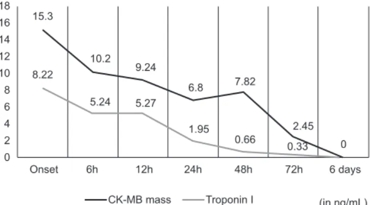
An echocardiogram was performed on day three at the ICU and showed considerable systolic dysfunction with an ejection fraction of 23% (Teichholz method, RV: 53-77%), systolic shortening of 10.6% (RV: 27-46%), end-systolic volume of 78 mL (RV: 25-66 mL), end-diastolic volume of 105 mL (RV: 51-154 mL), persistence of LV apical hypokinesis and mild diastolic dysfunction due to (type I) ventricular relaxation impairment, pulmonary arterial hypertension (estimated pulmonary artery systolic pressure - PASP=35 mmHg), and no relevant alterations in the heart valves. he levels of tissue necrosis markers decreased with the normalization of the CK-MB mass and troponin I concentration by the end of day three (Figure 2).
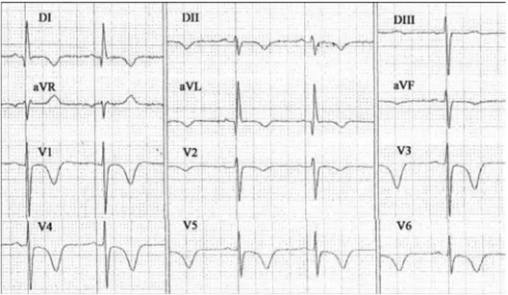
he patient was transferred to a general ward after a four-day stay in the ICU. he electrocardiogram on day four exhibited difuse T-wave inversion with asymmetry, which was only positive in leads DIII, aVR, and V2 (Figure 3).
An echocardiogram performed on day six showed substantial improvement of systolic function with an ejection fraction of 58% (Teichholz method) and normalization of the other parameters. hese data, along with the improvement of the clinical and laboratory parameters, allowed the patient to be discharged at the end of day six. However, the electrocardiogram performed at discharge still exhibited the same alterations of the ventricular repolarization that were observed on day four.
he patient was followed up as an outpatient using serial clinical and laboratory assessments. No signs of later efects on ventricular performance or relapses were observed.
Figure 1 - (A)Apical ballooning during ventricular systole shown using coronary angiography (dumbbell shape). (B)The Japanese octopus-trapping pot on which the disease name is based.
Figure 2 - Serial measurement of myocardial necrosis markers. Reference values: troponin I - <1.2 ng/mL; CK-MB mass - <6 ng/mL. The markers were normalized within 72 hours.
15.3
8.22
6.8
0.33 10.2
1.95
0 5.24
7.82 18
16 14 12 10 8 6 4 2 0
9.24
0.66
Troponin I (in ng/mL) 5.27
2.45
Onset 6h 12h 24h 48h 72h 6 days
CK-MB mass
LITERATURE REVIEW
Also known as stress-induced cardiomyopathy, LV transient apical ballooning syndrome, or broken heart syndrome, Takotsubo cardiomyopathy is a rare and recently described disease that is receiving increasing attention from the international medical community.(4)
hat syndrome was irst described in 1990 in Japan by Satoh et al.(1) In Japanese, “tako” means octopus, and
“tsubo” means earthenware pot; thus, the name of the
syndrome alludes to a ishing jar with a narrow neck and wide base used by ishermen to trap octopuses. he shape of the jar is similar to the shape of the heart when afected by Takotsubo syndrome in left ventriculography.
From an epidemiological perspective, takotsuba syndrome is most frequently observed in postmenopausal women (88.8% of cases). he patients usually present with the chest pain typical of acute coronary syndrome, which is preceded in most cases by an episode involving physical or emotional stress, such as a considerable argument, the death of a loved one, natural disasters, legal problems, accidents, surgical procedures, a stay in the ICU, and the use of illegal drugs.(5)
A European study estimated an overall prevalence of 1.2% of Takotsubo cardiomyopathy among the patients initially diagnosed with AMI, which increased to 4.9% among women.(6) Data from the United States indicate a prevalence 0.02% among all causes of hospital admission, and most of the incidence corresponds to older women who are smokers and alcohol users and exhibit a state of anxiety and hyperlipidemia.(7) In a prospective cohort study with 136 consecutive patients, a speciic trigger was identiied in 88% of the cases, the in-hospital death rate was 2%, and the recurrence rate
was 5%.(8) he overall mortality rate of the sample was higher than that of the control, particularly within the irst year of follow-up.(8)
Segmental contraction disorders without signiicant coronary injury have been previously described and are most frequently associated with myocarditis, coronary spasm, pheochromocytoma, and subarachnoid hemorrhage. he peculiar characteristics of Takotsubo syndrome include the following: transient dyskinesia of the LV anterior wall, increased ventricular base kinesis associated with chest pain, electrocardiographic alterations ranging from ST segment elevation to discrete ventricular repolarization disorders, and the absence of obstructive coronary disease.
he physiopathology of Takotsubo cardiomyopathy remains unknown. Neuroendocrine, hormonal, neuropsychological, and vascular causes have been proposed to explain the pathogenesis of the condition. he theory positing a catecholamine-mediated myocardial dysfunction is the most widely accepted.(6)
he following main diagnostic criteria for Takotsubo cardiomyopathy were established by the Mayo Clinic in 2008: (1) apical LV ballooning with basal compensatory hyperkinesia that does not follow the vascular territories on ventriculography or echocardiograms; (2) fresh ischemic alterations in the electrocardiogram; (3) lack of angiographic signs of signiicant obstruction of the epicardial coronary arteries or recent rupture of atherosclerotic plaque; and (4) no recent history of head injury or intracranial hemorrhage, pheochromocytoma, obstructive coronary disease, myocarditis, or hypertrophic cardiomyopathy.(4) Other data that contribute to the positive diagnosis include disproportionately low heart marker levels relative to the intensity of the ventricular dysfunction, quick improvement of the LV dysfunction, and a history of recent and intense physical or emotional stress.
he diferential diagnosis between acute coronary syndrome and Takotsubo cardiomyopathy is a priori based on coronary angiography and ventriculography. However, many authors are attempting to establish approaches to risk stratiication. Kosuge et al.(9) analyzed the electrocardiogram patterns upon admission and attempted to establish speciic T-wave patterns to distinguish between anterior wall AMI and Takotsubo syndrome. he results showed that the negative T waves exhibited greater maximum amplitudes and more difuse lead distribution. According to those authors, the presence of positive T waves on aVR and the lack of negative T waves on V1 exhibited 94.5% accuracy
in the diagnosis of Takotsubo cardiomyopathy.(9) hese indings might help identify the diferences in the electrophysiological mechanisms underlying the alterations and facilitate the diferential diagnosis of Takotsubo syndrome and AMI. However, the results still need to be conirmed.(7)
Noninvasive imaging assessment might provide important diagnostic information. For example, echocardiography is important in diagnosis because it identiies the medial-apical segmental alterations typical of Takotsubo cardiomyopathy and provides quantitative measures of the global LV systolic function.(3) As a function of its wide availability in the in-hospital setting, echocardiography is usually the irst test performed upon admission to the emergency unit. Because it is performed at the bedside, echocardiograms allow ventricular function to be monitored and the fast improvement of the abovementioned alterations to be identiied.
Cardiac magnetic resonance imaging might show increased signal intensity in the T2-weighted sequences on the apical ventricular wall, which is suggestive of edema, does not follow the vascular territories, and does not afect the basal areas. In addition, the gadolinium dynamic studies usually do not indicate late enhancement, which is compatible with myocardial necrosis, and might assist the diferentiation from the anterior wall AMI.(10)
he treatment of Takotsubo syndrome basically involves providing hemodynamic support and controlling the possible complications, including cardiogenic shock (6.5% of cases), intracavitary clot formation (3.8% of cases), heart failure (3.8% of cases), systemic embolism, and ventricular arrhythmias.(2)
he available data are not suicient to establish appropriate criteria for the indication of primary and secondary prophylaxis against thromboembolism in Takotsubo syndrome. According to an of-the-record recommendation based on observational studies, to avoid ventricular clot formation, the patients should be subjected to full anticoagulation over three months or until the recovery of the segmental function.(11) In the cases in which clot formation occurs, the patients should be subjected to anticoagulation over three to six months, independent of the status of the ventricular function.(11)
he prognosis of Takotsubo syndrome is good, and morphologic and functional recovery of the myocardium occurs within one month (18 days on average). he mortality rate varies from 0 to 8% with an average of 2%.
DISCUSSION
he patient’s complaints were initially attributed to high-risk acute coronary syndrome without ST segment elevation based on the clinical manifestations, present risk factors (age, hypertension, and diabetes), increased heart enzyme levels, and a lack of electrocardiographic alterations upon admission. Because of the initial hypothesis suggested by emergency care, early coronary angiography was performed, as recommended by the international guidelines. hus, the diagnosis of Takotsubo cardiomyopathy was incidental in our case report. he present case report clearly illustrates the ability of Takotsubo cardiomyopathy to mimic the signs and symptoms of myocardial injury and the importance of considering Takotsubo cardiomyopathy in the approach to patients with acute chest pain in the emergency setting.
he main aim of coronary angiography in this case is to rule out signiicant coronary injury and identify the signs typical of Takotsubo cardiomyopathy to establish a positive diagnosis. he following signs are signiicant: increased end-diastolic volume and severe difuse hypokinesia with ballooning of the medial-apical heart segment during ventricular systole, which causes the heart to be shaped like a trapped octopus in a ishing jar (Takotsubo in Japanese) as in the original description.
he management of patients with Takotsubo cardiomyopathy involves strictly conservative supportive treatment in the intensive care setting. he use of thrombolytic agents must be systematically avoided because it is not justiied by the etiopathogenesis of the disease. LV depression may be treated using diuretics, beta blockers, and angiotensin-converting enzyme (ACE) inhibitors. Beta blockers might also block the excessive release of catecholamines and the mechanism putatively underlying Takotsubo cardiomyopathy.(8)
In the present case report, although ventricular function was altered, and the echocardiogram indicated reduction of the ejection fraction, the patient did not progress into cardiogenic shock or exhibit relevant alterations of the heart valves in which inotropic support was not needed. Acetylsalicylic acid (AAS), which is an ACE inhibitor, and clopidogrel were prescribed on admission when the suspected diagnosis was AMI. The remainder of the treatment involved a short stay at the ICU.
the primary diagnostic criteria of takotsuba syndrome, and the ventricular functions fully recover approximately 18 days (varying from three to 50 days) after the onset of symptoms.(8) he patient did not exhibit any complications, the hospital stay lasted six days before the patient was discharged, and the clinical progression and echocardiographic indings indicated spontaneous resolution of the ventricular dysfunction.
CONCLUSION
Takotsubo cardiomyopathy is an important cause of chest pain that should be considered as a differential diagnosis because it might present clinically as acute coronary syndrome. Positive diagnosis of the syndrome might be easily established using characteristic imaging findings when the appropriated diagnostic tools are available. Therefore, the coronary angiography once more establishes itself as of major significance in this scenario.
he patient described in this report is consistent with the data described in the literature. he diagnosis of Takotsubo cardiomyopathy in patients presenting with clinical manifestations of acute coronary syndrome should be considered, particularly in postmenopausal women exposed to systemic stress conditions.
RESUMO
A síndrome de Takotsubo caracteriza-se por disfunção ventricular esquerda transitória, predominantemente me-dioapical, desencadeada caracteristicamente por estresse físico ou emocional. Relata-se aqui o caso de uma paciente de 61 anos de idade, admitida com tontura, sudorese pro-fusa e mal-estar súbito, após intenso estresse emocional. Exame físico e eletrocardiograma inicial foram normais, porém havia elevação de troponina I e CKMB massa. Suspeitou-se de infarto agudo do miocárdio sem supra-desnivelamento do segmento ST, indicando cineangioco-ronariografia de urgência. Foram evidenciados ventrícu-lo esquerdo com hipocinesia difusa grave, baventrícu-lonamento sistólico medioapical e coronárias sem lesões significati-vas. A paciente foi encaminhada aos cuidados intensivos, evoluindo satisfatoriamente com terapia de suporte. Con-forme visto, a cardiomiopatia de Takotsubo pode simular infarto agudo do miocárdio, sendo a cineangiocoronario-grafia importante para distinção na fase aguda. Neste caso, a paciente evoluiu com resolução espontânea da disfunção ventricular, sem sequelas.
Descritores: Disfunção ventricular esquerda; Terapia intensiva; Cardiomiopatia de Takotsubo; Hemodinâmica; Choque cardiogênico; Estresse psicológico; Relatos de casos
REFERENCES
1. Satoh H, Tateishi H, Uchida T, et al. Takotsubo-type cardiomyopathy due to multivessel spasm. In: Kodama K, Haze K, Hon M, editors. Clinical aspect of myocardial injury: from ischemia to heart failure. Tokyo: Kagakuhyouronsya; 1990. p. 56-64.
2. Vasconcelos JT, Martins S, Sousa JF, Portela A. Cardiomiopatia de Takotsubo: uma causa rara de choque cardiogênico simulando infarto agudo do miocárdio. Arq Bras Cardiol. 2005;85(2):128-30.
3. Lemos AE, Araújo AL, Lemos MT, Belém LS, Vasconcelos-Filho FJ, Barros RB. Síndrome do coração partido (síndrome de Takotsubo). Arq Bras Cardiol. 2008;90(1):e1-e3.
4. Golabchi A, Sarrafzadegan N. Takotsubo cardiomyopathy or broken heart syndrome: a review article. J Res Med Sci. 2011;16(3):340-5.
5. Kaballo MA, Yousif A, Abdelrazig AM, Ibrahim AA, Hennessy TG. Takotsubo cardiomyopathy after a dancing session: a case report. J Med Case Rep. 2011;5:533.
6. Previtali M, Repetto A, Panigada S, Camporotondo R, Tavazzi L. Left ventricular
apical ballooning syndrome: prevalence, clinical characteristics and pathogenetic mechanisms in a European population. Int J Cardiol. 2009;134(1):91-6. 7. Deshmukh A, Kumar G, Pant S, Rihal C, Murugiah K, Mehta JL. Prevalence
of Takotsubo cardiomyopathy in the United States. Am Heart J. 2012;164(1):66-71.e1.
8. Sharkey SW, Windenburg DC, Lesser JR, Maron MS, Hauser RG, Lesser JN, et al. Natural history and expansive clinical profile of stress (tako-tsubo) cardiomyopathy. J Am Coll Cardiol. 2010;55(4):333-41.
9. Kosuge M, Ebina T, Hibi K, Iwahashi N, Tsukahara K, Endo M, et al. Differences in negative T waves between Takotsubo cardiomyopathy and reperfused anterior acute myocardial infarction. Circ J. 2012;76(2):462-8.
10. Fernández-Pérez GC, Aguilar-Arjona JA, de la Fuente GT, Samartín M, Ghioldi A, Arias JC, et al. Takotsubo cardiomyopathy: assessment with cardiac MRI. AJR Am J Roentgenol. 2010;195(2):W139-45.