Hemodynamic and perfusion variables during
experimental septic shock treated with goal-directed
luid resuscitation
Evolução de variáveis hemodinâmicas e perfusionais durante o
choque séptico experimental tratado com ressuscitação volêmica
guiada por metas
INTRODUCTION
Hemodynamic optimization in a critical patient after systemic insults, such as surgery, severe trauma or sepsis, has frequently been discussed in the medical literature. It has been suggested that appropriate hemodynamic resuscitation, using clear parameters such as lactate, central/mixed venous oxygen saturation or base deicit, is the cornerstone of improved tissue perfusion and oxygenation, with consequential beneits with respect to clinical outcome.(1)
For instance, hemodynamic resuscitation in sepsis has been repeatedly
Marcelo Park1, André Loureiro
Rosário1, Guilherme de Paula Pinto
Schettino1, Luciano César Pontes
Azevedo1
1. Instituto Sírio-Libanês de Ensino e Pesquisa - São Paulo (SP), Brazil.
ABSTRACT
Objectives: Although luid
resuscitation guided by central venous
oxygen saturation (SvcO2) is currently
considered the gold standard in sepsis therapy, few studies have described hemodynamic and perfusion parameters during this procedure. his study aims to describe these parameters during septic shock without resuscitation and after 12 hours of goal-directed resuscitation.
Methods: hirteen anesthetized
pigs (35-45 kg) had peritonitis caused by fecal inoculation (0.75 g/kg). After developing persistent hypotension, both groups were given antibiotics and randomized either to the control group (n=7) or the experimental group (n=6). In the control group, hemodynamic control was optimized to maintain a central venous pressure of 8-12 mmHg, a urinary output above 0.5 mL/kg/hour and a mean arterial blood pressure above 65 mmHg. he experimental group received the above target therapy in
addition to maintaining a SvO2 above
65%. he interventions included lactated
Ringer’s solution and norepinephrine for both groups and dobutamine in the
SvO2 group. he animals were treated
for 12 hours or until death.
Results: Untreated sepsis was associated
with signiicant reductions in SvO2, PvO2,
cardiac output and central venous pressure in addition to increased arteriovenous
oxygen saturation and veno-arterial CO2
diferences. Following resuscitation, these parameters were corrected in both groups. Goal-directed resuscitation was associated with a better hemodynamic proile,
characterized by higher SvO2, cardiac
output and central venous pressure. Conclusions: Non-resuscitated sepsis showed a hemodynamic proile suggesting hypovolemia, with worsened perfusion and hemodynamics, which is reversed upon luid resuscitation. Goal-directed resuscitation is associated with signiicantly improved hemodynamic and perfusion parameters.
Keywords: Shock, septic/blood; Oxygen/ blood; Oximetry/methods; Lactates/blood; Resuscitation/methods; Hemodynamics/ physiology; Pigs
Study conducted at the Intensive Care Medicine and Anesthesiology Research Laboratory, Instituto Sírio-Libanês de Ensino e Pesquisa, São Paulo (SP), Brazil.
Conlicts of interest: None.
Submitted on August 2, 2011 Accepted on September 6, 2011
Corresponding author:
Marcelo Park
Av. Enéas de Carvalho Aguiar, 255 - Sala 5023
Zip Code: 05403-000 – São Paulo (SP), Brazil.
evaluated during this last decade. Essentially, this type of resuscitation consists of a multi-step protocol, including luid replacement, use of vasoactive agents, blood transfusion and administration of inotropes, that target pre-determined hemodynamic and metabolic parameters.(2,3) he most used parameter to guide reanimation is central venous oxygen saturation (SvcO2). It has been shown that maintaining a value of SvcO2 above 70% during the irst 6 hours of therapy (the so-called “golden hours”) is associated with survival beneits.(2) More recently, therapy guidelines have suggested that mixed venous oxygen saturation (SvO2) values above 65% may be a surrogate for SvcO2 during the “golden hours”.(2,3) After these studies and therapy guidelines were published, goal-directed luid resuscitation, including the use of SvcO2 values, started to be used in sepsis therapy in worldwide clinical practice. Although widely used for sepsis resuscitation, few studies have assessed the course of hemodynamic tissue perfusion markers used in this strategy. herefore, this study’s aim was to describe the time course of the main perfusion and hemodynamic parameters during a clinically relevant experimental model of non-resuscitated sepsis.
METHODS
his study was conducted at the Intensive Care Medicine and Anesthesiology Research Laboratory of the Instituto Sírio-Libanês de Ensino e Pesquisa and appropriately approved by the institution’s ethics committee. All experimental procedures were conducted in compliance with the Brazilian Instituto Nacional de Saúde’s guidelines for the use and care of experimental animals. his study was based on a previously described experimental fecal peritonitis model.(4) Part of the results, which used a diferent sample size and diferent parameters than those reported in this article, was accepted for publication in Shock.(5)
Preparation of the animals
hirteen male Agroceres pigs with an average body weight of 40 kg were fasted for 18 hours and had free access to water. he animals were pre-anesthetized with intramuscular midazolam and acepromazine. Following premedication, the pigs were anesthetized with thiopental and an orotracheal tube was inserted. After successfully intubating the animals, an infusion of thiopental (10 mg/kg/hour) and Fentanyl (10 mcg/ kg/hour) was maintained, and pancuronium (0.15 mg/ kg starting dose followed by an infusion of 0.25 mg/kg/
hour) for neuromuscular blockade during the surgical procedure was used. After stabilization, the animals were maintained with thiopental and pancuronium in the dosages described above. he animals were then connected to a mechanical ventilator (Evita XL, Dräger Medical, Lübeck, Germany) with a ixed positive end-expiratory pressure (PEEP) of 5 cm H2O and a FiO2 of 30%. A tidal volume of approximately 8 mL/kg and a respiratory rate adjusted for PaCO2 between 35 and 45 mmHg was maintained. In case of signiicantly reduced arterial oxygen saturation and/or PaO2 during the study, FiO2 was increased up to the minimum necessary to maintain arterial saturation above 92%. Multi-parameter monitoring was provided for the following variables: heart rate, arterial saturation and central venous and pulmonary artery pressure curves after a pulmonary artery catheter was inserted (DX2020, Dixtal Biomédica, Manaus, Brazil).
Next, vascular accesses were dissected. A pulmonary artery catheter was inserted via the right external jugular for continuous measurement of pressure curves, cardiac output and venous oxygen saturation, as well as right-ventricular end-diastolic pressure and ejection fraction (Vigilance VD®, Edwards Lifesciences LLC, Irvine, California, United States of America). A catheter was inserted via the right femoral artery for invasive blood pressure monitoring and arterial blood sample collection. he right femoral vein was catheterized for intravenous luid administration, drug infusion and venous blood collection. A catheter was inserted via cystostomy for urinary output assessment.
Immediately following installation of the monitoring devices, the animals underwent a median laparotomy. he incision was approximately 4 cm. he descending colon was identiied and a 2 cm incision was made. At this time, 0.75 g/kg of feces was removed and the intestine was sutured. Two large gauge catheters were installed into the abdominal cavity bilaterally to the parietocolic gutters, and the laparotomy was sutured. After 60 minutes of stabilization, an amount equivalent to 0.75 g/kg of feces was diluted into 200 mL of 37ºC saline solution and introduced into the abdominal cavity via the catheters. After luid resuscitation was started, all study animals were given intravenous amikacin (250 mg every 12 hours) and metronidazole (500 mg every 8 hours) for the duration of the experiment.
pulmonary capillary pressure, pulmonary artery pressure (PAP), mean blood pressure (MBP), SvO2, central temperature and urinary output. Arterial and venous blood gas, arterial lactate and hemoglobin/ hematocrit concentrations were measured with a blood gas analyzer (ABL 700 Radiometer, Copenhagen, Denmark) before sepsis induction and then every 3 hours until the end of the experiment or death.
Fluid resuscitation
For the study duration, the animals received a maintenance infusion every 24 hours of 100 mL of 10% glucose solution containing 25 mEq of potassium chloride and 75 mEq of sodium chloride, to maintain blood glucose, potassium and sodium levels as close to normal as possible.
During the surgery and the stabilization period, the animals were given a lactated Ringer solution. he protocol was started with a CVP above 8 mmHg and venous oxygen saturation above 70%. After peritonitis induction, the animals were given no volume replacement solution. Upon mean arterial blood pressure (MBP) reaching less than 65 mmHg for 30 minutes, the animals were divided into two groups:
a) Control group: In this group, luid replacement was started with a 500 mL lactated Ringer bolus every 30 minutes, targeted to reach and maintain the following hemodynamic parameters: MBP above 65 mmHg, CVP between 8 and 12 mmHg and urinary output above 0.5 mL/kg/hour. Norepinephrine was added after 30 minutes of hypotension if volume replacement was deemed unsatisfactory. Initially, the infusion was started at 0.05 mcg/kg/minute, and then it was increased by 0.025 mcg/kg/minute increments every 15 minutes, as necessary, to maintain MBP above 65 mmHg.
b) SvO2-guided resuscitation group: In this group, luid resuscitation was based on the mixed venous oxygen saturation values obtained from the pulmonary artery catheter with continued venous saturation, in addition to the hemodynamic goals mentioned above. herefore, appropriate resuscitation criteria were: MBP above 65 mmHg, urinary output above 0.5 mL/kg/hour, CVP 8-12 mmHg and SvO2 above 65%. hese hemodynamic targets were maintained for 12 hours or until death. Initially the animals were given 500 mL lactated Ringer solution every 30 minutes until reaching a CVP above 8 mmHg. In those animals still maintaining an SvO2 below 65%, dobutamine was started by an infusion of 2.5 mcg/ kg/minute with 2.5 mcg/kg/minute increments every
30 minutes for a maximal dose of 20 mcg/kg/minute. If CVP dropped during the dobutamine infusion, additional crystalloid solution boluses were given to reach the previously described CVP levels. Dobutamine infusion increments were limited by tachycardia above 180 bpm. Norepinephrine was used as necessary, as described above.
he animals were maintained with these parameters until death or for 12 hours (counted from the start of resuscitation), and then were euthanized with an overdose of potassium chloride after deepening anesthesia.
Statistical analysis
he data were tested for normality using the Shapiro-Wilk test and are presented as the mean ± standard deviation. For assessment of time course until shock, both groups were considered together and the statistical signiicance was assessed with repeated-measures analysis of variance (RM-ANOVA). After the start of shock and the randomization process, comparisons were made using a two-way analysis of variance (two-way
ANOVA). Post hoc ANOVA analyses were performed
using the Tukey Honest Signiicant Diferences (HSD) test. Correlation analyses were performed using the Pearson’s test. he R open source statistical package (Vienna, Austria, 2009) was used for statistical analysis and graphics plotting.
RESULTS
hirteen animals were studied, 6 in the SvO2
group and 7 in the control group. he median time to hypotension after sepsis induction was 11 hours (range: 7-21 hours).
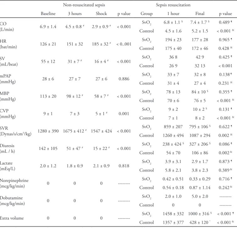
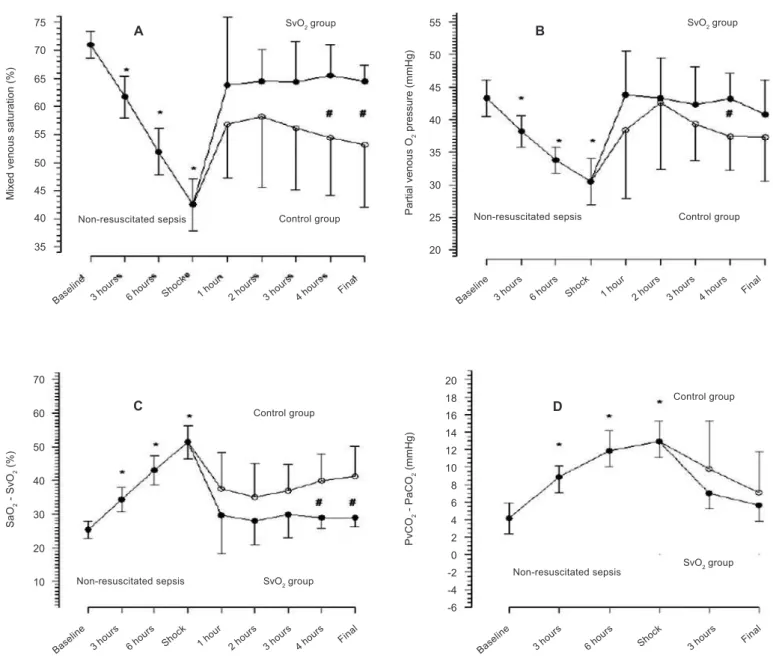
Figure 1 (A to D) shows hemodynamic and perfusion variable progression during non-resuscitated and treated sepsis. Peritonitis was associated with a signiicant worsening of perfusion and hemodynamic parameters during the observation time, including reduced SvO2 and PvO2 and increased arteriovenous oxygen saturation and venoarterial CO2 gradients. Table 1 identiies some of the study-related hemodynamic and perfusion variables and the efects of luid resuscitation. Non-resuscitated sepsis is characterized by signiicantly reduced cardiac output and systolic index, CVP and MBP. Following resuscitation, these parameters were
signiicantly improved; however, the SvO2 guided
Table 1 – Hemodynamic characteristics during the study
Non-resuscitated sepsis Sepsis resuscitation
Baseline 3 hours Shock p value Group 1 hour Final p value
CO
(L/min) 6.9 ± 1.4 4.5 ± 0.8 # 2.9 ± 0.9 # < 0.001
SvO2 6.8 ± 1.1 § 7.4 ± 1.7 § 0.489 ¶
Control 4.5 ± 1.6 5.2 ± 1.5 < 0.001 &
HR
(bat/min) 126 ± 21 151 ± 32 185 ± 32
# < 0..001 SvO2 194 ± 23 177 ± 28 0.965
¶
Control 175 ± 40 172 ± 46 0.428 &
SV
(mL/beat) 55 ± 12 31 ± 7
# 16 ± 4 # < 0.001 SvO2 36 8 42 9 0.425
¶
Control 26 9 32 13 < 0.001
mPAP
(mmHg) 28 ± 6 27 ± 7 27 ± 6 0.886
SvO2 33 ± 7 32 ± 8 0.138 ¶
Control 31 ± 4 27 ± 4 0.231 &
MBP
(mmHg) 113 ± 20 98 ± 12 # 58 ± 7 # < 0.001
SvO2 78 ± 13 84 ± 10 § 0.355 ¶
Control 70 ± 6 76 ± 5 < 0.001 &
CVP
(mmHg) 9 ± 1 7 ± 3 5 ± 1
# 0.001 SvO2 9 ± 2 10 ± 2
§ 0.131 ¶
Control 7 ± 1 8 ± 2 < 0.001 &
SVR
(Dynas/s/cm5/kg) 1280 ± 390 1675 ± 412 # 1547 ± 424 < 0.001
SvO2 859 ± 207 795 ± 106 § 0.622 ¶
Control 1260 ± 494 1087 ± 294 0.002 &
Diuresis
(mL / h) 142 ± 105 51 ± 47 # 15 ± 22 # < 0.001
SvO2 238 ± 424 § 327 ± 206 § 0.086 ¶
Control 54 ± 70 106 ± 86 0.002 &
Lactate
(mEq/L) 2.0 ± 1.2 1.8 ± 0.9 2.1 ± 0.9 0.818
SvO2 3.9 ± 3.1 2.9 ± 1.7 0.873 ¶
Control 5.8 ± 2.1 3.8 ± 2.3 0.389 &
Norepinephrine
(mcg/kg/min) 0 0 0
---SvO2 0.42 ± 0.51 0.33 ± 0.29 0.716 ¶
Control 0.54 ± 0.18 0.87 ± 1.14 0.242 &
Dobutamine
(mcg/kg/min) 0 0 0
---SvO2 2.0 ± 1.0 5.0 ± 2.0
---Control 0 0
---Extra volume 0 0 0 --- SvO2 1458 ± 332 1000 ± 316
§ < 0.001 ¶
Control 1357 ± 377 428 ± 120 ® < 0.001 &
CO – cardiac output; HR – heart rate; SV – stroke volume; MBP – mean systemic blood pressure; CVP – central venous pressure; mPAP – mean pulmonary artery pressure; SVR – systemic vascular resistance. Intra and inter-group two-way ANOVA. No factor-time interaction. #
Post-hoc Tukey HSD analysis: p < 0.05 vs. baseline. § Post-hoc Tukey HSD analysis: p < 0.05 vs. control group. ® Post-hoc Tukey HSD analysis: p < 0.05 vs. 1 hour.
CVP, MBP, SvO2, PvO2 and arteriovenous oxygen saturation gradient.
Figure 2 shows the correlation between several perfusion variables and cardiac output. Of the analyzed parameters, venoarterial CO2 gradient showed the
Figure 1 - Progression of the assessed variables during non-resuscitated sepsis and after resuscitation for SvO2 and
control groups. A) SvO2 progression (one-way RM-ANOVA for non-resuscitated sepsis data p < 0.001; two-way
ANOVA for SvO2 and control comparison, intra-group comparison p = 0.973, inter-group comparison p < 0.001 and
factor-time interaction p = 0.951). B) Partial oxygen venous pressure (one-way RM-ANOVA for non-resuscitated sepsis data p < 0.001; two-way ANOVA for SvO2 and control comparison, intra-group p = 0.726, inter-group p = 0.048 and factor-time interaction p = 0.914). C) The difference between arterial and mixed venous oxygen saturation (one-way RM-ANOVA for non-resuscitated sepsis data p < 0.001 and factor-time interaction non-resuscitated p = 0.907) and
D) The venoarterial CO2 gradient (one-way RM-ANOVA for sepsis p = 0.452; inter-group p = 0.465 and factor-time
interaction p = 0.716). *Post-hoc analysis, Tukey HSD p < 0.05 vs. baseline. # Post-hoc analysis, Tukey HSD p < 0.05 between groups.
Base line
3 ho urs
6 ho urs
Shock 1 ho ur
2 ho urs
3 h ours
4 ho urs Final
Base line
3 ho urs
6 ho urs
Shock 1 ho ur
2 ho urs
3 h ours
4 ho urs Final
Base line
3 ho urs
6 ho urs
Shock 1 ho ur
2 ho urs
3 h ours
4 ho urs Final
Base line
SvO2 group SvO2 group
SvO2 group SvO2 group
75 70 65 60 55 50 45 40 35 55 50 45 40 35 30 25 20 20 18 16 14 12 10 8 6 4 2 0 -2 -4 -6 70 60 50 40 30 20 10 A B D C
Control group Control group
Control group Control group
Non-resuscitated sepsis Non-resuscitated sepsis
Non-resuscitated sepsis Non-resuscitated sepsis Mi xe d ve n o u s sa tu ra ti o n (% ) Pa rt ia l ve n o u s O2 p re ssu re (mmH g ) PvC O2 - Pa C O2 (mmH g ) Sa O2 - SvO 2 (% )
3 ho urs
6 ho urs
Shock 3 hou rs
DISCUSSION
he time course of hemodynamic and perfusion parameters during non-resuscitated sepsis has been relatively poorly documented in the literature. In this study, we have shown that a non-resuscitated sepsis hemodynamic proile is primarily characterized by hypovolemia, possibly associated with myocardial
dysfunction. his proile has a signiicant impact on the perfusion parameters that are commonly measured in the ICU. Fluid, antibiotics and vasoactive drug therapy signiicantly improve these parameters.
In this study, the hemodynamic and inlammatory changes induced by sepsis were signiicant. Early non-resuscitated sepsis proiles in humans have been characterized by hypovolemia secondary to reduced Figure 2 – Correlation between perfusion parameters and cardiac output. A) Correlation between venoarterial CO2 gradient and cardiac output. B) Correlation between arterial lactate and cardiac output. C) Correlation between SvO2 and cardiac output. D) Correlation between PvO2 and cardiac output.
Cardiac output (L/min) R = 0.31
p < 0.001
R = 0.02 p < 0.364
R = 0.18 p < 0.001 R = 0.18
p < 0.001
C A
D B
2 4 6 8 10
2 4 6 8 10
2 4 6 8 10
2 4 6 8 10
Cardiac output (L/min)
Cardiac output (L/min)
Cardiac output (L/min)
SvO
2
(%
)
L
a
ct
a
te
(mmo
l/
L
)
PvO
2
(mmH
g
)
V
e
n
o
a
rt
e
ri
a
l
C
O2
g
ra
d
ie
n
t
(mmH
g
)
2
4
6
8
1
0
1
2
1
4
1
6
1
8
2
4
6
8
3
0
4
0
5
0
6
0
7
0
8
0
2
0
3
0
4
0
5
0
6
oral ingestion and increased insensitive losses due to vomiting, diarrhea or diaphoresis. In these patients, venodilation and leakage of luid into the interstitial space due to increased endothelial capillary permeability may result in reduced cardiac preload, reduced cardiac output and inappropriate systemic oxygen supply. After luid replacement, cardiac output is increased and systemic vascular resistance is reduced, characterizing the hyperdynamic pattern commonly associated with resuscitated septic shock.(6,7) Clinical indings on hypovolemia were reproduced in this study, as shown by signiicant hypotension, tachycardia and cardiac performance indicators after sepsis (Figure 1 and Table 1). After resuscitation, the hemodynamic and perfusion data were similar to those commonly described in the hyperdynamic state proile.
Global tissue hypoxia has been described as one of the main components of early sepsis hemodynamics. It develops when systemic oxygen supply is insufficient to meet tissue requirements. Therefore, low SvO2 (< 65%) or ScvO2 (< 70%) and high lactate concentration suggests global tissue hypoxia, as a higher supplied-oxygen fraction is being extracted by tissues, resulting in less venous oxygen measurements. This feature, reproduced in this study, is considered to be an early phase of the disease, or hyperdynamic sepsis.(8-11) Similarly, the correlation between high SvO2 and cardiac output has previously been established.(12) As anticipated, in our study, goal-directed resuscitation was associated with better SvO2 values but required more volume replacement and use of inotropes.
The venoarterial CO2 gradient has been described as a potential hemodynamic parameter for sepsis resuscitation. Although commonly mentioned as a tissue hypoxia marker, recent studies have suggested that it is more closely associated with flow changes, such as cardiac output.(13-17) This gradient increase in the context of low cardiac output can be explained using the CO2 stagnation principle. This principle states that, due to the delayed transit time, any CO2 increase beyond the normal in efferent vessels may lead to venous blood hypercapnia.(14) In our study, a CO2 gradient increase was clearly shown during non-resuscitated sepsis, as was its drop following therapy. This can be explained by the low cardiac output and the signs of hypovolemia that developed in the animals during the untreated phase. For this variable, no significant differences were seen by the type of fluid resuscitation.
his study has some strengths that should be emphasized. First, our model shows several similarities with human sepsis, including the hemodynamic proile before therapy, the administration of antibiotics, the use of perfusion parameters-guided vasoactive drugs (which is unusual in other sepsis experimental trials) and a hemodynamic therapy algorithm similar to that used in clinical practice. However, this study has limitations. Type II analysis errors may have occurred due to the relatively small sample and disease heterogeneity. hese factors may have inluenced the results.
CONCLUSION
his study shows that non-resuscitated sepsis has a hemodynamic proile suggesting hypovolemia, with low cardiac output, low CVP, reduced SvO2 and PvO2 and increased CO2 venoarterial diference. he treatment of sepsis with antibiotics, luids, inotropes and vasopressors signiicantly improves these parameters. Early goal-directed luid resuscitation is associated with improved hemodynamic parameters when compared to a group not resuscitated using this method.
Acknowledgements: his study was supported by grants from Fundação de Amparo à Pesquisa do Estado de São Paulo (FAPESP) and Instituto Sírio-Libanês de Ensino e Pesquisa.
RESUMO
Objetivos: Apesar da ressuscitação volêmica guiada por
sa-turação venosa central de oxigênio (SvcO2) ser considerada
atu-almente padrão ouro no tratamento da sepse, poucos estudos caracterizaram o peril evolutivo de variáveis hemodinâmicas e perfusionais durante esta abordagem terapêutica. Este estudo teve por objetivo descrever evolutivamente estes parâmetros du-rante o choque séptico experimental sem ressuscitação e após 12 horas de ressuscitação guiada por metas.
Métodos: Treze porcos (35-45 kg) anestesiados foram sub-metidos a peritonite por inoculação fecal (0,75g/kg). Após de-senvolverem hipotensão persistente, ambos os grupos receberam antibióticos e foram randomizados em dois grupos: controle (n=7), com suporte hemodinâmico otimizado para pressão ve-nosa central entre 8-12mmHg, diurese acima de 0,5ml/kg/h e
pressão arterial média maior que 65mmHg; e SvO2 (n=6), com
os objetivos acima e SvO2 acima de 65%. As intervenções
inclu-íram ringer lactato e noradrenalina nos 2 grupos e dobutamina
no grupo SvO2. Os animais foram tratados durante doze horas
Resultados: A sepse não tratada associou-se a uma signiicante
redução da SvO2, PvO2, débito cardíaco e pressão venosa central
e aumento da diferença arterio-venosa da saturação de oxigênio e
veno-arterial de CO2. Após ressuscitação, esses parâmetros foram
corrigidos em ambos os grupos. A ressuscitação guiada por metas associou-se a um melhor peril hemodinâmico caracterizado por
maiores SvO2, débito cardíaco e pressão venosa central.
Conclusões: A sepse não ressuscitada apresenta um peril
hemodinâmico sugestivo de hipovolemia, com piora perfusional e hemodinâmica revertida após ressuscitação volêmica. A ressus-citação guiada por metas associa-se a uma signiicante melhora dos parâmetros hemodinâmicos e perfusionais.
Descritores: Choque séptico; Oxigênio/sangue; Oximetria/ métodos; Lactatos/sangue; Ressuscitação/métodos; Hemodinâmica/ isiologia; Porcos
REFERENCES
1. da Silva Ramos FJ, Azevedo LC. Hemodynamic and perfusion endpoints for volemic resuscitation in sepsis. Shock. 2010;34 Suppl 1:34-9. Review.
2. Rivers E, Nguyen B, Havstad S, Ressler J, Muzzin A, Knoblich B, Peterson E, Tomlanovich M; Early Goal-Directed herapy Collaborative Group. Early goal-directed therapy in the treatment of severe sepsis and septic shock. N Engl J Med. 2001;345(19):1368-77.
3. Dellinger RP, Levy MM, Carlet JM, Bion J, Parker MM, Jaeschke R, Reinhart K, Angus DC, Brun-Buisson C, Beale R, Calandra T, Dhainaut JF, Gerlach H, Harvey M, Marini JJ, Marshall J, Ranieri M, Ramsay G, Sevransky J, hompson BT, Townsend S, Vender JS, Zimmerman JL, Vincent JL; International Surviving Sepsis Campaign Guidelines Committee; American Association of Critical-Care Nurses; American College of Chest Physicians; American College of Emergency Physicians; Canadian Critical Care Society; European Society of Clinical Microbiology and Infectious Diseases; European Society of Intensive Care Medicine; European Respiratory Society; International Sepsis Forum; Japanese Association for Acute Medicine; Japanese Society of Intensive Care Medicine; Society of Critical Care Medicine; Society of Hospital Medicine; Surgical Infection Society; World Federation of Societies of Intensive and Critical Care Medicine. Surviving Sepsis Campaign: international guidelines for management of severe sepsis and septic shock: 2008.Crit Care Med. 2008;36(1):296-327. Erratum in Crit Care Med. 2008;36(4):1394-6.
4. de Azevedo LC, Park M, Noritomi DT, Maciel AT, Brunialti MK, Salomão R. Characterization of an animal model of severe sepsis associated with respiratory dysfunction. Clinics (Sao Paulo). 2007;62(4):491-8. 5. Rosario AL, Park M, Brunialti M, Mendes M, Raposo M,
Fernandes D, Salomão R, Laurindo FR, Schettino GP,
Azevedo LC. SvO2-guided resuscitation for experimental
septic shock: efects of luid infusion and dobutamine on hemodynamics, inlammatory response and cardiovascular oxidative stress. Shock, In press 2011.
6. Rivers EP, Coba V, Visbal A, Whitmill M, Amponsah
D. Management of sepsis: early resuscitation. Clin Chest Med. 2008;29(4):689-704, ix-x.
7. Rady MY, Rivers EP, Nowak RM. Resuscitation of the critically ill in the ED: responses of blood pressure, heart rate, shock index, central venous oxygen saturation, and lactate. Am J Emerg Med. 1996;14(2):218-25.
8. Astiz ME, Rackow EC, Kaufman B, Falk JL, Weil MH. Relationship of oxygen delivery and mixed venous oxygenation to lactic acidosis in patients with sepsis and acute myocardial infarction. Crit Care Med. 1988;16(7):655-8. 9. Rivers EP, McIntyre L, Morro DC, Rivers KK. Early and
innovative interventions for severe sepsis and septic shock: taking advantage of a window of opportunity. CMAJ. 2005;173(9):1054-65.
10. Bilkovski RN, Rivers EP, Horst HM. Targeted resuscitation strategies after injury. Curr Opin Crit Care. 2004;10(6):529-38. Review.
11. Otero RM, Nguyen HB, Huang DT, Gaieski DF, Goyal M, Gunnerson KJ, et al. Early goal-directed therapy in severe sepsis and septic shock revisited: concepts, controversies, and contemporary indings. Chest. 2006;130(5):1579-95. 12. Perner A, Haase N, Wiis J, White JO, Delaney A. Central
venous oxygen saturation for the diagnosis of low cardiac output in septic shock patients. Acta Anaesthesiol Scand. 2010;54(1):98-102.
13. Mecher CE, Rackow EC, Astiz ME, Weil MH. Venous hypercarbia associated with severe sepsis and systemic hypoperfusion. Crit Care Med. 1990;18(6):585-9.
14. Lamia B, Monnet X, Teboul JL. Meaning of arterio-venous
PCO2 diference in circulatory shock. Minerva Anestesiol.
2006;72(6):597-604.
15. Ho KM, Harding R, Chamberlain J. A comparison of central venous-arterial and mixed venous-arterial carbon dioxide tension gradient in circulatory failure. Anaesth Intensive Care. 2007;35(5):695-701.
16. Vallet B, Teboul JL, Cain S, Curtis S. Venoarterial CO(2) diference during regional ischemic or hypoxic hypoxia. J Appl Physiol. 2000;89(4):1317-21.