he impact of positive end-expiratory pressure on
cerebral perfusion pressure in adult patients with
hemorrhagic stroke
Repercussão da pressão positiva expiratória inal na pressão de
perfusão cerebral em pacientes adultos com acidente vascular
hemorrágico
INTRODUCTION
he skull is a rigid, constant volume sphere containing cerebral tissues (1,400 mL), cerebrospinal luid (150 mL) and blood (75 mL).(1-3) Preserved
intracranial volume maintains normal intracranial pressure (ICP) values.(4,5)
According to the Monro Kellie theory, volume changes in one compartment must be compensated by volume changes in a diferent compartment in or-der to maintain a constant intracranial volume and pressure.(1,6,7) he body
is able to make physiological changes to maintain a normal ICP. However, when these coping mechanisms are exhausted, ICP increases.(4,6,8)
Intracranial hypertension (ICH) is common in intensive care unit (ICU) patients admitted for diferent reasons. hese may include central nervous system or systemic conditions from traumatic, infective or metabolic cau-ses.(7,9,10) ICH is currently deined as pressure values persistently above 20
mmHg.(1,11) Under normal conditions, the vascular cerebral resistance
regu-Wildberg Alencar Lima1, Antônio
Roberto Leite Campelo2, Rodrigo
Luís Mousinho Gomes1, Daniella
Cunha Brandão1
1. Department of Physiotherapy and Intensive Care, Real Hospital Português de Beneicência em Pernambuco – Recife (PE), Brazil.
2. Department of Pulmonology, Real Hospital Português de Beneicência em Pernambuco – Recife (PE), Brazil.
ABSTRACT
Positive intrathoracic pressure may cause hemodynamic changes, whi-ch can be transmitted to the cranial compartment, changing intracranial pressure and cerebral perfusion pres-sure. his can be increased when high positive end-expiratory pressure values are used.
Objective: To measure the impact of diferent positive end-expiratory pressure levels on intracranial pressure, cerebral perfusion pressure and mean blood pressure.
Method: his study was conducted in a neurological intensive care unit and included 25 adult hemorrhagic stroke patients who were mechanically ventilated on airway pressure control mode. Patients were subjected to va-rious positive end-expiratory values ranging between 0 and 14 cmH2O.
he order of these values were rando-mized, and the variables were assessed ive minutes after each new positive end-expiratory pressure level was ini-tiated.
Results: Incremental positive end--expiratory pressures led to increased intracranial pressure (p < 0.001), ho-wever, no statistically signiicant chan-ges were observed in mean blood pres-sure or cerebral perfusion prespres-sure.
Conclusion: In this population of patients with hemorrhagic stroke, po-sitive end-expiratory pressure values up to 14 cmH2O did not alter cerebral perfusion pressure or mean blood pres-sure. Increased intracranial pressures were noted, although these elevations were not clinically signiicant.
Keywords: Intracranial pressure; Positive-pressure respiration; Stroke
Study conducted at the Neurological Intensive Care Unit, Real Hospital Português de Beneicência em Pernambuco – Recife (PE), Brazil.
Conlicts of interest: None.
Submitted on January 11, 2011 Accepted on August 15, 2011
Corresponding author: Wildberg Alencar Lima
Rua Emiliano Braga, 1019 – Apto. 108 - Ipê Rosa – Iputinga
Zip Code: 50670-380 - Recife (PE), Brazil.
Phone: +55 81 3454-5881 / + 55 81 9959-3737
lation mechanisms are able to maintain constant blood low under a wide range of cerebral perfusion pressures (CPP).(1,4,8) Failure of the regulating mechanisms will
lead to reduced tissue perfusion and worsened ischemic cell injury.(1,4,8,12)
Monitoring of ICH patients should include conti-nuous vital sign assessment and maintenance, with spe-cial attention to hypotension, hypoxia, brain edema, hemorrhage, herniation and hemodynamic changes. he efectiveness of the therapy should also be continu-ally reassessed.(1,9)
Acute brain injury patients have a high likelihood of pulmonary involvement, leading to reduced arte-rial oxygen pressure (PaO2) and carbonic gas (CO2) retention, requiring mechanical ventilation assistance (MVA).(1,13-17) Approximately 20% of intracranial
he-morrhage patients will eventually develop a respiratory infection, pulmonary edema and acute lung injury re-quiring more aggressive MVA aimed at reverting hypo-xemia.(14) hese developments are especially common
in patients receiving positive end-expiratory pressure (PEEP).(18-24) MVA may impact hemodynamics,(25)
lea-ding to increased ICP and reduced CPP. his is because the compression of the alveolar capillary vessels by dis-tended alveoli may lead to increased right-ventricular afterload, thus reducing the venous return. However, few articles assessing the impact of high PEEP values on ICP and CPP are available in the literature.(26-31)
Hi-gher PEEP values could be necessary, even in normal complacent patients, aimed at RFC (residual functional capacity) optimization after airway suction, as this pro-cedure may lead to reduced tidal volume and pulmona-ry complacency on the pressure control mode.(32)
his study was designed to assess the efects of PEEP on ICP and CPP in adult patients during the acute phase after a hemorrhagic stroke (HS) without ICH.
METHODS
his was a prospective trial conducted at the neuro-logical ICU of Real Hospital Português de Beneicência em Pernambuco, Brazil. his study was approved by the institution’s ethics committee under the registration number #402. An informed consent form was signed by each patient’s legal representative.
he inclusion criteria were adult patients with HS secondary to systemic arterial hypertension and inva-sive ventricular drainage and ICP monitoring catheter, without intracranial hypertension. Patients with intra-cranial hypertension (ICP > 20 mmHg), hemodynamic
instability (MBP < 70 MMhG), and an SpO2 < 90% were excluded. Patients could also be withdrawn from the study upon the request of their legal representative, however, all patients completed the study.
he study would be discontinued if during the pro-tocol ICP increased above 20 mmHg, arterial blood pressure increased by 20 mmHg, SpO2 dropped below 90%, respiratory rate (RR) was increased, or capnome-try was above than 46 mmHg.
Orotracheally intubated patients referred from sur-gery, receiving manual AMBU ventilation, were adap-ted to a mechanical ventilator (Inter5, Intermed, São Paulo, Brazil) on pressure control mode when they ar-rived in the ICU. hese patients were also equipped with an implanted ventricular catheter and their vital signs were monitored using a multi-parameter monitor (7000 Siemens). After 30 minutes of stabilization in the ICU, the patient was maintained at a 30º bed head elevation and the protocol was started to assess the im-pact of PEEP on ICP. To assess the pulmonary mecha-nics, the ventilation mode was changed to volume con-trol with the following parameters: tidal volume (TV) = 8 mL/kg body weight, peak low (PF) = 6 x volume minute, inspired O2 fraction (FiO2) = 40%, respiratory rate (RR) = 16 inspirations per minute (ipm), sensitivi-ty = 1 cmH2O. he following variables were monitored: ICP, blood pressure (BP), heart rate (HR), respiratory rate (RR), peak airway pressure (Pp), peripheral oxygen saturation (SpO2), capnometry, and respiratory system plateau pressure (Ppl). A 3 second pause was used for measuring Ppl and the assessments were performed with PEPP = 5 cmH2O.
During the assessment protocol, pressure control mode was used with the following ventilation para-meters: Pp = 30 cmH2O, inspiratory time: 1 second, FiO2 = 40%, RR = 16 ipm, and sensitivity = 1 cmH2O. PEEP ranged between 0 and 14 cmH2O, using even values. To eliminate the likely physiological accommo-dation due to progressive PEEP increases, the orders of the PEEP values were randomized using one sealed envelope per patient. For each PEEP value, the patient was ventilated for ive minutes and then ICP, BP, HR, CPP, RR, SpO2 and capnometry were measured. he ICP monitoring catheter was kept closed for drainage and open for monitoring since the arrival from the sur-gery room, and only open for drainage if ICP increased above 20 mmHg.
in-vasively controlled via radial artery puncture with the catheter connected to a pressure transducer that was connected to the monitor.
After the parameters were assessed with seven di-ferent PEEP values, ventilation mode was changed to volume control to reassess pulmonary mechanics with the initial parameters.
Statistical analysis
he results are expressed as mean ± standard devia-tion. he Kolmogorov-Smirnov test was used to assess the distribution of continuous variables. he Student’s t test or ANOVA was used to evaluate pairwise repea-ted measures. he ANOVA-identiied diferences were examined using the Least Signiicant Diference (LSD) test. A 5% level was considered signiicant, with a β error of 20%. he data were analyzed using Excel 2000 and SPSS v8.0 software.
RESULTS
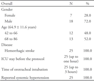
All patients included in this study complied with the protocol and no patients met the discontinuation cri-teria. he sample’s characterization is shown in table 1.
HR, RR, SpO2 and capnometry were not changed at any PEEP level, p > 0.5 (Table 2). ICP was signii-cantly increased for the following PEEP values: 10, 12 and 14 cmH2O as compared with 0 cmH2O; 8, 10, 12 and 14 cmH2O as compared with 2 cmH2O; 8, 10, 12 and 14 cmH2O as compared with 4 cmH2O; 12 and 14 cmH2O as compared with 6 cmH2O; 12 and 14
cmH2O as compared with 8 cmH2O; and 12 and 14 cmH2O as compared with 10 cmH2O (Figure 1).
Table 3 shows CPP and MBP mean distributions as related to the diferent PEEP values. No statistically signiicant diferences were identiied for CPP and MBP means.
Table 1 – Patients’ characteristics
Overall N %
Gender
Female 7 28.0
Male 18 72.0
Age (64.9 ± 11.6 years)
42 to 66 12 48.0
68 to 86 13 52.0
Disease
Hemorrhagic stroke 25 100.0
ICU stay before the protocol 25 (up to one hour) 100.0
Time of orotracheal intubation 25 (up to 3 hours) 100.0 Reported systemic hypertension 25 100.0
ICU – intensive care unit.
Table 2 – Analysis of respiratory variables according to the positive end-expiratory pressure level
PEEP RR* SpO2* Capnometry*
0 16 99.1 + 1.66 34.44 + 1.08
2 16 98.8 + 1.61 34.6 + 1.0
4 16 99 + 1.49 34.64 + 0.95
6 16 98.9 + 1.28 34.72 + 0.89
8 16 99 + 1.63 34.76 + 0.83
10 16 99.2 + 1.03 35 + 0.7
12 16 99.4 + 0.69 35.16 + 0.68 14 16 99.5 + 0.84 35.28 + 0.73
*p > 0.05. PEEP –positive end-expiratory pressure; RR – respiratory rate; SpO2 – peripheral oxygen saturation.
Figure 1 – Assessment of intracranial pressure according to the positive end-expiratory pressure levels.
PEEP – positive end-expiratory pressure; ICP – intracranial pressure. ICP pressure was diferent between the irst arrow and other arrows in the same line. p < 0.05.
Table 3 – Analysis of hemodynamic variables and cerebral perfusion pressures according to the positive end-expiratory pressure level
PEEP MBP* CPP** HR *
0 100.6 + 16.2 90.1 + 17.5 78 + 11.63 2 99.4 + 15.6 88.9 + 16.1 77.63 + 11.47 4 99.0 + 13.9 88.6 + 15.0 79 + 11.84 6 98.1 + 14.1 87.2 + 14.4 77.81 + 10.79 8 98.2 + 15.4 86.9 + 15.7 76.54 + 10.73 10 98.3 + 21.7 86.7 + 22.3 78.09 + 11.86 12 101.0 + 15.6 88.8 + 16.4 78.72 + 11.08 14 100.5 + 16.8 88.0 +17.0 77 + 12.19
A comparison between initial and final mean sta-tic respiratory system complacency, with a PEEP of 5 cmH2O, showed a significant increase from the ini-tial value of 49.6 ± 11.2 to 61.4 ± 14.2, p < 0.001.
DISCUSSION
Based on the results, we conclude that in this po-pulation of patients with respiratory system compla-cency within the physiological range, higher PEEP values significantly increased ICP. However, this fin-ding lacks clinical relevance because even the highest PEEP value tested was below the 20 mmHg bounda-ry. MBP and CPP were not changed. It is interesting to note that incremental increases in PEEP augment final complacency, as compared to the initial values, in a statistically significant fashion.
Theoretically, PEEP values above 10 cmH2O could be harmful to the intracranial compartment because they may reduce systemic venous return.(26)
This transmission of PEEP into the thoracic com-partment is influenced by the chest wall, the lung’s properties and the patient’s hemodynamics. A study conducted by Chapin et al.(27) showed that in the
set-ting of low chest wall complacency, PEEP increases the intrathoracic pressure with possible meaningful hemodynamic changes. However, this would not be seen with reduced pulmonary complacency.
How was dissipated the increased intrapulmonary pressure, with PEEP values ranging between 0 and 14 cmH2O, not causing clinically meaningful ICP increase? Apparently, intrathoracic pressure is not di-rectly transmitted into the cerebral compartment, gi-ven that respiratory system complacency, hemodyna-mic stability, cerebral elasticity and the actual PEEP values could also influence this relationship. The-refore, the sum of these factors will determine the transmission of alveolar pressure into pleural pres-sure, which is reflected in the hemodynamic system. Cuypers et al.(28) have shown that increased central
venous pressure in cats only leads to a transient in-crease in ICP, suggesting that inin-creased right atrium pressure would not be a determinant of increased ICP. Thus, reduced venous return caused by alveolar capillary vessel compression due to PEEP would not be sufficient enough to increase ICP.
Our results regarding the impact of PEEP on ICP are different from those described by Aidinis et al.,(29) who assessed induced lung injury in
ani-mals. The data from our study are partially
diffe-rent from those of Apuzzo et al.(30) and Burchiel et
al.,(31) but were similar to the findings in normal
cerebral elasticity patients. In contrast to the con-clusions by Huynh et al.,(33) our results showed no
increase in CPP.
Similar to Cooper et al.,(34) whose study design
was similar to ours, we observed increases in ICP that were not clinically meaningful. This differed from the findings of Georgiadis et al.,(35) who found
no ICP increase.
Our patients were laying in supine decubitus with a 30º bed head elevation. In this position, the chest is lower than the intracranial compartment, easing the venous return, preventing excessive increases in ICP. Schwarz et al.(36) assessed the effects of
positio-ning the head above the chest in hemorrhagic stroke patients and observed that a 30º bed head elevation leads to reduced ICP. Toung et al.(37) used an animal
study to evaluate the effects of bed head elevation on the impact of PEEP on cerebrospinal fluid pressure. The results showed that the ICP was not changed when PEEP values of 15 cmH2O were used. A raised bed head is likely to have influenced the lack of in-creased ICP in our study.
In assessing the impact of PEEP on CPP, our re-sults corroborate the findings of McGuire et al.,(38)
who found no CPP changes during the use of high PEEP values. Considering that the heart is contai-ned within a pressurized chamber, during MVA the positive intrathoracic pressure leads to a reduced left ventricular transmural pressure. This increases the ejection volume, contributing to stable CPP even with increased ICP.(39)
Our results agree with previous studies(35-38) with
respect to the lack of MBP changes secondary to in-creased PEEP. Positive intrathoracic pressure may reduce the cardiac output due to reduced venous return,(40) however, this would be minimized by the
improved left ventricle function, explaining MBP stability.
Static complacency, below physiological values, minimizes negative PEEP cardiovascular system effects.(27) Our patients had static complacency below
he-modynamic effects. Based on our study design, the patients were ventilated on pressure control mode, limiting the maximal respiratory system pressure. Even when increasing PEEP, Pp was not changed. This finding was different from that of other stu-dies,(29,30) with dissimilar results showing increased
maximal respiratory system pressure upon PEEP in-crease.
As Pp was limited, PEEP increases would redu-ce the pressure delta and TV, which could result in CO2 retention. However, our results showed no sta-tistically significant capnographic changes. Our hy-pothesis is that this is due to the short time in each PEEP value, which would be insufficient to cause changes. Another possible explanation is that PEEP increases improved the complacency and may have caused increased TV.
A limitation of our study is that the values were not directly obtained from blood gas measurements, but were indirectly monitored by capnometry and SpO2. In addition, we did not assess the tidal volume to the patient during the protocol.
CONCLUSION
Our findings show that, in our population sam-ple, the use of high PEEP values caused no clinically meaningful changes to ICP, CPP and MBP. Our re-sults suggest that in patients with acute lung injury or respiratory distress syndrome, PEEP values up to 14 cmH2O could be used for gas exchange optimi-zation, and may also prove beneficial in improving these patients’ residual capacity following suction or ventilator circuit disconnection. However,
additio-nal longer and multicenter studies would be useful in confirming these findings.
RESUMO
A pressão positiva intratorácica pode levar a alterações hemodinâmicas com repercussão no compartimento intra-craniano, alterando a pressão intracraniana e a pressão de perfusão cerebral. Esse efeito pode se tornar mais intenso quando utilizados elevados valores de pressão positiva ex-piratória final.
Objetivo: Medir o impacto que diferentes valores de pres-são positiva expiratória inal causam na prespres-são intracrania-na, na pressão de perfusão cerebral e pressão arterial média.
Método: O estudo foi desenvolvido em uma unidade de terapia intensiva neurológica envolvendo 25 pacientes adul-tos com acidente vascular cerebral hemorrágico, ventilados mecanicamente no modo com controle pressórico de vias aéreas. Foram instituídos valores de pressão positiva expi-ratória final variando de 0 a 14 cmH2O, de forma aleatória através de sorteio, utilizando valores pares. A monitorização das variáveis estudadas ocorreu após cinco minutos em cada patamar de pressão positiva expiratória final.
Resultados: O incremento da pressão positiva expira-tória final aumentou a pressão intracraniana, (p < 0,001) sem causar alteração estatisticamente significativa na pres-são arterial média ou na prespres-são de perfupres-são cerebral.
Conclusão: Na população estudada, de pacientes com acidente vascular cerebral hemorrágico, os achados mos-traram que valores de pressão positiva expiratória final até 14 cmH2O, não alteram a pressão de perfusão cerebral e a pressão arterial média, aumentando a pressão intracrania-na, porém sem relevância clínica.
Descritores: Pressão intracraniana; Pressão positiva expiratória final; Acidente cerebral vascular
REFERENCES
01. Teive HAG, NovaK EM. Hipertensão intracraniana: tratamento básico. In: Teive HAG, NovaK EM, editores. Condutas em emergências neurológicas: um guia prático de orientação terapêutica. São Paulo: Lemos Editorial; 2001. 02. Shapiro K. Increased intracranial pressure. In: Levin DL,
Morriss FC, editors. Essentials of pediatric intensive care. 2nd ed. New York: Churchill Livingstone; 1997.
03. Han CY, Backous DD. Basic principles of cerebrospinal luid metabolism and intracranial pressure homeostasis. Otolaryngol Clin N Am. 2005;38(4):569-76.
04. Luerssen TG, Wolla CE. Pathophysiology and management of increased intracranial pressure in children. In: Andrews
BT, Hammer GB. Pediatric neurosurgical intensive care. Park Ridge: American Association of Neurological Surgeons; 1997. 05. Greenberg MS. Handbook of neurosurgery. 5th ed. New
York: hieme; 2001.
06. Abaine I, Leone M, Martin C. Head injury in patients with multiple trauma. In: Vincent JL, editor. Yearbook of intensive care and emergency medicine. Berlin: Springer-Verlag; 2001.
07. Pillai S, Praharaj SS, Rao GS, Kolluri VR. Cerebral perfusion pressure management of severe difuse head injury: efect on brain compliance and intracranial pressure. Neurol India. 2004;52(1):67-71.
2001;2(1):28-40.
09. Barbosa AP, Cabral SA. Novas terapias para hipertensão intracraniana. J Pediatr (Rio J). 2003;79(Supl 2):S139-48. 10. Cooper DJ, Murray L. Trauma. In: Vincent JL, editor.
Yearbook of intensive care and emergency medicine. Berlin: Springer-Verlag; 2001.
11. Seppelt I. Intracranial hypertension after traumatic brain injury. Indian J Crit Care Med. 2004;8(2)120-6.
12. Chesnut RM, Marshall LF, Klauber MR, Blunt BA, Baldwin N, Eisenberg HM, et al. he role of secondary brain injury in determining outcome from severe head injury. J Trauma. 1993;34(2):216-22.
13. Emmerich JC. Monitorização respiratória: fundamentos. 2a ed. Rio de Janeiro: Revinter; 2001.
14. Solenski NJ, Haley EC Jr, Kassel NF, Kongable G, Germanson T, Truskowski L, Torner JC. Medical complications of aneurysmal subarachnoid hemorrhage: a report of the multicenter, cooperative aneurysm study. Participants of the Multicenter Cooperative Aneurysm Study. Crit Care Med. 1995;23(6):1007-17.
15. Mayer SA, Copeland D, Bernardini GL, Boden-Albala B, Lennihan L, Kossof S, Sacco RL. Cost and outcome of mechanical ventilation for life-threatening stroke. Stroke. 2000;31(10):2346-53.
16. García AH, Domínguez YS, Alfonso ARE, Montiel IP. Manejo ventilatorio de los pacientes con patología aguda del sistema nervioso central. Rev Cub Med Int Emerg. 2004;3(2):53-68.
17. Muizelaar JP, Marmarou A, Ward JD, Kontos HA, Choi SC, Becker DP, et al. Adverse efects of prolonged hyperventilation in patients with severe head injury: a randomized clinical trial. J Neurosurg. 1991;75(5):731-9. 18. Slutsky AS. Mechanical ventilation. American College
of Chest Physicians’ Consensus Conference. Chest. 1993;104(6):1833-59. Review. Erratum in Chest. 1994;106(2):656.
19. II Consenso Brasileiro de Ventilação Mecânica. J Pneumologia. 2000;26 (Supl 2).
20. Ventilation with lower tidal volumes as compared with traditional tidal volumes for acute lung injury and the acute respiratory distress syndrome. he Acute Respiratory Distress Syndrome Network. N Engl J Med. 2000;342(18):1301-8. 21. Tobin MJ, Jubran A, Laghi F. Patient-ventilator interaction.
Am J Respir Crit Care Med. 2001;163(5):1059-63. 22. Tobin MJ. Advances in mechanical ventilation. N Engl J
Med. 2001;344(26):1986-96. Review.
23. Fontes M. Progress in mechanical ventilation. Curr Opin Anaesthesiol. 2002;15(1):45-51.
24. Ranieri VM, Guiliani R, Cinnella G, Pesce C, Brienza N, Ippolito EL, et al. Physiologic efects of positive end-expiratory pressure in patients with chronic obstructive pulmonary disease during acute ventilatory failure and controlled mechanical ventilation. Am Rev Respir Dis. 1993;147(1):5-13.
25. Barbas CSV, Bueno MAS, Amato MBP, Hoelz C, Junior MR. Interação cardiopulmonar durante a ventilação mecânica. Rev Soc Cardiol Estado de São Paulo. 1998;3:406-19. 26. West JB, Dollery CT, Naimark A. Distribution of blood low
in isolated lung; relation to vascular an alveolar pressures. J Appl Physiol. 1964;19:713-24.
27. Chapin JC, Downs JB, Dougral ME, Murphy EJ, Ruiz BC. Lung expansion, airway pressure transmission, and positive end-expiratory pressure. Arch Surg. 1979;114(10):1193-7. 28. Cuypers J, Matakas F, Potolicchio SJ Jr. Efect of central
venous pressure on brain tissue pressure and brain volume. J Neurosurg. 1976;45(1):89-94.
29. Aidinis SJ, Laferty J, Shapiro HM. Intracranial responses to PEEP. Anesthesiology. 1976;45(3):275-86.
30. Apuzzo JL, Wiess MH, Petersons V, Small RB, Kurze T, Heiden JS. Efect of positive end expiratory pressure ventilation on intracranial pressure in man. J Neurosurg. 1977;46(2):227-32.
31. Burchiel KJ, Steege TD, Wyler AR. Intracranial pressure changes in brain-injured patients requiring positive end-expiratory pressure ventilation. Neurosurgery. 1981;8(4):443-9.
32. Almgren B, Wickerts CJ, Heinonen E, Högman M. Side efects of endotracheal suction in pressure- and volume-controlled ventilation. Chest. 2004;125(3):1077-80. 33. Huynh T, Messer M, Sing R, Miles W, Jacobs DG, homason
MH. Positive end-expiratory pressures alters intracranial and cerebral perfusion pressure in severe traumatic brain injury. J Trauma. 2002;53(3):488-92; discussion 492-3. 34. Cooper KR, Boswell PA, Choi SC. Safe use of PEEP
in patients with severe head injury. J Neurosurg. 1985;63(4):552-5.
35. Georgiadis D, Schwarz S, Baumgartner RW, Veltkamp R, Schwab S. Inluence of positive end-expiratory pressure on intracranial pressure and cerebral perfusion pressure in patients with acute stroke. Stroke. 2001;32(9):2088-92. 36. Schwarz S, Georgiadis D, Aschof A, Schwab S. Efects
of body position on intracranial pressure and cerebral perfusion in patients with large hemispheric stroke. Stroke. 2002;33(2):497-501.
37. Toung TJ, Miyabe M, McShane AJ, Rogers MC, Traystman RJ. Efect of PEEP and jugular venous compression on canine cerebral blood low and oxygen consumption in the head elevated position. Anesthesiology. 1988;68(1):53-8. 38. McGuire G, Crossley D, Richards J, Wong D. Efects
of varying levels of positive end-expiratory pressure on intracranial pressure and cerebral perfusion pressure. Crit Care Med. 1997;25(6):1059-62.
39. Marini JJ, O’Quin R, Culver BH, Butler J. Estimation of transmural cardiac pressures during ventilation with PEEP. J Appl Physiol. 1982;53(2):384-91.