Disponible en castellano/Disponível em língua portuguesa SciELO Brasil www.scielo.br/rlae 1 RN, M.Sc. in Obstetric and Neonatal Nursing, e-mail: [email protected]; 2 PhD in Nursing, Faculty, University of São Paulo, School of Nursing, Brazil, e-mail: [email protected]
RANDOMIZED CONTROLLED CLINICAL TRIAL ON TWO PERINEAL TRAUMA SUTURE
TECHNIQUES IN NORMAL DELIVERY
Sandra Ferreira Silva de Almeida1 Maria Luiza Gonzalez Riesco2
Almeida SFS, Riesco MLG. Randomized controlled clinical trial on two perineal trauma suture techniques in normal delivery. Rev Latino-am Enfermagem 2008 março-abril; 16(2):272-9.
The aim was to compare healing and perineal pain with the use of continuous and interrupted suture techniques in women after normal delivery. A randomized controlled trial was carried out at a hospital birth center in Itapecirica da Serra, Sao Paulo, Brazil. A total of 61 women participated with episiotomy or second degree perineal tear, allocated in two groups according to the continuous (n=31) or interrupted (n=30) suture techniques. The main outcomes evaluated were edema, ecchymosis, hyperemia, secretion, dehiscence, fibrosis, frequency and degree of pain (evaluated by numerical scale from 1 to 10). Data were collected during hospitalization and after discharge (four and 41 days after birth). Healing occurred by first intention in 100% of cases in both suture techniques. There were no statistically significant differences for the occurrence of morbidities, except for perineal pain due to palpation at four days after delivery, which was more frequent among women with interrupted suture.
DESCRIPTORS: delivery; perineum; pain; suture techniques; obstetrical nursing; clinical trials
ENSAYO ALEATORIO CLÍNICO CONTROLADO PARA DOS TÉCNICAS DE SUTURA PERINEAL
EN PARTO NORMAL
El objetivo fue comparar la cicatrización y el dolor perineal utilizando técnicas de sutura continua y separada, en mujeres que realizaron parto normal. El estudio fue controlado aleatorio, realizado en un centro para parto normal en Itapecerica de la Sierra, Sao Paulo. Participaron 61 mujeres con episiotomía o desgarro perineal de segundo grado, distribuidas en dos grupos (sutura continua n=31 y sutura separada n=30). Las principales medidas evaluadas fueron edema, equimosis, hiperemia, secreción, dehiscencia de herida, fibrosis, frecuencia y magnitud del dolor (evaluada por escala numérica de 1 a 10). Los datos fueron recolectados durante la hospitalización y después del alta (de 4 a 41 días post-parto). La cicatrización fue por primera intención en 100% de los casos, para las dos técnicas de sutura. No se encontró diferencia estadísticamente significativa en los casos de morbilidad, con excepción del dolor perineal a la palpación al cuarto día post-parto, el que fue más frecuente en mujeres con sutura separada.
DESCRIPTORES: parto; perineo; dolor; técnicas de sutura; enfermería obstétrica; ensayos clínicos
ENSAIO CLÍNICO CONTROLADO ALEATÓRIO SOBRE DUAS TÉCNICAS DE SUTURA DO
TRAUMA PERINEAL NO PARTO NORMAL
O objetivo foi comparar a cicatrização e a dor perineal com a utilização das técnicas de sutura contínua e separada em mulheres com parto normal. Realizou-se estudo controlado aleatório, em centro de parto normal, em Itapecerica da Serra, São Paulo. Participaram 61 mulheres com episiotomia ou rotura perineal de segundo grau, alocadas em dois grupos, segundo a técnica de sutura contínua (n=31) ou separada (n=30). Os principais desfechos avaliados foram edema, equimose, hiperemia, secreção, deiscência, fibrose, freqüência e magnitude da dor (avaliada pela escala numérica de 1 a 10). Os dados foram coletados na internação e após a alta (quatro e 41 dias pós-parto). A cicatrização foi por primeira intenção em 100% dos casos, nas duas técnicas de sutura. Não houve diferença estatisticamente significante para a ocorrência de morbidades, exceto na dor perineal à palpação, com quatro dias de pós-parto, que foi mais freqüente entre as mulheres com sutura separada.
INTRODUCTION
M
aternal morbidity caused by perineal trauma in vaginal delivery is a common problem ofglobal occurrence. Annually, 350,000 women in the
United Kingdom need postpartum perineal repair. It
is estimated that the majority will experience perineal
pain and a fifth will have long-term problems, such
as dyspareunia(1).
Episiotomy is frequently performed and its
incidence is variable, with rates ranging from 9.7% in
Sweden to 100% in Taiwan, considering both
nulliparous and multiparous women. In Latin American
hospitals, the rate of episiotomy among nulliparous
women varies from 69% to 96%, with an average of
94% in the 14 countries studied(2).
In Brazil, these rates exceed 76%, reaching
95.2% among nulliparous women(3-4). Although
episiotomy is a routine intervention in most services,
there is a downward tendency due to the advantages
of its selective use. Its prevalence diminishes sharply
when it is done in view of a protocol of indications.
In such cases, the occurrence of perineal lacerations
is common and requires suture. In 2001, 26.5% of
women in the Hospital General of Itapecirica da Serra
(HGIS) underwent episiotomy after normal
delivery(5).
There is little scientific interest in the study
of episiotomy indications and complications as well
as a lack of research on its surgical repair,
denominated perineorrhaphy, despite its high
prevalence and potential for morbidities, such as
edema, haematoma, pain, infection and dehiscence.
The factors associated with morbidity and
related to perineal trauma repair consist in the choice
of the suture material, the repair technique and the
surgeon’s competence, giving rise to research fields
in the birth care area(6).
Despite the existence of publication on the
types of threads most indicated for perineal suture
and on local pain after it(7-8), studies on the remaining
morbidities associated to perineal trauma after normal
delivery are rare(1,9-12), especially in Brazilian
literature.
A literature review on skin suture techniques
in perineal trauma after delivery found only four
randomized trials, concluding that the continuous
suture technique is associated to lesser pain in the
short run. However, authors point to the need for more
studies comparing suture techniques(13).
After consideration of this scientifically little
explored problem and taking into account the number
of women who experience perineal trauma after
delivery, the need for repair and local pain, this study
was proposed to contribute to the reduction of perineal
morbidity related to the suture technique.
The aim was to compare the characteristics
of the healing process, the frequency and degree of
perineal pain with the use of continuous and interrupted
techniques of perineal trauma in women after normal
delivery.
METHODS
A randomized controlled trial (RCT) was
adopted to analyze the effect of two different suture
techniques of episiotomy and second-degree perineal
tear in the healing process and in the occurrence of
local pain.
The study was performed in the Birth Center
(BC) of the General Hospital of Itapecirica da Serra, in
Itapecirica da Serra, SP, Brazil. At the BC, normal
deliveries are attended by nurse-midwives, episiotomy
is performed based on the institution’s protocol and all
parturients are accompanied by someone of their choice.
Women who met the following criteria were
chosen: presented no leucorrhoea or infectious lesion
in the genital area, physiometry, retention of ovular
remains or cervical and vaginal cul-de-sac tears during
birth; did not undergo blood transfusion; did not use
antibiotics, imunossupressors, anti-inflammatories or
intimate hygiene products other than soap and water.
The sample was composed of 61 puerperas
-31 in the group in which the continuous suture
technique was used and 30 in the group in which the
interrupted technique was used. Randomization was
performed through an electronic table and applied at
the moment of delivery to 95 women. A total of 34
women were excluded from the study and replaced
by others, according to the randomization table.
The estimator used to determine the sample
size (n) was the result of a study on perineal pain in
the first ten days after delivery. The average frequency
of women with local pain in this study was 51%, varying
between 21% and 63% between the first and tenth
days of puerperium(14). In order to obtain a confidence
interval of 95% in the present study, we estimated
that 51% of the women would experience perineal pain,
The suture was performed with polyglactin
910*, a thread of synthetic origin with tensile strength
in vivo of 100% on the first day, 81% on the third,
57% on the fifth, 53% on the seventh and 0% after
fourteen days. Complete absorption occurs by
hydrolysis within approximately 35 days.
For a better result of the suture, “dead space”
between the wound edge and exaggerated tension
must be avoided, so that an adequate hemostasis of
the bleeding vessels in the incision or tear is promoted.
Likewise, a minimum of suture levels is
recommended, approximating separately the
subcutaneous level in case of need. After the suture,
hygiene of the perineum must be done with
physiological solution.
In both groups, topic
polivinil-iodine-pirrolidone was used for the antisepsis of the vulvar
skin, perineum and internal face of the thighs, and
the following surgical tools were used: needle holder
and rat tooth surgical tweezers in all suture points.
The interrupted and continuous suture
techniques are described below:
Interrupted technique: 1. Start the suture
by the apex of the wound of vaginal mucous
membrane with needle ½ circle, taper point of four
or five centimeters; 2. Fasten the stitch with three
knots, with two loops in the first, two inverted loops
in the second and a single loop in the third knot, leaving
the thread end with approximately one centimeter;
3. Proceed the suture of the mucous membrane with
continuous locking stitches up to the hymeneal ring;
4. Fasten the last stitch the same way as the first and
cut the thread; 5. In the muscle level, approximate
the edges with interrupted stitches, fastening each
stitch with three knots in the same way as the previous
ones; 6. Approximate the skin edges with interrupted
stitches using a 3/8 circle reverse cut needle of three
centimeters; 7. Fasten each stitch and cut the thread,
leaving the ends with one centimeter.
Continuous technique: 1. and 2. Identical
to the technique described above; 3. Proceed with
the suture of the mucous membrane, with continuous
non-locking stitches up to the hymeneal ring; 4.
Proceed to the muscle plan with non-locking
continuous suture; 5. At the end of the muscle layer,
change the needle to a 3/8 circle reverse cut needle
of three centimeters and perform intradermic suture,
also continuous, initiating by the inferior part towards
the fourchette where the suture is fastened with three
knots, fixated by two loops in the first, two inverted
loops in the second and a single loop in the third one;
6. Cut the thread, leaving the ends with one centimeter
of length.
The perineal suture was performed by one
of the researchers and ten nurse-midwives from the
BC team, between July 2001 and April 2002. Before
starting the research, the nurse-midwives were
submitted to theoretical-practical training, with
accompanied suture performance and evaluation
through the verification of the correct use of the
proposed suture techniques.
The data collection was carried out in four
steps: the first, denominated Greenberg period,
corresponded to the first hour after finishing the
perineal suture and was performed by the professional
who assisted the delivery. The second step was
performed by one of the researchers after consulting
the patient’s file in order to obtain data related to the
parturient period of hospitalization. The third one,
denominated first return visit, occurred
approximately four days after birth and was carried
out by the same researcher or by the outpatient clinic
nurse-midwife through an interview with the puerpera,
perineal exam and blood sample collection for
hemoglobin and hematocrit dosage. The fourth step,
denominated second return visit, was also carried
out by the same researcher, about 41 days after the
delivery, with a new interview and perineal exam.
The first and second return visits occurred at the
outpatient clinic of the HGIS or at the women’s home,
when the puerperal woman did not return on the
scheduled date.
The independent variables were the
continuous and interrupted perineal suture techniques.
The dependent variables were: perineal healing
process (edema, ecchymosis, hyperemia, secretion,
dehiscence and fibrosis); perineal hygiene; suture
conditions; frequency and degree of spontaneous
perineal pain, and pain due to palpation, to sitting,
walking, urinating, evacuating, sexual activity and use
of analgesics.
For the signs indicative of healing, swollen
perineum (edema), altered skin color (ecchymosis
or hyperemia), indication of secretion in the suture
site, separated suture edges in any of the levels
(dehiscence) and engorgement of the cicatricial line
(fibrosis) were considered. Perineal hygiene was
considered adequate when the perineum had no
accumulated dirt and inadequate when there was
accumulated dirt or bad odor not attributed to the
infectious process. In the evaluation of suture
c o n d i t i o n s , t h e c a t e g o r i e s c o n s i d e r e d w e r e :
p r e s e r v e d , p a r t i a l l y u n f a s t e n e d o r t o t a l l y
unfastened.
When the puerpera reported painful
sensation, in the first, third and fourth steps of the
study, according to the description above, the degree
of pain was evaluated according to a numerical scale
from 1 to 10, 1 indicating the lowest degree and 10
the highest. The use of analgesics distinguished the
use of pain relief medication from hospitalization until
the second return visit.
The other variables analyzed were: maternal
characteristics (age; vaginal deliveries and previous
perineal scar; hematological conditions in the first
return visit); perineal suture characteristics (type of
trauma; professional who performed it and duration
of suture; sutured layers, hemostasis method and
bleeding after the suture); characteristics of the
newborn (gestational age; vitality; weight).
The SPSS program was used for statistical
analysis. Central tendency and dispersion measures
were computed for descriptive analysis of the
quantitative continuous variables, and absolute and
relative frequencies were computed for the qualitative
variables. Chi-square (χ2) and Fisher’s exact tests were used for inferential analysis. A 5% significance
level was considered (p-value=0.05).
The project was approved by the institution’s
Research Ethics Committee and the women’s
participation was voluntary after free and informed
consent. The participation of midwives in the data
collection was also voluntary.
RESULTS
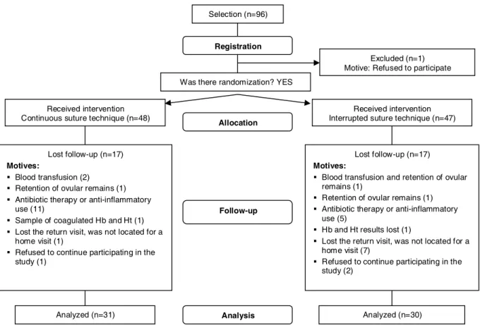
A total of 96 women who met the inclusion
criteria were selected for the research, 95 were
allocated in the study groups and n was 61. The results
of 31 women with continuous suture and 30 with
interrupted suture were analyzed (Figure 1).
Selection (n=96)
Excluded (n=1) Motive: Refused to participate
Lost follow-up (n=17)
Motives:
Blood transfusion (2) Retention of ovular remains (1) Antibiotic therapy or anti-inflammatory
use (11)
Sample of coagulated Hb and Ht (1) Lost the return visit, was not located for a
home visit (1)
Refused to continue participating in the study (1)
Received intervention Continuous suture technique (n=48)
Lost follow-up (n=17)
Motives:
Blood transfusion and retention of ovular remains (1)
Retention of ovular remains (1) Antibiotic therapy or anti-inflammatory
use (5)
Hb and Ht results lost (1)
Lost the return visit, was not located for a home visit (7)
Refused to continue participating in the study (2)
Received intervention Interrupted suture technique (n=47)
Analyzed (n=30)
Allocation
Analysis Registration
Was there randomization? YES
Follow-up
Analyzed (n=31)
The results regarding maternal conditions and characteristics of perineal suture are presented in Tables 1 and 2, respectively.
Table 1 - Maternal conditions according to the suture technique
Table 4 - Frequency and degree of perineal pain
according to suture technique
n o i t i d n o C l a n r e t a M e u q i n h c e T e r u t u S s u o u n i t n o C ) % ( 1 3 = n d e t p u r r e t n I ) % ( 0 3 = n ) s r a e y ( e g
A Average(s.d.) 24.2(5.3) 24.2(6,8)
) L d / g ( n i b o l g o m e
H Average(s.d.) 11.3(1.4) 11.0(1,3)
) L d / L m ( t i r c o t a m e
H Average(s.d.) 34.2(3.9) 33.3(4,1)
y r e v i l e d l a n i g a v s u o i v e r
P 16(51.6) 15(50.0)
r a c s l a e n i r e p s u o i v e r
P 12(38.7) 15(50.0)
Table 2 - Characteristics of perineal suture according to technique and suture
s c i t s i r e t c a r a h C e u q i n h c e t e r u t u S s u o u n i t n o C ) % ( 1 3 = n d e t p u r r e t n I ) % ( 0 3 = n a m u a r t l a e n i r e
P Episiotomy 15(48.4) 17(56.7)
2nddegreetear 16(51.6) 13(43.3)
e f i w d i m -e s r u
N BC’steam 12(38.7) 21(70)
r e h c r a e s e
R 19(61.3) 9(30)
) n i m ( n o i t a r u d e r u t u
S Average(s.e.) 16.8(6.2) 21.4(9.5)
d o h t e m c i t a t s o m e
H Vesselilgaiton 4(66.7) 5(50)
e r u s s e r p l a u n a
M 1(16.7) 4(40)
e r u s s e r p + n o it a g i
L 1(16.7) 1(10)
s r e y a l e l c s u
M One 25(80.6) 12(40)
e r o m r o o w
T 6(19.4) 18(60)
e r u t u s s u o e n a t u c b u
S 11(35.5) 17(56.7)
e r u t u s r e t f a g n i d e e l
B 3(9.7) 6(20)
Tables 3 and 4 show the results regarding healing, frequency and degree of perineal pain in each of the study steps. Healing occurred by first intention in 100% of cases, with both suture techniques.
The median of the numerical scale from 1 to 10 was considered for the degree of spontaneous pain and palpation pain, according to the description provided in the Methods section.
Table 3 - Characteristics of perineal healing according to suture technique
e g a t
S Characteristics
e u q i n h c e t e r u t u S e u l a v -p s u o u n i t n o C ) % ( 1 3 = n d e t p u r r e t n I ) % ( 0 3 = n g r e b n e e r
G Edema 14(45.2) 12(40.0) 0.684*
n o i t a z i l a t i p s o
H Edema 8(25.8) 8(26.7) 0.939*
s i s o m y h c c
E 1(3.2) 1(3.3) 0.999** a i m e r e p y
H 2(6.5) 2(6.7) 0.999**
t i s i v n r u t e r t s
1 Edema 1(3.2) 6(20.0) 0.053**
s i s o m y h c c
E 3(9.7) 2(6.7) 0.999** a i m e r e p y
H 2(6.5) 1(3.3) 0.999** e n e i g y h e t a u q e d
A 31(100) 25(83.3) 0.024** e r u t u s d e v r e s e r
P 31(100) 27(90.0) 0.354**
t i s i v n r u t e r d n
2 Cicatricialifbrosis 11(35.5) 14(46.7) 0.375*
* χ2 test
** Fisher’s exact test
e g a t
S Perinealpain
e u q i n h c e t e r u t u S e u l a v -p s u o u n i t n o C ) % ( 1 3 = n d e t p u r r e t n I ) % ( 0 3 = n g r e b n e e r
G Spontaneous[Med] 3(9.7)[2] 3(10.0)[3] 0.999*
n o i t a z i l a t i p s o
H Analgesicuse 18(58.1) 19(63.3) 0.674**
n r u t e r t s
1 Spontaneous[Med] 15(48.4)[5] 18(60.0)[5] 0.323**
] d e M [ n o it a p l a
P 20(64.5)[5] 27(90.0)[5] 0.018** g
n it t i
S 14(45.2) 20(66.7) 0.091** g n i k l a
W 4(12.9) 6(20.0) 0.508* g n it a n i r
U 4(12.9) 5(16.7) 0.731* g n it a u c a v
E 6(19.4) 1(3.3) 0.104* e s u c i s e g l a n
A 9(29.0) 11(36.7) 0.525**
n r u t e r d n
2 Palpaiton[Med] 4(12.9)[4] 7(23.3)[3] 0.289** g
n it t i
S - 2(6.7) 0.238*
g n it a u c a v
E - 1(3.3) 0.492*
e s u c i s e g l a n
A 19(61.3) 17(56.7) 0.714*
y t i v it c a l a u x e
S 12(37.8) 11(36.7) 0.869* ] d e M [ a i n u e r a p s y
D 5(41.7)[5] 5(45.5)[7] 0.999* * Fisher’s exact test
** χ2 test
According to inferential analysis, there was a
higher frequency of adequate perineal hygiene and lower frequency of perineal palpation pain with
continuous suture in the first return visit, with
statistically significant association for both variables (Tables 3 and 4). The results showed no statistically
significant differences between the two perineal suture
techniques for the other variables analyzed.
All newborns presented Apgar scores ≥9 at the fifth minute of life; 98.4% were full-term according
to gestational age based on Capurro’s method. Their
weight varied between 2,140 and 4,180 grams, with an average of 3,274.8.
DISCUSSION
The local inflammatory signs and symptoms
such as edema, pain, redness and heat are expected in the initial phase of the healing process and subside
as local reactions and absorption of the suture material
evolve. However, perineal trauma morbidities, such as haematoma, ecchymosis, infection and dehiscence,
hinder complete maternal recovery. The puerpera’s
characteristics and general conditions, such as age, protein deficiency, deficit of tissue oxygen and drugs
action, influence the time and quality of healing(15).
The variables that can affect the healing process, not
controlled by the study inclusion criteria, were analyzed and indicated sample homogeneity (Table 1).
The type of trauma, manipulation of the site
important aspects for the reduction of pain and the
good evolution of the healing process. Studies indicate
better results in cases of spontaneous laceration when
compared to episitomy, with less suture layers(16).
The technical details of the suture appoint the
advantages of continuous suture (Table 2). In this
technique, large layers of muscle tissue are seized
with approximation in one single level. The suture
depends on the subcutaneous anatomic
characteristics in the perineum; when this level is slim,
its approximation occurs with the skin suture.
Although the application of some hemostasis
method produces greater manipulation, it might be
necessary because the perineal trauma bleeding is a
morbidity factor. Bleeding after the suture, hardly
frequent with continuous suture, indicates the
hemostatic effect of this technique. The partially
unfastened suture in the skin, verified in three cases
with interrupted suture during the first return visit,
can be attributed to the lack of an inverted slipknot in
all knots. After these occurrences, the importance of
inverting the slipknot with the use of a more smooth
and slippery suture thread, like the one adopted in
this study, was stressed among the nurse-midwives
and the problem did not happen again (Table 2).
Despite the benefits reported in literature,
including diminished pain and increased mobility of
women submitted to continuous suture of the perineal
trauma(13), professionals admit experiencing some
personal uneasiness when exposed to the
non-traditional suture technique for the first time(1,11). In
this study, despite the training provided, some nurses
initially reported being afraid to perform continuous
suture. Due to this fact, the majority of sutures with
this technique was performed by the researcher (Table
2). This aspect, which can constitute bias, was
appointed in other studies, suggesting that the
interrupted technique is more commonly taught and
easier to be performed by inexperienced
professionals(1,6,13).
The delay in trauma repair was smaller with
the continuous technique, with the advantage that, the
lesser time spent in the suture, the smaller the risk of
infection and the puerpera’s discomfort (Table 2).
The development of the healing process and
the result observed in the second return visit
postpartum did not indicate statistically significant
differences between continuous and interrupted
perineal suture techniques. Precocious edema is more
frequent in the Greenberg period and can be
associated to manipulation during birth and to the
anesthetics accumulated in the tissue. Its persistence
can be attributed to local inflammatory reaction and
to the quantity of material used in trauma repair
(Table 3). It is important to consider that polyglactin
910 is a thread with short exudative phase, early
proliferative phase and absorption by minimal tissue
reaction(17).
Perineal ecchymosis, resulting from blood
outflow to interstitial space, was more evident in the
first return visit, without statistically significant
differences between groups. This sign tends to resolve
spontaneously by re-absorption without further
consequences for good tissue healing. It is worth
mentioning that, immediately after the suture, local
bleeding was present in nine cases.
Report of comparison of the esthetic results
of the perineal suture with the traditional and
continuous techniques, six weeks after delivery,
indicates that scars were totally indistinguishable
between both suture types, though skin closeness in
the first technique is done by direct approximation of
the edges while, in the second, tissue edges are
incompletely united by a deeper intradermic suture.
In view of the results of eight years of clinical practice
with continuous suture, in which there was no infection
related to suture, the predisposition to perineal wound
infection with the use of this technique was discarded(11).
Likewise, none of the puerperas presented infection of
the perineal wound in the present study, although there
were some cases of hyperemia. In all cases, healing
occurred in the first intention.
Local hygiene is important for good healing
and lochia is a means of culture for bacteria. The
women in this study were oriented to keep perineal
hygiene with water and soap. However, during the
first return visit, a statistically significant difference
was observed between the two groups (p=0.024), with
good conditions of perineal hygiene in all women with
continuous suture. The knot on the skin left by the
interrupted suture causes discomfort and fear of
accidentally unfastening the suture, which can favor
accumulation of dirt, predisposing to infection.
As presented in the Introduction, there is little
research on perineal morbidity related to the perineal
trauma suture technique during delivery. The clinical
trials found in literature mainly analyze spontaneous
pain in the short run (frequency and analgesics use),
the need for re-suture and dyspareunia as
The RCT(1) performed with 1,542 women in England
indicated that continuous suture reduces perineal pain in
one out of six women, on the tenth day after delivery. The
benefits of this technique were also evidenced in other
periods of the puerperium (2 days, 3 and 12 months after
delivery), with lesser pain when walking, sitting, urinating
and evacuating. Only in the case of complaints related to
dyspareunia did the results of both continuous and
interrupted techniques show no differences. These findings
are similar to those obtained in the systematic review
previously performed with four RCT(13).
In the present study, the higher frequency of
perineal pain with median degree 5 on a scale from 1
to 10 was registered during the first return visit
-spontaneous pain, palpation pain and pain when
sitting. Palpation pain was more frequent among
puerperas with interrupted suture technique and was
the only result with statistically significant difference
(p=0.018). Dyspareunia was reported as pain of higher
degree among the same group of women, with
median 7 (Table 4).
We consider the main limitation of this study
to be the lack of concealing, which cannot be performed
for obvious reasons. However, the participation of more
than one observer in outcome assessment might control
the occurrence of measuring bias.
CONCLUSION
The continuous and interrupted perineal
suture techniques with Vicryl of rapid absorption
(polyglactin 910) showed to be equally adequate and
safe for the repair of perineal trauma and local
healing.
C o n t i n u o u s s u t u r e w a s l e s s r e l a t e d t o
perineum pain due to palpation four days after
delivery, as well as activities such as sitting,
w a l k i n g , u r i n a t i n g a n d e v a c u a t i n g d u r i n g t h e
puerperium. At 41 days after delivery, none of
the puerperas with continuous suture reported pain
r e l a t e d t o t h e s e a c t i v i t i e s . T h e r e w a s n o
s t a t i s t i c a l l y s i g n i f i c a n t d i f f e r e n c e f o r t h e
occurrence of other morbidities. Healing occurred
by first intention in 100% of cases in the two suture
techniques. The favorable results obtained can
also be attributed to the good suture technique
performed by the BC’s nurse-midwives, with both
methods studied.
We consider that the work involving perineal
trauma care, techniques and suture threads must be
stimulated among health professionals. Additionally,
the knowledge available in other areas, such as
surgery, must also be used for the improvement of
current practice.
ACKNOWLEDGEMENTS
To t h e S t a t e o f S ã o Pa u l o Re s e a r c h
Foundation for financial support and to
Ethicon-Jonhson&Jonhson, represented by Anita N. Ribeiro,
for the incentive and concession of the suture
thread.
REFERENCES
1. Kettle C, Hills RK, Jones P, Darby L, Gray R, Johanson R.
Continuous versus interrupted repair with standard or rapidly absorbed sutures after spontaneous vaginal birth: a
randomised controlled trial. Lancet 2002; 359:2217-23. 2. Graham ID, Carroli G, Davies C, Medves JM. Episiotomy
rates around the world: an update. Birth 2005; 32(3):219-23.
3. Oliveira SMJV, Miquilini EC. Freqüência e critérios para
indicar a episiotomia. Rev Esc Enferm USP 2005; 39(3):288-95.
4. Davim RMB, Enders BC, Reis MN. Estudo retrospectivo quanto à prática da episiotomia e a ocorrência de lacerações
perineais em uma maternidades-escola. Nursing 2003; 62(6)38-42.
5. Schneck CA, Riesco MLG. Intervenções no parto de mulheres atendidas em um centro de parto normal intra-hospitalar. Rev Min Enf 2006; 10(3):240-6.
6. Grant A. Commentary: repair of episiotomies and perineal tears. BJOG 1986; 93(5):417-9.
7. Greenberg JA, Lieberman E, Cohen AP, Ecker JL. Randomized comparison of chromic versus fast-absorbing polyglactin 910 for postpartum perineal repair. Obstet Gynecol 2004; 103(6):1308-13.
8. Leroux N, N, Bujold E. Impact of chromic catgut versus polyglactin 910 versus fast-absorbing polyglactin 910 sutures for perineal repair: a randomized, controlled trial. Am J Obstet Gynecol 2006; 194(6):1585-90.
9. Grant A, Gordon B, Mackrodat C, Fern E, Truesdale A, Ayres S. The Ipswich childbirth study: one year follow up of alternative methods used in perineal repair. BJOG 2001; 108(1):34-40.
10. Mahomed K, James D, Grant A, Ashurst H. The Southmead perineal suture study: a randomized comparison of suture materials and suturing techniques for repair of perineal trauma. BJOG 1989; 96(11):1272-80.
12. Isager-Sally L, Legarth J, Jacobsen B, Bostofte E. Episiotomy repair - immediate and long-term sequelae. A prospective randomized study of three different methods of repair. BJOG 1986; 93(5):420-5.
13. Kettle C, Johanson RB. Continuous versus interrupted sutures for perineal repair (Cochrane Review). In: The Cochrane Library, Issue 3, 2007. Oxford: Update Software. 14. Alexandre CW, Kimura AF, Tsunechiro MA, Oliveira SMJV. A interferência da dor nas atividades e necessidades da puérpera. Nursing 2006; 93(9):664-8.
15. Bevilacqua RG, Chapchap P, Almeida CG. Cicatrização. In: Algöwer M. Manual de cirurgia. 4ª ed. São Paulo: EPU; 1988. p.1-20.
16. Myers-Helfgott MG, Helfgott A. Routine use of episiotomy in modern obstetrics. Obstet Gynecol Clin North Am 1999; 26(2):305-25.
17. Carvalho PSP, Okamoto T, Carvalho ACP, Rodrigues SO. Estudo comparativo em ratos da inflamação provocada por três fios de sutura absorvíveis. Rev Ciênc Bioméd 1985; 6:31-41.