www.reumatologia.com.br
REVISTA BRASILEIRA DE
REUMATOLOGIA
* Corresponding author.
E-mail: [email protected] (S. Kutilek).
0482-5004/$ - see front matter. © 2013 Elsevier Editora Ltda. All rights reserved. http://dx.doi.org/10.1016/j.rbre.2013.06.001
Original article
Serum homocysteine levels in children and adolescents with
impaired bone health
Petra Rehackova
a, Sylva Skalova
b, Stepan Kutilek
c,*
a Center for Clinical and Basic Research, Pardubice, Czech Republicb Department of Pediatrics, Faculty of Medicine, Hradec Králové, Charles University, Prague, Czech Republic c Department of Pediatrics, Pardubice Hospital, Faculty of Health Studies, University of Pardubice, Czech Republic
a r t i c l e i n f o
Article history: Received 4 April 2013 Accepted 24 June 2013
Keywords: Homocysteine Bone mineral density Bone turnover markers Fractures
a b s t r a c t
Introduction: Association between high serum homocysteine (S-Hcy) levels and low bone mineral density (BMD) and increased fracture risk in postmenopausal women has been documented. Data concerning S-Hcy and bone health in children are scarce.
Objective: Our aim was to evaluate S-Hcy in children and adolescents with impaired bone health and look for correlations with clinical and laboratory data.
Patients and methods: We assessed S-Hcy levels in 37 children and adolescents (22 boys and 15 girls; mean age 13.9 ± 3.5 years) with prevalent low-energy trauma fractures (mean 3.3 ± 2.3 per patient) and/or low spinal L1-L4 BMD (below -2SD Z-score; DXA Lunar GE). We also evaluated S-ALP, serum CrossLaps, osteocalcin (S-OC), body height, weight, body mass index (BMI) and serum levels of folate and vitamin B12. At the time of assessment, the chil-dren were not taking any drugs known to inl uence bone metabolism. The age-dependent parameters were expressed as Z-scores ± SD.
Results: S-Hcy Z-score was signii cantly higher (1.3 ± 1.5; P < 0.0001) and L1-L4 BMD Z-score was signii cantly lower (-1.7 ± 1.3; P < 0.0001), respectively, in comparison with reference values. S-ALP did not differ from reference values (P = 0.88), while S-CrossLaps and S-osteo-calcin were higher (1.2 ± 1.8 and 0.4 ± 0.5; P = 0.0001 and P = 0.001, respectively). S-Hcy was inversely correlated to L1-L4 BMD (r = -0.33; P = 0.05) and S-ALP (r = -0.36; P = 0.04) and not related to number of prevalent fractures (r = 0.01), S-osteocalcin (r = -0.22) or S-CrossLaps (r = 0.003).
Conclusion: These results suggest increased bone turnover and negative inl uence of elevat-ed S-Hcy on bone formation and BMD in children and adolescents with recurrent fractures.
Níveis séricos de homocisteína em crianças e adolescentes com comprometimento da saúde óssea
Palavras-chave: Homocisteína
Densidade mineral óssea Marcadores da remodelação óssea Fraturas
r e s u m o
Introdução: Foi documentada uma associação entre níveis séricos elevados de homocisteína (S-Hci) e baixa densidade mineral óssea (DMO) e aumento do risco de fratura em mulhe-res na pós-menopausa. São escassos os dados concernentes à S-Hci e à saúde óssea em crianças.
Objetivo: Avaliar S-Hci em crianças e adolescentes com comprometimento da saúde óssea e procurar por relações com dados clínicos e laboratoriais.
Pacientes e métodos: Avaliamos os níveis de S-Hci em 37 crianças e adolescentes (22 meni-nos e 15 meninas; média de idade, 13,9 ± 3,5 ameni-nos) com fraturas prevalentes por trauma de baixa energia (média 3,3 ± 2,3 por paciente) e/ou baixa DMO espinhal/L1-L4 (escore Z abaixo de -2 DP; DXA Lunar GE). Também avaliamos S-ALP, CrossLaps sérico (S-Hci-CrossLaps), os-teocalcina (S-OC), altura, peso corporal, índice de massa corporal (IMC) e níveis séricos de folato e vitamina B12. Por ocasião da avaliação, as crianças não estavam tomando qualquer medicação que sabidamente inl uenciasse o metabolismo ósseo. Os parâmetros dependen-tes de idade foram expressos como escores Z ± DP.
Resultados: O escore Z para S-Hci foi signii cativamente mais alto (1,3 ± 1,5; P < 0,0001) e o escore Z de para DMO/L1-L4 foi signii cativamente mais baixo (-1,7 ± 1,3; P < 0,0001), res-pectivamente, em comparação com os valores de referência. S-ALP não diferiu dos valores de referência (P = 0,88), enquanto S-CrossLaps e S-osteocalcina foram mais elevados (1,2 ± 1,8 e 0,4 ± 0,5; P = 0,0001 e P = 0,001, respectivamente). S-Hci estava inversamente correla-cionada com DMO/L1-L4 (r = -0,33; P = 0,05) e S-ALP (r = -0,36; P = 0,04) não tendo relação com o número de fraturas prevalentes (r = 0,01), S-osteocalcina (r = -0,22) ou S-CrossLaps (r = 0,003).
Conclusão: Esses resultados sugerem aumento na remodelação óssea e uma inl uência ne-gativa da S-Hci elevada na formação óssea e na DMO em crianças e adolescentes com fraturas recorrentes.
© 2013 Elsevier Editora Ltda. Todos os direitos reservados.
Introduction
Homocysteine is an amino acid which is biosynthesized from methionine by the removal of its terminal C methyl group. Furthermore, homocysteine can be recycled into methionine or converted into cysteine. The homocysteine metabolism is dependent on vitamin B6, B9, B12 status and also on serum folate concentration.1,2 High serum
homocys-teine level (S-Hcy) is associated with alterations in vascular morphology, loss of endothelial anti-thrombotic function, and induction of a procoagulant environment, and has been linked to cardiovascular and neurodegenerative disease, dia-betes, thrombosis, and Raynaud’s phenomenon.2-4
Associations between high S-Hcy levels and low bone min-eral density (BMD) and increased fracture risk in postmeno-pausal women have been repeatedly documented.5-13 To date,
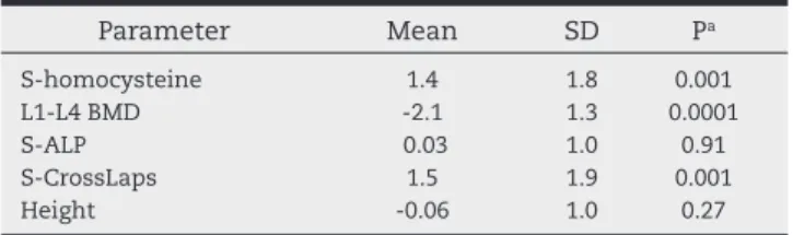
there are scarce data concerning S-Hcy and bone health in children and adolescents. In our recent pilot study involving 19 children (12 boys and 7 girls; mean age 14.9 ± 3.3 years) with recurrent fractures and low BMD, we observed elevated S-Hcy (Table 1) with high inverse and signii cant correlations between S-Hcy and BMD (r = -0.66; P = 0.01) and S-Hcy and se-rum alkaline phosphatase activity (S-ALP) (r = -0.56; P = 0.03), respectively.14 S-Hcy was not related to number of prevalent
fractures (r = 0.11) or S-CrossLaps (r = -0.14).14
Therefore, we further analysed S-Hcy levels together with indices of bone turnover and several clinical and biochemi-cal parameters in a group of 37 children and adolescents with low BMD and/or prevalent low-energy trauma fractures.
Patients and methods
Patients
We evaluated the charts of children and adolescents who at-tended our Pediatric Bone Clinic at The Department of
Pe-Table 1 – Patient data from previous pilot study (n = 19),14
expressed as Z-scores ± SD.
Parameter Mean SD Pa
S-homocysteine 1.4 1.8 0.001 L1-L4 BMD -2.1 1.3 0.0001
S-ALP 0.03 1.0 0.91
S-CrossLaps 1.5 1.9 0.001
Height -0.06 1.0 0.27
BMD, bone mineral density; S-ALP, serum alkaline phosphatase activity.
diatrics in Pardubice in the years 2006-2012. The inclusion criteria were: (i) age between i ve and 20 years; (ii) at least two prevalent low-energy trauma fractures in personal his-tory; or (iii) low spinal L1-L4 BMD (below -2 SD Z-score); or (iv) a combination of both.
Four children with less than two prevalent fractures and low BMD were included. In these four children, the BMD mea-surement was performed due to the following reasons: in one boy with no prevalent fracture, the BMD measurement was performed due to presence of bone fragility risk factors (life-long immobilisation after intracranial hemorrhage); in two girls without fractures, BMD was measured due to estimation of low bone density on the X-ray of the extremities performed by a surgeon after soft tissue injuries; and in one girl due to presence of one low-energy trauma femoral fracture.
The subjects were not eligible for evaluation if they were taking any drugs known to inl uence bone metabolism (al-facalcidol, calcitriol, dihydrotachysterol, bisphosphonates, glucocorticosteroids, anabolic steroids, antiepileptics, thia-zides, furosemide, thyroid hormones, growth hormone, hep-arin, warfarin) at the time of the assessment or at any time before. The subjects with diabetes mellitus, celiac disease, inl ammatory bowel disease, autoimmune disorders, Cush-ing syndrome, chronic renal failure, hypercalciuria, urolithi-asis, hyper- and hypothyroidism, hyper- and hypoparathyri-odism were also excluded.
All subjects were on a standard central European diet, con-sisting mostly of meat and carbohydrates. None was on a diet poor in vitamin B, nor was receiving doses of vitamin B exce-eding the recommended daily allowances. One obese boy was treated for hypertension with enalapril. Others were normo-tensive. Therefore, we included 37 children and adolescents (22 boys and 15 girls; age range 7-20 years; mean age 13.9 ± 3.5 years).
Procedures
We evaluated the following data in all 37 patients: S-HCy, markers of bone formation: total S-ALP and serum osteocal-cin (S-OC), marker of bone resorption serum Crosslaps, serum calcium (S-Ca), serum phosphate (S-P), serum levels of folate and vitamin B12, body height, weight, body mass index (BMI), and L1-L4 spinal BMD. S-cholesterol was also evaluated. The biochemical parameters were assessed from a single morning blood draw in fasting patients.
Materials and methods
Body height was measured on the day of the relevant blood draw to the nearest ± 0.5 cm on a calibrated stadiometer.
Body weight was measured on the same day on a calibrated scale to ± 0.5 kg.
The BMI was calculated using the equation BMI = weight (kg)/height2 (m).
BMD was measured at spine (L1-L4) with dual energy X-ray absorptiometry (DXA) Lunar GE at the day of the blood draw. Measurement precision, expressed as coefi cient of variation, was 1.0%.
S-homocysteine level was evaluated by chemilumines-cence (Immulite 2500 immunoassay system, Siemens
Health-care Diagnostics, Germany) and expressed in μmol/L. The interassay variation was 2.06% in samples with S-Hcy concen-tration 7.43 μmol/L; 1.99% with S-HCy 10.31 μmol/L, and 1.72% with S-Hcy 22.25 μmol/L, respectively. S-Ca and S-P were as-sessed by colorimetric assay and expressed in mmol/L. S-ALP was measured by colorimetric assay and expressed in μkat/L.
Serum CrossLaps and S-OC were assessed by means of electrochemiluminescence immunoassay – ECLIA on Elecsys-Cobas analyzers and expressed in ng/L and ng/mL, respec-tively.
Serum folic acid and serum vitamin B12 (S-B12) were as-sessed by chemiluminiscence on Access analyzer (Beckman Coulter) and expressed in μg/L and ng/L, respectively.
Statistics
To eliminate the inl uence of age, the obtained results of body height, weight, BMI, S-HCy, S-ALP, S-CrossLaps, and S-OC were calculated as standard deviationscores (SDS) or Z-scores by the equation SDS = (actual individualvalue – mean value for age and sex)/standard deviationfor age and sex. The BMD refe-rence data (concerning European paediatric population) were supplied by the manufacturer within the DXA software pac-kage. Previously published results served as reference data: Czech anthropometric parameters from a 2001 survey,15
pre-viously obtained HCy values of healthy Czech paediatric po-pulation,16 S-ALP values of Czech children,17 and S-CTx levels
of healthy British population.18 The normal S-OC range was
determined in 77 children aged 7-19 years without evidence of bone, hepatic, renal, gastrointestinal or endocrine disorders. For statistical analysis, Sigmaplot 2.0 and Systat programme were used. The statistical analysis was performed by unpaired
t-test. The linear regression analysis was performed to com-pare the relationship among respective parameters. For all re-sults, P < 0.05 was required for statistical signii cance.
Results
The mean number of prevalent low-energy trauma fractures was 3.3 ± 2.3 (SD) per patient.The mean S-homocysteine level was 10.7 μmol/L ± 2.9 SD; range 6.7-20 μmol/L. There was a sig-nii cant positive correlation with age (r = 0.47, P < 0.01) (Fig. 1). Seven patients (19%) had S-homocysteine level above +2 SD, 30 patients (81%) had S-homocysteine levels in the range of ± 2 SD.
When converted to Z-scores and compared with reference values, the S-Hcy Z-score was signii cantly higher and L1-L4 BMD Z-score was signii cantly lower, respectively, in compari-son with reference values. S-ALP Z-score did not differ from reference values, while S-CrossLaps were higher, same as S-OC (Table 2).
The mean levels of S-folate and B12 were 7.5 μg/L ± 4.2 SD and 413 ng/L ± 143 SD, which fall within normal reference ranges of 2.3-17 μg/L and 180-914 ng/L, respectively.
Mean body height, body weight, and BMI Z-score did not differ from reference values (Table 2), however in i ve sub-jects the BMI Z-score exceeded + 2 SD. The body height, weight and BMI Z-scores positively correlated with L1-L4 BMD Z-score (r = 0.44, 0.53 and 0.50, respectively; P = 0.02).
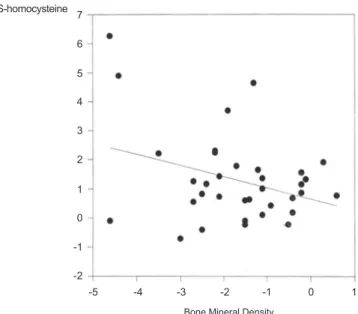
Hcy was inversely correlated to L1-L4 BMD (Fig. 2) and S--ALP (Fig. 3), respectively, and not related to number of preva-lent fractures (r = 0.11), S-osteocalcin (r = -0.22) or S-CrossLaps (r = 0.003).
There was signii cant inverse correlation between S-Hcy and vitamin B12 (r = -0.36, P = 0.05) and no correlation be-tween S-Hcy and serum level of folic acid (r = 0.05).
Furthermore, we found positive correlations between S-os-teocalcin and S-CTx (r = 0.36, P = 0.05), S-ALP and S-OC (r = 0.58, P = 0.01), and S-CTx and S-ALP (r = 0.39, P = 0.05), respectively.
Discussion
In our previous pilot study of 19 children with prevalent fra-ctures and low BMD, the obtained results suggested
negati-ve inl uence of elevated S-Hcy on bone formation and BMD, while bone resorption was increased in this group of pediat-ric patients.14 In an expanded group of 37 children with
im-paired bone status, we obtained very similar results. S-Hcy was elevated and inversely correlated with S-ALP and BMD. Furthermore, we found indices of increased bone turnover in our patients, as S-OC and S-Crosslaps were elevated in comparison to the reference data, and there were positi-ve mutual correlations among the bone turnopositi-ver markers. The low BMD in our patients was not inl uenced by stun-ted growth, as the body height did not differ from reference value, however our results coni rm dependence of BMD on basic anthropometric parameters. As there is a proven
as-Table 2 – Patient data from currently presented study (n = 37) expressed as Z-scores ± SD.
Parameter Mean SD Pa
S-homocysteine 1.31 1.49 < 0.0001 L1-L4 BMD -1.74 1.32 < 0.0001
S-ALP 0.02 1.01 0.88
S-CrossLaps 1.21 1.79 0.0001 S-osteocalcin 0.38 0.53 0.001
Height -0.05 1.31 0.82
Weight 0.18 1.62 0.50
BMI 0.48 1.88 0.12
BMD, bone mineral density; S-ALP, serum alkaline phosphatase activity; BMI, body mass index.
a Compared to reference data.
Fig. 2 – S-homocysteine (Z-score) vs. bone mineral density (Z-score). R = -0.33, P = 0.05.
7
6
5
4
3
2
1
0
-1
-2
-5 -4 -3 -2
Bone Mineral Density
-1 0 1
S-homocysteine
Fig. 3 – S-homocysteine (Z-score) vs. S-alkaline phosphatase activity (Z-score). R = -0.36, P = 0.04.
7
6
5
4
3
2
1
0
-1
-2
S-ALP
0 1 2 3 4 5 6 7 8 9
S-homocysteine
Fig. 1 – S-homocysteine (μmol/L) vs. age (years). R = 0.47, P < 0.01.
22
20
18
16
14
12
10
8
6
6 8 10 12 14 16 18 20 22
age
sociation between low bone density and fractures in chil-dren,19,20 hyperhomocysteinemia might represent an
impor-tant risk factor for children´s bone health. Furthermore, in adults, an association between high S-Hcy and low BMD was reported by several authors,6,11 same as high S-HCy in
os-teoporotic postmenopausal women with no relationship to BMD values.7 In addition, further research revealed that high
homocysteine levels are associated with an increased risk of hip fracture in elderly population.10
From the pathophysiological point of view, osteoporosis and higher fracture risk in patients with high S-Hcy are cur-rently explained by accumulation of homocysteine in bone, resulting in a distinct reduction of cancellous bone and a drop in bone strength.9 Furthermore, homocysteine
stimu-lates osteoclast activity.21 A reduced methylation capacity of
bone cells might contribute as well.22
As we did not i nd any relationship between the num-ber of prevalent fractures and S-Hcy in our patient group, we may hypothesize that the study was not powered enough to detect signii cant relationship between these two pa-rameters. Concerning the cardiovascular risk factors, the S-cholesterol and BMI were not signii cantly elevated in our patients, and only one subject was hypertensive.
Anyway, our results further coni rm the negative effect of elevated S-Hcy on bone health. even in pediatric popula-tion. The developmental aspects of various disease states of the elderly (i.e. osteoporosis, high blood pressure, hypecho-lesterolemia, obesity) are currently being given special at-tention, as these disorders usually originate in childhood. It seems likely that the effect of homocysteine on bone quality is this case as well.
Conclusion
Our results further suggest that elevated S-Hcy could be a risk factor of impaired bone health in children and adoles-cents.
Conl icts of interest
The authors declare no conl icts of interest.
R E F E R E N C E S
1. Rauh M, Verwied S, Knerr I, Dörr HG, Sönnichsen A, Koletzko B. Homocysteine concentrations in a German cohort of 500 individuals: reference ranges and determinants of plasma levels in healthy children and their parents. Amino Acids. 2001;20:409-18.
2. Stanger O, Herrmann W, Pietrzik K, Fowler B, Geisel J, Dierkes J, et al.; DACH-LIGA Homocystein e.V. DACH-LIGA homocystein (german, austrian and swiss homocysteine society): Consensus paper on the rational clinical use of homocysteine, folic acid and B-vitamins in cardiovascular and thrombotic diseases: guidelines and recommendations. Clin Chem Lab Med. 2003;41:1392-403. 3. Riddell LJ, Chisholm A, Duncan A, Mann JI. Homocysteine levels in healthy New Zealanders and those with vascular disease.N Z Med J. 1999;112:438-42.
4. Kutilek S, Nemec V, Bockayova E. Elevated serum homocysteine levels in paediatric patients with primary Raynaud´s phenomenon Rev Bras Reumatol. 2012;52:125-30.
5. Bozkurt N, Erdem M, Yilmaz E, Erdem A, Biri A, Kubatova A, et al. The relationship of homocysteine, B12 and folic acid with the bone mineral density of the femur and lumbar spine in Turkish postmenopausal women. Arch Gynecol Obstet. 2009;280:381-7.
6. Bucciarelli P, Martini G, Martinelli I, Ceccarelli E, Gennari L, Bader R, et al. The relationship between plasma homocysteine levels and bone mineral density in post-menopausal women. Eur J Intern Med. 2010;21:301-5. 7. Haliloglu B, Aksungar FB, Ilter E, Peker H, Akin FT, Mutlu
N, et al. Relationship between bone mineral density, bone turnover markers and homocysteine, folate and vitamin B12 levels in postmenopausal women. Arch Gynecol Obstet. 2010;281:663-8.
8. Krivosíková Z, Krajcovicová-Kudlácková M, Spustová V, Stefíková K, Valachovicová M, Blazícek P, et al. The association between high plasma homocysteine levels and lower bone mineral density in Slovak women: the impact of vegetarian diet. Eur J Nutr. 2010;49:147-53.
9. Levasseur R. Bone tissue and hyperhomocysteinemia. Joint Bone Spine. 2009;76:234-40.
10. Leboff MS, Narweker R, LaCroix A, Wu L, Jackson R, Lee J, et al. Homocysteine levels and risk of hip fracture in postmenopausal women. J Clin Endocrinol Metab. 2009;94:1207-13.
11. Ouzzif Z, Oumghar K, Sbai K, Mounach A, Derouiche EM, El Maghraoui A. Relation of plasma total homocysteine, folate and vitamin B12 levels to bone mineral density in Moroccan healthy postmenopausal women. Rheumatol Int. 2010;32:123-8.
12. Shiraki M, Kuroda T, Shiraki Y, Tanaka S, Higuchi T, Saito M. Urinary pentosidine and plasma homocysteine levels at baseline predict future fractures in osteoporosis patients under bisphosphonate treatment. J Bone Miner Metab. 2011;29:62-70.
13. Yilmaz N, Eren E. Homocysteine oxidative stress and relation to bone mineral density in post-menopausal osteoporosis. Aging Clin Exp Res. 2009;21:353-7. 14. Kutilek S, Rehackova P, Nemec V, Bockayová E. Serum
homocysteine levels in children with fractures and low bone mineral density – a pilot study. Osteol Bull. 2012;17:65-8.
15. Kobzova J, Vignerova J, Blaha P, Krejcovsky L, Riedlová J. The 6th nationwide anthropological survey of children and
adolescents in the Czech Republic in 2001. Cent Eur J Publ Health. 2004;12:126-30.
16. Nemec V, Bockayova E, Kutilek Š. Serum homocysteine levels in Czech children and adolescents. Acta Medica. 2012;55:87-90.
17. Kutilek S, Bayer M. Total serum alkaline phosphatase activity and its relationship to age and growth in children. Osteol Bull. 2003;8:52-5.
18. Crofton PM, Evans N, Taylor MRH, Holland CV. Serum CrossLaps: Pediatric reference intervals from birth to 19 years of age. Clin Chem. 2002;48:671-3.
19. Clark EM, Ness AR, Bishop NJ, Tobias JH. Association between bone mass and fractures in children: A prospective cohort study. J Bone Miner Res. 2006;21:1489-95.
20. Clark EM, Tobias JH, Ness AR. Association between bone density and fractures in children: a systematic review and meta-analysis. Pediatrics. 2006;117:291-7.
22. Herrmann M, Tami A, Wildemann B, Wolny M, Wagner A, Schorr H, et al. Hyperhomocysteinemia induces a tissue