Cop
yright
© ABE&M t
odos os dir
eit
os r
eser
vados
.
Diagnosis and management
of Paget’s disease of bone
Diagnóstico e tratamento da doença de Paget óssea
Luiz Griz1, Daniele Fontan1, Patricia Mesquita1, Marise Lazaretti-Castro2,
Victoria Zeghbi Cochenski Borba3, João Lindolfo Cunha Borges4,
Thyciara Fontenele1, Juliana Maia1, Francisco Bandeira1
ABSTRACT
Objective: To conduct a literature review on the diagnosis and management of Paget’s disease
of bone. Materials and methods: This scientiic statement was generated by a request from the
Brazilian Medical Association (AMB) to the Brazilian Society of Endocrinology and Metabolism (SBEM) as part of its Clinical Practice Guidelines program. Articles were identiied by searching in PubMed and Cochrane databases as well as abstracts presented at the Endocrine Socie-ty, Brazilian Society for Endocrinology Annual Meetings and the American Society for Bone and Mineral Research Annual Meeting during the last 5 years. Grading quality of evidence and strength of recommendation were adapted from the irst report of the Oxford Centre for Evi-dence-based Medicine. All grades of recommendation, including “D”, are based on scientiic evi-dence. The differences between A, B, C and D, are due exclusively to the methods employed in generating evidence. Conclusion: We present a scientiic statement on Paget’s disease of bone
providing the level of evidence and the degree of recommendation regarding causes, clinical presentation as well as surgical and medical treatment. Arq Bras Endocrinol Metab. 2014;58(6):587-99
Keywords
Paget’s disease of bone; diagnosis; treatment
RESUMO
Objetivo: Conduzir uma atualização das últimas evidências cientíicas a respeito da
apresen-tação, diagnóstico e manejo clínico da doença de Paget óssea. Materiais e métodos: Este
do-cumento foi concebido pelo Departamento de Metabolismo Ósseo da Sociedade Brasileira de Endocrinologia e Metabologia (SBEM) a partir daquele oriundo do Programa de Diretrizes da Associação Médica Brasileira (AMB). Realizamos uma revisão dos artigos mais relevantes obti-dos nos bancos de daobti-dos PubMed e Cochrane, além de abstracts apresentaobti-dos nos encontros anuais da Endocrine Society, Sociedade Brasileira de Endocrinologia e da American Society for Bone and Mineral Research dos últimos cinco anos e classiicamos as evidências em níveis de recomendações de acordo com a força cientíica por tipo de estudo, adaptando o primei-ro relato do “Oxford Centre for Evidence-based Medicine”. Todos os graus de recomendação, incluindo-se o “D”, foram baseados em evidência cientíica, sendo as diferenças entre o A, B, C e D devidas exclusivamente ao desenho empregado na geração da evidência. Conclusão:
Apresentamos uma atualização cientíica a respeito da doença de Paget óssea, classiicando e graduando em níveis de recomendações as principais evidências cientíicas sobre as suas causas, as variadas formas de apresentação, seu diagnóstico e tratamento. Arq Bras Endocrinol Metab. 2014;58(6):587-99
Descritores
Doença de Paget óssea; diagnóstico; tratamento
1 Department of Endocrinology,
Diabetes and Bone Diseases, Agamenon Magalhães Hospital, Ministry of Health/SUS/ University of Pernambuco (UPE), Recife, PE, Brazil
2 Federal University of Sao Paulo
(Unifesp), Department of Medicine, Division of Endocrinology, Sao Paulo, SP, Brazil
3 Federal University of Paraná
(UFPR), Division of Endocrinology and Metabolism, Curitiba, PR, Brazil
4 Catholic University of Brasilia
(UCB), School of Medicine, Brasilia, DF, Brazil
Division of Endocrinology, Diabetes and Bone Diseases, Agamenon Magalhaes Hospital, Ministry of Health, University of Pernambuco Medical School, Recife, PE, Brazil A Scientiic Statement, from the Department of Bone Metabolism, The Brazilian Society for Endocrinology and Metabolism
Correspondence to:
Juliana Maia
Rua Marechal Rondon, 120, ap. 1602 52061-050 – Recife, PE, Brazil maia.juliana@gmail.com
Received on Aug/17/2013 Accepted on Mar/18/2014
DOI: 10.1590/0004-2730000002941
INTRODUCTION
P
aget’s disease of bone is a metabolic bone disease characterized by very high rates of bone remodelingCop
yright
© ABE&M t
odos os dir
eit
os r
eser
vados
.
being most commonly encountered in white Euro peans, and those of European descent over 55 years of age (2,3). In Brazil, a prevalence study in a city originally colonized by Europeans identiied rates comparable to those encountered in southern Europe (3). The etiology of the disease remains controversial, but genetic factors are involved as well as environmental factors (1,4). The diagnosis is made primarily by characteristic radiological indings and the most common complications are pa thologic fractures, bone deformities and osteoarthrosis.
MATERIALS AND METHODS
This scientiic statement was generated by a request from the Brazilian Medical Association (AMB) to the Brazilian Society for Endocrinology as part of its Clinical Practice Guidelines program. Through the Brazilian Society for Endocrinology’s Department of Bone Metabolism, a task force was established. A draft of this report was submitted for comment to the membership of the Brazilian Medical Association and Brazilian Society of Endocrinology. This report represents the completion of this process.
Grading quality of evidence and strength of recom mendation were adapted from the irst report of the Oxford Centre for EvidenceBased Medicine, detailed described elsewhere (5) and summarized in table 1. Grades of recommendation are reported, as follows:
A: More consistent experimental or observational
trials.
B: Less consistent experimental or observational
trials.
C: Case reports (noncontrolled trials).
D: Opinion without critical evaluation, based on
consensus, physiological studies or animal mo dels.
Articles were identiied by searching in PubMed and Cochrane databases, as well as abstracts presented at the Endocrine Society, American Society for Bone and Brazilian Society for Endocrinology Annual Meetings during the last 5 years. References are listed numeri cally in order of appearance in the text, followed by the levels of evidence.
EPIDEMIOLOGY
The highest prevalence of Paget’s disease of bone is found in England, the United States, Australia and New Zealand, mainly among patients older than 55 years (4). In other locations, such as Asia, it is a rare disorder (6,7) (C4), as it is in Scandinavia (4) and most of Latin America (8)(C4).
PDB has been linked to white ancestry in European and other countries, being less common in people who are not of European origin. A study published in 2006 on reported cases of Paget’s disease in Latin America over the past 30 years showed that a total of 1,149 cases of Paget’s disease had been previously published in Latin America, more than half of them in Argentina and Brazil (9) (C4).
One report from Italy showed a 0.74% radiographic prevalence (n = 8 of 1,068 patients evaluated) of pelvic PDB in rural regions of Calabria, located in the sou thern part of the Italian peninsula, with a male: female ratio of 5:3 and a mean age of 71.6 ± 13.1 years (10) (C4). The region of Campania, also in southern Italy, was shown to be an area with a high prevalence of Pa get’s disease (11) (C4).
PDB rarely appears before the age of 40, but its prevalence tends to double every decade starting at the age of 50, rising to approximately 10% after ninth de cade (12).
Seitz and cols. retrospectively evaluated the bone biopsies and medical records of 754 patients historically proven to have PDB and found the peak incidence to occur between the ages of 70 and 80 years (13) (B3b).
A study from Spain evaluated 4,528 radiographs, in cluding all those of the lumbar vertebrae, pelvis, sacrum and femoral head in 13 centers studied, reporting a 1% incidence of PDB (95% CI: 0.7 to 1.3) in subjects over 55 years age and an estimated prevalence ranging from 1.1% (95% CI: 0.8 to 1.4) to 1.6% (95% CI: 1.1 to 2.1), with pelvic involvement reported in 6090% of patients considered to have PDB. The prevalence was slightly higher in men than in women and signiicantly greater
Table 1. Grades of recommendation and strength scientiic evidence
A 1 A Systematic review of randomized controlled trials Systematic reviews of prospective cohort studies 1 B Individual RCT with narrow conidence interval
Prospective cohort studies
B 2 A Systematic review of retrospective cohort studies 2 B RCT with > 20% dropout
Retrospective cohort studies
3 A Systematic review of case-control studies 3 B Individual case-control studies
C 4 Case series
D 5 Expert opinion
Cop
yright
© ABE&M t
odos os dir
eit
os r
eser
vados
.
in individuals over 75 years of age. A considerable geo graphical variation in the prevalence was observed (p = 0.004) in Spain, with 73% of the patients unaware of their disease when the radiographs were taken (14) (B3b).
One study, from the US, found a diagnosis of PDB in 236 residents of Olmsted County, Minnesota, with a mean age of 69.9 years at diagnosis, 55% of those af fected being men (15) (C4).
Paget’s disease of bone is extremely rare in Asia (16), especially in Korea (17), and among ethnic Chi nese (18)(C4). It is also very rare in Japan. A review of the literature was carried out of all cases reported in Japan from January 1990 to December 2002. Most cases (72.1%) were reported by the Department of Or thopedic Surgery and a prevalence of 2.8 cases per mil lion capita was detected, conirming the rareness of the disease in Japanese (19) (B3b).
There is little information available on the existence of the PDB in the Arab world. A recent study reported four cases of Arab patients with PDB, with variable pre sentations, characterizing the existence of the disease in the country studied (20) (C4).
A number of studies have reported an unexplained downward trend in the prevalence of PDB, some postu lating that the disease will become increasingly rare in the future (21,22) (B3b). A review of approximately 2,000 pelvic radiographs, estimated the prevalence of the disease in individuals of European descent, over 55 years of age, in two New Zealand cities (Dunedin and Auckland). The prevalence rate increased with age (p = 0.022), being hi gher in men (p = 0.014), but showing no signiicant gen der difference in either of the two cities. The Dunedin data was compared to that of another study in the year 1983 in the same city, with the prevalence now being roughly half the previous level (p = 0.012). In Auckland, the prevalence of an isolated raised plasma alkaline phosphatase level (> 150 U/L, normal range < 120 U/L) was estimated in over 80,000 blood samples processed at a community lab oratory. The prevalence of “biochemical Paget’s disease”, as measured, was very similar to that seen in a radiographic study of the same city involving subjects under 80 years of age, but not for dose over 80 (23)(B3b).
There are also reports, as in an Australian study, of a decrease in incidence and severity of the disease in newly diagnosed cases, but for unknown reasons (24), with more cases of monostotic disease, whose incidence has doubled in the last 30 years. Some reports suggest that these indings are more evident in women, as in a study conducted in England (25)(B3b).
The prevalence of PDB in Italy was assessed based on radiography, scintigraphy, and biochemical data from two Italian cities, Siena (central Italy) and Turin (northern Italy). At the end of the radiological survey, 16 of 1,778 cases of pelvic PDB (8 men and 8 women) were observed in Siena, and 41 of 6,609 cases (27 men and 14 women) in Turin. Disease prevalence was 0.89% in Siena, and 0.62% in Turin. Since pelvic involvement is normally described in 60 to 90% of the patients with PDB, the overall estimated prevalence ranged from 1.0 to 1.5% in Siena, and 0.7 to 1% in Turin. No decrease in the prevalence of PDB was evident after comparison of prevalence rates during different time periods. Bio chemical analyses showed that 296 of 7,449 subjects had elevated levels of alkaline phosphatase and normal liver enzymes, 87 of whom had a conirmed diagno sis of PDB. The estimated biochemical prevalence was 1.5%. The scintigraphy study showed an estimated prevalence of PDB in 194 of 7,906 cases (2.4%), which was signiicantly higher than radiological and bioche mical estimates. This study suggests that PDB in Italy has an estimated prevalence of at least 1%, comparable to what has been observed in the United States, and some European countries, but lower than that reported in Great Britain and New Zealand. No secular trend indicating decreased prevalence of PDB was observed in this study (26) (B3b).
Figure 1 illustrates the extensive geographical varia tion in prevalence of PDB worldwide.
Figure 1. Worldwide prevalence studies of Paget’s disease of bone.
Prevalence studies. Case series: 145.
Data from references 3,14,21,24,26-33.
EPIDEMIOLOGY OF PDB IN BRAZIL
Cop
yright
© ABE&M t
odos os dir
eit
os r
eser
vados
.
(C4) and in 2002 a case series from Recife retrospectively analyzed the characteristics of 89 cases (35)(C4).
The colonization of Pernambuco was strongly inlu enced by immigrants from Portugal and Holland, many of whom were Jews. In Recife, the occupation by the Dutch and Jews continued after the initial colonization by the Portuguese for political and administrative rea sons, which may explain the high frequency of PDB in the state of Pernambuco(8,36) (C4).
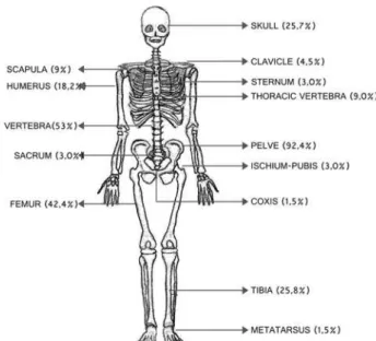
Bandeira and cols. reported an analysis of 108 cases diagnosed in two centers in Recife between 1984 and 2005 and found that about 90% of the patients were of European ancestry. The mean age at diagnosis was 66.2 years; 49.1% were men; the polyostotic form of the disease was the most common; lightcolored eyes were observed in 22.2% of the patients (data on Brazilian general population is not available); 23.1% of the pa tients were shown to have a family history of PDB. The most affected skeletal locations in this study were the pelvis, lumbar vertebrae, femur and cranium. It was also found that zoledronate was the most effective drug in reducing alkaline phosphatase in these patients (8) (C4).
A Brazilian study about the epidemiology of PDB in Brazil evaluated patients aged 45 years or older at tending the Osteoporosis Center of the Department of Endocrinology and Diabetes of Pernambuco between January of 2006, and December of 2009. The agere lated period of prevalence and incidence density were both calculated, separately for men and women for each year. A total of 7,752 patients were evaluated, 53 of whom had PDB. The mean age of patients was 69.53 ± 8.51 years. The overall prevalence of PDB was 6.8 per 1,000 patients (p = 0.013, 95% CI 5.1 to 8.9), and the incidence density of PDB was 50.3 per 10,000 person years (p = 0.026 95% CI 35.8 to 68.8). The prevalence and incidence both increased in both sexes during this period. These data shows that both the prevalence and incidence of PDB in Recife are comparable to the cor responding rates in southern Europe (3)(B3b).
A case series from the city of Florianopolis, retro spectively evaluated data from patients enrolled in 6 centers between 1995 and 2009. A total of 134 pa tients with PDB were identiied, with a mean age at diagnosis of 63.2 + / 10.5 years, of whom 67.2% were women and 91.1% white. A positive family history was observed in only 8.2% of patients. Polyostotic disease was found in 75.0%, bone pain in 77.9%, and bone deformities in 15.9%. Higher levels of alkaline phos phatase showed a signiicant association with both the
Figure 2. Sites of bone involvement in 108 cases of polyostotic Paget's
disease of bone (PDB). Data from reference 8.
polyostotic form of the disease and involvement of the cranium. The pelvic bones were the most frequently affected (53.7%). Treatment with zoledronic acid pro duced the best results, with only 2.9% of the patients failing to show anadequate response (37) (C4).
ETIOLOGY
The etiology of the disease remains controversial, with evidence that genetic along with environmental factors are involved. SQSTM1 (sequestosome 1 encoder) is the most important gene that has been associated with the disease up to now (38) (B3b).
Various loci of susceptibility have been linked to the disease, including SQSTM1 and TNFRSF11A (encoder
of the RANK) on chromosome 18q2122.SQSTM1,
Cop
yright
© ABE&M t
odos os dir
eit
os r
eser
vados
.
causing function loss in the TNFRSF11B gene, encod ing the OPG, could also lead to an activating effect in the signalization of NFkB (39) (C4) and there are also evidence that TNFRSF11A gene allelic variants interact with SQSTM1 mutations to cause the severity of the disorder (40) (B3b).
Not only genetic causes have been proposed as the etiology of PDB, biological hybridization studies (in
situ), along with immunohistochemistry, have also
suggested the possibility of infection of the osteoclasts by a virus, mainly paramyxovirus, as being the cause of PDB (4) and a study published in 2010 suggested that measles virus nucleocapsid gene expression and the
SQSTM1 mutation both contribute to the increased os
teoclast activity in PDB (41) (B3b) (42) (C4).
A French study found that half of PDB familial forms carried a SQSTM1 mutation (43) (B3b). Other loci that predispose to PDB were recently identiied by genomic association studies that have identiied variants at seven loci predisposing to the disease. These alone increase the risk of PDB from 1.3 to 1.7 times, but they have combined effects that affect about 86% of the PDB at risk population with negative SQST1 (42) (C4).
Recent studies also suggest that proinlammatory cytokines are involved in the pathophysiology of Paget’s disease of bone. A recent control case study evaluated the genomic DNA for functionally active polymor phism of the genes of proinlammatory cytokines (interleukin1α, interleukin1 β, interleukin 6), and tu mor necrosis factor α, involving 144 PDB patients and 115 healthy controls. The frequency of genotypes and alleles of the polymorphisms examined demonstrated practically identical results in both cases and controls. Regarding proinlammatory genes, patients with PDB genotype C/C gene of interleukin 6 (IL6) showed signiicantly (p < 0.001) greater hearing loss and pri mary hyperparathyroidism. There were no signiicant differences in the other clinical features. This study does not support the hypothesis that the proinlam matory genes examined represent an important genetic risk factor for PDB. However, data suggests a role for the IL6 gene in modifying the clinical characteristics of the disease (44) (B3b).
A study from New Zealand evaluated the rela tionship between family history, phenotype, and the state of SQSTM1 mutation in patients with a family history, and/or severe phenotype of PDB. The sever ity of the phenotype was signiicantly associated with
the SQSTM1 mutation status, but not with family his
tory (p < 0.005). SQSTM1 mutations were found in 10.5% of the patients with early onset, and/or severe disease, but without a family history of the disorder (38) (B3b).
An earlier study of genomic association had al ready identiied variants in loci CSF1, OPTN and
TNFRSF11A, as risk factors for Paget’s disease of bone,
and an extension of this study identiied three new loci and recently conirmed these associations with PDB in 2,215 affected individuals (cases), and 4,370 con trols from seven independent population groups. The new associations were with rs5742915 within PML
on 15q24, rs10498635 within RIN3 on 14q32 and
rs4294134 within NUP205 in 7q33 and also conirms
the association of TM7SF4 with PDB. These seven loci account for the familial risk for PDB in approximately 13% of cases (45) (B3b).
There are studies investigating the relationship between genetic polymorphisms and sporadic PDB, with the aim of identifying polymorphisms represent ing susceptibility to the disease. A recent study in vestigated the association between polymorphisms in three candidate genes and the functional development of PDB, TNFSF11 (activator receptor of the nuclear factor Kb ligand, RANKL), VCP (valosinacontaining protein), and IL6 (interleukin6), in 196 patients with sporadic PDB, and 212 Belgian control subjects, and revealed that VCP SNP (rs565070) was associated with PDB in this population study (p = 0.5). Through the use of genetic testing in the study, no association linking TNFSF11 or IL6 with PDB was conirmed. More data are therefore needed because when the VCP data is combined with data from other regions, involving susceptible genes in previous studies (i.e. the
TNFRS11A, CSF1, OPTN and TM7SF4 genes), the
independent effect of each gene region was conirmed and the accumulated population attributable risk was 72.7% (46) (B3b).
Cop
yright
© ABE&M t
odos os dir
eit
os r
eser
vados
.
STAMP, which had no signiicance in association with the genomic GWAS, but because of its effect on osteo clasts, can be considered a strong candidate gene. The cumulative risk attributable to these four loci calculated for the two populations studied was shown to be ap proximately 67%, indicating that the major part of the genetic risk for PDB comes from genetic variants close to these four genes (47) (B2b).
Patients with PDB without mutation of the
SQSTM1 gene seem to be susceptible to genetic poly
morphisms in regions of the genes CaSR, ESR1,
TNFRSF11B (OPG), TNFRSF11A (RANK), CSF1
(M-CSF), OPTN, TM7SF4 (DC-STAMP) VCP,
NUP205, RIN3, PML, and GOLGA6A, resulting in an
increased risk for developing PDB. The nature of these genes suggests that the regulation of osteoclastogenesis has a key role in the pathogenesis of PDB. Moreover, the involvement of SQSTM1 and VCP in autophagy and the formation of protein aggregates suggest that the disruption of these processes may represent a risk factor (48,49).
There are also reports of a high prevalence of vita min D deiciency in patients with PDB. One possibly reason for this is that osteoclast precursors have a high responsiveness and sensitivity to 1.25 (OH) 2D3, re sulting in increased expression of coactivators of vita min D receptors (VDR) in PDB (50) (C4).
In recent years there has been signiicant progress in the study of the epidemiology of PDB. However, we still lack a combined evaluation of genetic and envi ronmental factors to enable us to fully understand their interaction with the etiology of the disease (D5).
CLINICAL MANIFESTATIONS
Most patients with Paget’s disease of bone (PDB) are asymptomatic (51), having been incidentally diagnosed through indings from imaging or because of high se rum levels of alkaline phosphatase (52) (C4).
The clinical spectrum of PDB is highly variable and depends on the sites affected, the type and magnitude of the complications, and the metabolic activity (53). Although the disease may affect any part of the skeletal structure, the pelvis, spine, cranium and long bones are the most often affected (15,54) (C4).
Bone pain is the most common symptom. In two recently published studies pain was found to be pres ent in 4045% of the patients. It is usually deep, pre cisely located and persists when the patient is at rest,
constant, exacerbated both at night and by weight overload. It may occur suddenly as a result of pagetic injury, or more frequently, from complications caused by the breakdown of bone structure, leading to condi tions such as degenerative arthritis, nerve compression or sarcomatous degeneration (a rare occurrence present in only 1% of cases) (15,55) (C4).
Bone deformities are the second most common mani festation with a prevalence ranging from 12 to 36%. They occur most commonly in the femur and tibia, causing bending, which is characteristically anterolateral in the fe mur and anterior in the tibia. These deformities can lead to changes in gait and mechanical stress, increasing the likelihood of joint degeneration. Involvement of the cra nium begins with circumscribed osteoporosis, followed years later by increases in volume, regions of sclerosis, an enlarged diploe and frontal bossing (15,55) (C4).
We suggest evaluation for PDB in all individuals over 50 years of age who present an unexplained eleva tion of serum alkaline phosphatase, as well as bone pain or deformities (C).
LABORATORY AND IMAGING PROCEDURES
PDB is associated with increased bone turnover, indi cated by the elevation of biochemical markers for bone formation and resorption. This occurs because a high correlation between formation and resorption is main tained with PDB. The increase in these markers is pro portional to the intensity, size, and number of lesions, and may be more pronounced in cases involving the cranium. Serum alkaline phosphatase, a bone formation marker, has been used for the diagnosis and monitoring of patients affected by the disease. However, none of the biochemical markers of bone remodeling are enti rely speciic to bone alone. Serum alkaline phosphatase has a sensitivity of 78%, and a speciicity of almost 100%. However it may be normal in up to 20% of patients with monostotic disease (4,56,57). In a study by Bandeira and cols. serum alkaline phosphatase was shown to be elevated in 92% of the cases. The mean increase was sig niicantly more pronounced in patients with the polyos totic form than in those with monostotic form of the disease (5.9 ± 2.8 vs. 2.2 ± 1.9 times the ULN) (58).
Cop
yright
© ABE&M t
odos os dir
eit
os r
eser
vados
.
est diagnostic accuracy (59). Recent data has demon strated a signiicant reduction in CTX after use of oral ibandronate. After six months of treatment, the mean decrease in CTX was 65.24 ± 9.28%, with reductions greater than 80% in seven of the patients. One patient with normal sCTX showed a reduction of 97.5% by the end of the treatment (60) (B2b). As with betaCTX, there was evidence of a greater reduction in the ratio of urinary αCtelopeptide of type 1 collagen tocreatinine (mg/nmol of creatinine) with the infusion of zoledron ic acid, when compared to risedronate (61) (B3b).
Serum calcium and phosphorus levels are normal in most patients. Hypercalcemia and hypercalciuria may occur in the case of immobilization or fracture. The inding of hypercalcemia normally points to a second ary disorder such as hyperparathyroidism.
Bone scintigraphy followed by radiography of the affected areas determines the extent of involvement of the bone in Paget’s disease. Sites of increased uptake occur as a result of the high rate of bone formation and blood low. As a more sensitive method, bone scan with Tc 99MDP can be positive even before the lytic changes seen on plain radiography. For this reason, about 1015% of lesions detected by scintigraphy ap pear normal on plain radiographs. Comparing results from both methods, bone scintigraphy and radiography show alterations 56 to 86% of the time, with 223% of the cases showing alterations only in the scintigrams, and 1120% only in the radiographs. The late stages of the disease may show a normal uptake of the radiophar maceuticals, owing to the declinein metabolic activity and alterations in the indings of plain radiography. The characteristic indings of plain radiography are hyperos tosis, osteosclerosis and bone expansion. CT and MRI provide little additional information when dealing with uncomplicated cases of the disease. They can be useful when complications associated with PDB are suspected, such as fractures or sarcomatous degeneration of page tic bone. Due to the excellent resolution, MRI is the method of choice for the staging of sarcomatous de generation (62). It can also be useful in the evaluation of neurological complications, such as compression of spinal nerve roots and cranial nerves (6265) (C4).
COMPLICATIONS
Clinical manifestations of the disease are usually related to the presence of complications, which can be classi ied according to the particular system involved: ske
letal (bone pain, osteoarthrosis, fractures, deformities and hypercalcemia), cardiovascular (high output heart failure, vascular calciications, and valvular stenosis), neurological (deafness, increased intracranial pressure, and cranial nerve dysfunction), metabolic (hyperurice mia, hypercalciuria, hypercalcemia and nephrolithiasis) and neoplastic (osteosarcoma and giant cell tumors) (6668) (D5).
Osteoarthritis is a common complication, most of ten affecting the knee and hip joints, resulting in the modiication of bone biomechanics that causes bone and cartilage degeneration. Rheumatoid arthritis and its variants, as well as arthropathy from crystal deposi tion, have also been associated with the disease (69) (D5). Deformities and fractures are the result of ab normal bone formation, and associated with high morbidity due to the high incidence of associated pain (70) (D5). The involvement of cranial bones can cause neurological complications such as hearing loss (either neurosensorial or conductive), headache, dizziness, and more rarely, vascular dementia and hydrocephalus. In volvement of the jaw bones may lead to periodontal disease and dental malocclusion (66,67) (D5).
Malignant transformation of pagetic bone involv ing osteosarcoma or giant cell tumor is rare, occurring in less than 1% of cases. It classically affects individuals with the polyostotic form of the disease, and manifests itself accompanied by an increase in bone pain, swell ing, and more rarely, pathologic fracture (7174) (D5). Hypercalcemia is often associated with prolonged immobilization or dehydration. Cardiovascular chan ges, although described, are rarely evident in clinical practice (75) (D5). Because PDB is a chronic disease, and diagnosed belatedly, it is important to be aware of the signs and symptoms that indicate the need for fur ther radiological investigation.
TREATMENT
Pharmacological treatment seeks to promote pain relief and reduce the rate of bone remodeling. Restoration of typical bone turnover normalizes the rate of bone deposition, reducing bone vascularization, and slowing progression of the disease.
Cop
yright
© ABE&M t
odos os dir
eit
os r
eser
vados
.
these cases is usually indicated by a concomitant eleva tion of serum alkaline phosphatase (7678) (D5).
The asymptomatic form of the disease is often de tected by imaging studies, motivated by suspicion of other diseases, or by observation of persistently elevat ed levels of serum alkaline phosphatase. Therapeutic decisions involving affected patients should take into account the location of the disease in sites that are susceptible to complications, such as the cranium and spine, in addition to abnormally high alkaline phos phatase levels (two to four times above the upper limit considered normal), and the presence of comorbidi ties (79).
Other indications for treatment of asymptomatic patients include planned surgery at an active pagetic site in order to reduce the risk of bleeding (including blood loss during surgery), along with the rare possi bility of developing hypercalcemia associated with the immobilization of patients with the polyostotic form of the disease (76) (D5).
We suggest the initiation of pharmacological treat ment for all symptomatic patients, for preoperative as ymptomatic patients requiring bone surgery, those with hypercalcemia, and cases involving locations liable to present complications (C).
PHARMACOLOGICAL TREATMENT MODALITIES
Several treatment modalities have been employed in an effort to care for patients with Paget’s disease. They involve agents that target osteoclasts, the primary cells responsible for the disease, and that act by suppressing bone resorption within days or weeks. Most researchers conclude that these drugs are best considered as tools for helping to control the disease, rather than being a deinitive therapy (80) (A1b).
The irst therapy used for Paget’s disease (in the 1970s) involved salmon calcitonin, followed later by human cal citonin. The medication acts directly on calcitonin recep tors located on the osteoclasts. Owing to its short dura tion of action, partial response, and acquired resistance, it is used only in those with intolerance to bisphosphonates (81) (D5). Although radiological improvement has been reported during treatment, recurrence is common after the medication is discontinued. Side effects are common, including lushing, nausea and vomiting (22) (D5).
The eficacy of parenteral salmon calcitonin was evaluated in a trial involving 85 patients. Alkaline phos phatase levels and urinary hydroxyproline excretion de
creased by approximately 50% after the irst few months of therapy. However, in 22 of the 85 patients, despite the continued treatment, these parameters returned to pretreatment levels. Nineteen patients were considered treatmentresistant, presenting elevated amounts of an tibodies to calcitonin (82) (B2b). The usual initial dose is 50 to 100 units per day (as tolerated), and the main tenance dose is normally 50 units daily, or 50 to 100 units every three days. The intranasal formulation, easy to administer, can be as effective as parenteral therapy, but has not yet been approved in the United States for this speciic purpose (83,84) (D5).
Currently, the most widely used agents for treating the disease are the bisphosphonates, a broad class of medications that work by blocking osteoclastic bone resorption. Nitrogenous bisphosphonates (alendro nate, risedronate, pamidronate, and zoledronic acid) are the drugs of choice (60,79) (A1b).
The irst bisphosphonate used for the treatment of Paget’s disease (in 1971) was the etidronate form, a nonnitrogenous bisphosphonate. The recommended dosage is 5 mg/kg per day (mean dose 400 mg/day) for six months. In general, patients with very active forms of the disease experience moderate clinical and biochemical improvement, followed by rapid relapse after stopping the medication. In addition, there is a tendency to become resistant to the medication after repeated courses of therapy (85) (D5).
Clodronate has greater potency than etidronate, and does not lead to mineralization defects. It should be administered intravenously at dose levels of 300 mg daily for 5 days. However, it is generally less effective than pamidronate (86) (B2b).
In the nonnitrogenous class of bisphosphonates, tiludronate is recommended in doses of 400 mg/day for 3 months, normalizing alkaline phosphatase in 35% of pa tients. It is more effective than etidronate, and does not cause bone demineralization. In a randomized, placebo controlled trial, 149 patients used tiludronate at doses of 400 and 800 mg/day for three months, presenting a sig niicant reduction in bone markers and pain (87) (A1b).
Alendronate is used at dose levels of 40 mg daily for six months. It is generally a welltolerated drug, ef fective in normalizing serum alkaline phosphatase. It should not be used in patients with creatinine clearance below 35 ml/min (88) (A1b).
Cop
yright
© ABE&M t
odos os dir
eit
os r
eser
vados
.
line phosphatase (79% vs. 44%), and urinary deoxypyrid inoline (75% vs. 51%) than a group treated with 400 mg of etidronate (p < 0.001 in both cases). Alendronate was well tolerated, and had a safety proile similar to that of etidronate (89) (A1b). In an open trial lasting two years, 72 patients with Paget’s disease were assigned to receive either 60 mg of pamidronate every 3 months or 40 mg of alendronate daily for 3 months. The study concluded that alendronate and pamidronate have similar eficacy in achieving biochemical remission (90) (A1b).
Risedronate is used in 30mg doses daily for two months, but should not be administered to patients with a creatinine clearance of less than 30 ml/min. In an American multicenter study, 62 patients received risedro nate, 30 mg daily for 2 months, and 61 patients received etidronate, 400 mg daily for 6 months. Serum levels of alkaline phosphatase were controlled in 73% of the pa tients treated with risedronate, compared with 15% of the patients who received etidronate (P < 0.001). The aver age time for normalization was 91 days for the patients treated with risedronate, and 360 days for the patients treated with etidronate (P < 0.001). Relapse rates were 3% in the risedronate group and 15% in the etidronate group (P < 0.05). Pain reduction was statistically signii cant in the risedronate group, but not in the etidronate group. Both drugs were well tolerated (91) (A1b).
Pamidronate is well tolerated and can be used with a clearance above 30 ml/min. It is administered intrave nously in 30 mg doses daily for three days. One drawback to its use is the development of resistance, which may inluence the effectiveness of retreatment (92) (A1b). It may lead to a fall in serum alkaline phosphatase by 70% and about 6080% will normalize it. The response is bet ter in patients with higher values at baseline (93).
Ibandronate has been used safely and effectively in treating Paget’s disease with 2 mg intravenous doses (94) (B2b). Recent data from a series of cases shows a signiicant reduction in the levels of sCTX, and in the algic aspect following oral use of ibandronate with 150 mg doses per month for six months. After six months of treatment, there was a mean reduction in sCTX of 65.24 ± 28.9%, and reduction of more than 80% in 58.3% of the patients studied. One patient with normal sCTX showed a reduction of 97.5% at the end of the treatment period. The mean reduction in alka line phosphatase was 49.21 ± 37.9%, with all patients presenting normal levels after the treatment. There was a signiicant clinical response in all patients, with a marked improvement in bone pain (60) (C4).
Zoledronic acid is the most potent bisphosphonate approved for use in cases of Paget’s disease. Adminis tered in a single intravenous dose of 5 mg, it is not recommended for patients with a clearance below 35 ml/min. Sustained remissions are achieved in most pa tients, lasting up to two years (61) (A1b). This inding was conirmed when the study was extended for 6.5 years (95). Zoledronic acid can lead to a more rapid and prolonged remission during the treatment of Paget’s disease when compared to risedronate. When evaluated for six months, using a single 5 mg infusion adminis tered over 15 minutes, the effective response being considered normalization of alkaline phosphatase (or a decrease of at least 75%), it resulted in a 96% reduction in alkaline phosphatase, compared with a 74.3% reduc tion with risedronate when administered in daily 30 mg doses for 3 months. Normalization of alkaline phos phatase levels was more frequently achieved in patients treated with the zoledronic acid (88.6% vs. 57.9%) than in with those receiving risedronate (96) (A1b).
We indicate the use of nitrogenated bisphosphonates (alendronate, risedronate, pamidronate, and zoledronic acid) for the treatment of Paget’s disease, emphasizing that zoledronic acid is the most potent bisphosphonate for use with this disease. (C)
MONITORING DISEASE ACTIVITY
Alkaline phosphatase, being a marker for bone remo deling, is commonly used as a parameter for measuring the biochemical response to treatment with bisphos phonates. Normalization of the alkaline phosphatase level is associated with biochemical remission, histolo gical evidence of normal bone turnover, and its eleva ted level is related to the increase in disease activity. The measurement of alkaline phosphatase levels should be conducted after the irst three to six months of treat ment, in order to evaluate the initial response, followed by two annual measurements as a marker of bone acti vity (92) (D5).
Cop
yright
© ABE&M t
odos os dir
eit
os r
eser
vados
.
In a case series study, signiicant reductions in the levels of alkaline phosphatase and serum CTX were demonstrated after six months of treatment with orally administered ibandronate at dosages of 150 mg per month for six months. After 6 months of treatment, the mean reduction in CTX was 65.24 ± 28.9%, de ceasing more than 80% in 7 patients. One patient with normal CTX showed a reduction of 97.5% at the end of the treatment period. The average reduction in alka line phosphatase was 49.21 ± 37.9%, with all patients presenting normal levels after the treatment, suggesting that the followup of patients with PDB should also include CTX levels (60) (B2b).
Remission is considered to have been achieved when normal levels of alkaline phosphatase are attained, and partial remission when there is a decrease in levels greater than 75% after three to six months of treatment. Treatment should be resumed when alkaline phospha tase levels begin to rise again (when treatment involves normalization), or when there is a 25% increase com pared to posttreatment levels (98) (D5).
We suggest the measurement of serum alkaline phosphatase after three and six months from the start of treatment in order to monitor the initial response, followed by biannual measurements of markers indicat ing disease activity (B).
NATURAL HISTORY OF PDB
Several clinical guidelines cast doubt on the belief that the treatment of Paget’s disease alters the history of disease complications. Results from the PRISM study showed that most treatment approaches have limited impact on quality of life, pain and hearing loss, and hi ghlighted the need for further studies to examine whe ther the effects of bisphosphonates on bone remodeling can actually translate into a clinical improvement and lower risk of complications in the individuals affected (99) (B2b). On the other hand the followup was too short to show the expected beneicial effects on chro nic complications of the disease. Longterm studies are therefore needed in order to assess the impact of treat ment on the natural history and progression of PDB.
CONCLUSIONS
Paget’s disease of bone has a variable geographic distri bution worldwide, being most commonly encountered in white Europeans, and those of European descent over
55 years of age. In Brazil, a prevalence study in a city originally colonized by Europeans identiied rates com parable to those encountered in southern Europe. The etiology of the disease still remains controversial, with evidence that genetic factors are involved, particularly those relates to SQTM1 and genetic polymorphisms in sections of the following genes: CaSR, ESR1, TNFRS-F11B (OPG), TNFRSF11A (RANK), CSF1 (M-CSF), OPTN, TM7SF4 (DC-STAMP), VCP, NUP205, RIN3,
PML and GOLGA6A (1,4). Environmental factors,
along with viral and proinlammatory cytokines may also be involved. The clinical presentation is quite ex tensive, with bone pain, fractures, skeletal deformities and secondary arthrosis comprising part of the pictu re. The diagnosis is made primarily by characteristic radiological indings and high alkaline phosphatase. The most common complications include pathologic fractures, bone deformities and osteoarthrosis. Malig nant transformation of pagetic bone is rare. Treatment should be recommended for all symptomatic or asymp tomatic patients, during the preoperative phase of bone surgery, or when hypercalcemia or support bones are involved. Nitrogen contained bisphosphonates are the medications of choice, with zoledronic acid being the most potent bisphosphonate approved for pharmaco logical treatment. Newer compounds such as deno sumab, a monoclonal antibody against RANKL may show promises for the treatment of PDB, although data are still lacking. Serial measurements of serum alkaline phosphatase should be used to monitor disease activity, combined with clinical monitoring of the patient.
Disclosure: no potential conlict of interest relevant to this article was reported.
REFERENCES
1. Falchetti A, Di Stefano M, Marini F, Del Monte F, Mavilia C, Strigoli D, et al. Two novel mutations at exon 8 of the sequestosome 1 (SQSTM1) gene in an Italian series of patients affected by Paget’s disease of bone (PDB). J Bone Miner Res. 2004;19(6):1013-7. 2. Britton C, Walsh J. Paget disease of bone - an update. Aust Fam
Physician. 2012;41(3):100-3.
3. Reis RL, Poncell MF, Diniz ET, Bandeira F. Epidemiology of Paget’s disease of bone in the city of Recife, Brazil. Rheumatol Int. 2012;32(10):3087-91.
4. Griz L, Caldas G, Bandeira C, Assunção V, Bandeira F. Paget’s dise-ase of bone. Arq Bras Endocrinol Metab. 2006;50(4):814-22. 5. Oxford Centre for Evidence-based Medicine. Levels of evidence
and grades of recommendations. Available at: http://cebm.jr2. ox.ac.uk/docs/levels.html. Accessed on: Nov 16, 2014.
Cop
yright
© ABE&M t
odos os dir
eit
os r
eser
vados
.
7. Bhadada S, Bhansali A, Unnikrishnan AG, Khadgawat R, Singh SK, Mithal A, et al. Does Paget’s disease exist in India?: A series of 21 patients. J Assoc Physicians India. 2006;54:530-4.
8. Bandeira F, Assunção V, Diniz ET, Lucena CS, Griz L. Characteris-tics of Paget’s disease of bone in the City of Recife, Brazil. Rheu-matol Int. 2010;30(8):1055-61.
9. Rojas-Villarraga A, Patarroyo PA, Contreras AS, Restrepo JF, Iglesias-Gamarra A. Paget disease of bone in Colombia and Latin America. J Clin Rheumatol. 2006;12(2):57-60.
10. Rendina D, Gianfrancesco F, De Filippo G, Merlotti D, Esposito T, Aloia A, et al. Epidemiological, clinical, and genetic characteris-tics of Paget’s disease of bone in a rural area of Calabria, Sou-thern Italy. J Endocrinol Invest. 2010;33(8):519-25.
11. Rendina D, Gennari L, De Filippo G, Merlotti D, de Campora E, Fazioli F, et al. Evidence for increased clinical severity of familial and sporadic Paget’s disease of bone in Campania, southern Italy. J Bone Miner Res. 2006;21(12):1828-35.
12. Colina M, La Corte R, De Leonardis F, Trotta F. Paget’s disease of bone: a review. Rheumatol Int. 2008;28(11):1069-75.
13. Seitz S, Priemel M, Zustin J, Beil FT, Semler J, Minne H, et al. Paget’s disease of bone: histologic analysis of 754 patients. J Bone Miner Res. 2009;24(1):62-9.
14. Guañabens N, Garrido J, Gobbo M, Piga AM, del Pino J, Torrijos A, et al. Prevalence of Paget’s disease of bone in Spain. Bone. 2008;43(6):1006-9.
15. Wermers RA, Tiegs RD, Atkinson EJ, Achenbach SJ, Melton LJ 3rd. Morbidity and mortality associated with Paget’s disease of bone: a population-based study. J Bone Miner Res. 2008;23(6):819-25. 16. H’ng MW, Ho YY. Paget’s disease of the bone in a Chinese woman.
Australas Radiol. 2005;49(6):505-7.
17. Lee CH, Han SH, Yoon BY, Lee YW. Monostotic Paget’s disease of the tibia in Korea. Clin Rheumatol. 2004;23(4):381-2.
18. Wang WC, Cheng YS, Chen CH, Lin YJ, Chen YK, Lin LM. Paget’s disease of bone in a Chinese patient: a case report and review of the literature. Oral Surg Oral Med Oral Pathol Oral Radiol Endod. 2005;99(6):727-33.
19. Hashimoto J, Ohno I, Nakatsuka K, Yoshimura N, Takata S, Zam-ma M, et al. Prevalence and clinical features of Paget’s disease of bone in Japan. J Bone Miner Metab. 2006;24(3):186-90.
20. Alshaikh OM, Almanea H, Alzahrani AS. Paget disease of the bone: does it exist in Saudi Arabia? Ann Saudi Med. 2011;31(3):305-10. 21. Bastin S, Bird H, Gamble G, Cundy T. Paget’s disease of bone
-be-coming a rarity? Rheumatology (Oxford). 2009;48(10):1232-5. 22. Poór G, Donáth J, Fornet B, Cooper C. Epidemiology of Paget’s
disease in Europe: the prevalence is decreasing. J Bone Miner Res. 2006;21(10):1545-9.
23. Doyle T, Gunn J, Anderson G, Gill M, Cundy T. Paget’s disea-se in New Zealand: evidence for declining prevalence. Bone. 2002;31(5):616-9.
24. Walsh JP. Paget’s disease of bone. Med J Aust. 2004;181(5):262-5. 25. Haddaway MJ, Davie MW, McCall IW, Howdle S. Effect of age and gender on the number and distribution of sites in Paget’s disease of bone. Br J Radiol. 2007;80(955):532-6.
26. Gennari L, Di Stefano M, Merlotti D, Giordano N, Martini G, Tamo-ne C, et al. Prevalence of Paget’s disease of boTamo-ne in Italy. J BoTamo-ne Miner Res. 2005;20(10):1845-50.
27. Altman RD, Bloch DA, Hochberg MC, Murphy WA. Prevalence of pelvic Paget’s disease of bone in the United States. J Bone Miner Res. 2000;15(3):461-5.
28. Saraux A, Brun-Strang C, Mimaud V, Vigneron AM, Lafuma A. Epi-demiology, impact, management, and cost of Paget’s disease of bone in France. Joint Bone Spine. 2007;74(1):90-5.
29. Cooper C, Schafheutle K, Dennison E, Kellingray S, Guyer P, Ba-rker D. The epidemiology of Paget’s disease in Britain: is the pre-valence decreasing? J Bone Miner Res. 1999;14(2):192-7.
30. Cooper C, Harvey NC, Dennison EM, Staa van TP. Update on the epidemiology of Paget’s disease of bone. J Bone Miner Res. 2006;21(2):P3-8.
31. van Staa TP, Selby P, Leufkens HG, Lyles K, Sprafka JM, Cooper C. Incidence and natural history of Paget’s disease of bone in En-gland and Wales. J Bone Miner Res. 2002;17(3):465-71.
32. Guyer PB, Chamberlain AT. Paget’s disease of bone in South Afri-ca. Clin Radiol. 1988;39(1):51-2.
33. Acotto CG, Mautalen CA. European origin of patients with Paget’s di-sease of bone in the Buenos Aires area. Euro J Epid. 2001;17:409-11. 34. Spina L, Carvalho F, Eksterman L, Torres S, Janini M, Farias M.
Doença de Paget com manifestação nos maxilares. Arq Bras En-docrinol Metab. 2000;44(3):267-72.
35. Bandeira F, Alencar S, Caldas G, Griz L, Macedo G, Marinho C, et al. Paget’s disease of bone revisited - a study on 89 patients. Arq Bras Endocrinol Metab. 2002;46(1):S364.
36. Kaufman TN. A presença judaica em Pernambuco – Brasil. 4a Edi-ção. Recife: Ensol; 2005.
37. Werner de Castro GR, Heiden GI, Zimmermann AF, Morato EF, Ne-ves FS, Toscano MA, et al. Paget’s disease of bone: analysis of 134 cases from an island in Southern Brazil: another cluster of Paget’s disease of bone in South America. Rheumatol Int. 2012;32(3):627-31. 38. Cundy T, Naot D, Bava U, Musson D, Tong PC, Bolland M. Familial
Paget disease and SQSTM1 mutations in New Zealand. Calcif Tis-sue Int. 2011;89(3):258-64.
39. Janssens K, Van Hul W. Molecular genetics of too much bone. Hum Mol Genet. 2002;11(20):2385-93.
40. Merlotti D, Gianfrancesco F, Gennari L, Rendina D, Stefano M, Mossetti G, et al. TNFRSF11A gene allelic variants are associa-ted with Paget’s disease of bone and interact with sqstm1 mu-tations to cause the severity of the disorder. J Bone Miner Res. 2010;25(Suppl 1):S11.
41. Kurihara N, Zhou H, Dempster D, Windle J, Brown J, Roodman D. Measles virus nucleocapsid gene expression and the SQTM1 mutation both contribute to the increased osteoclast activity in Paget’s disease. J Bone Miner Res. 2010;25(Suppl 1):S11. 42. Ralston SH, Albagha OM. Genetic determinants of Paget’s
disea-se of bone. Ann N Y Acad Sci. 2011;1240:53-60.
43. Michou L, Collet C, Morissette J, Audran M, Thomas T, Gagnon E, et al. Epidemiogenetic study of French families affected by Paget’s disease of bone. J Bone Miner Res. 2011;26(Suppl 1):S439. 44. Gallone S, Di Stefano M, Fenoglio P, Rubino E, Criasia A,
Pines-si L, et al. Proinlammatory cytokine gene polymorphisms and susceptibility to Paget’s disease of bone: an association study. Cytokine. 2011;56(3):560-3.
45. Albagha OM, Wani SE, Visconti MR, Alonso N, Goodman K, Brandi ML, et al. Genome-wide association identiies three new susceptibility loci for Paget’s disease of bone. Nat Genet. 2011;29;43(7):685-9.
46. Chung PY, Beyens G, de Freitas F, Boonen S, Geusens P, Vanhoe-nacker F, et al. Indications for a genetic association of a VCP poly-morphism with the pathogenesis of sporadic Paget’s disease of bone, but not for TNFSF11 (RANKL) and IL-6 polymorphisms. Mol Genet Metab. 2011;103(3):287-92.
47. Chung PY, Beyens G, Boonen S, Papapoulos S, Geusens P, Karpe-rien M, et al. The majority of the genetic risk for Paget’s disease of bone is explained by genetic variants close to the CSF1, OPTN, TM7SF4, and TNFRSF11A genes. Hum Genet. 2010;128(6):615-26. 48. Cundy T, Reid IR. Paget’s disease of bone. Clin Biochem.
2012;45(1-2):43-8.
Cop
yright
© ABE&M t
odos os dir
eit
os r
eser
vados
.
50. Kurihara N, Ishizuka S, Demulder A, Roodman GD. Paget’s disease-a VDR coativator disease? J Sterois Biochem Mol Biol. 2004;89-90(1-5):321-5.
51. Rousiere M, Michou L, Cornelis F, Orcel P. Paget’s disease of bone. Best Pract Res Clin Rheumatol. 2003;17:1019-41.
52. Papapoulos SE. Paget’s disease of bone: clinical, pathogene-tic and therapeupathogene-tic aspects. Baillieres Clin Endocrinol Metab. 1997;11:117-44.
53. Whyte MP. Clinical practice. Paget’s disease of bone. N Engl J Med. 2006;355:593.
54. Langston AL, Campbell MK, Fraser WD, Maclennan G, Selby P, Ralston SH. Clinical determinants of quality of life in Paget’s dise-ase of bone. Calcif Tissue Int. 2007;80:1-9.
55. Seton M, Moses AM, Bode RK, Schwartz C. Paget’s disease of bone: the skeletal distribution, complications and quality of life as perceived by patients. Bone. 2011;48:281.
56. Hosking D, Meunier PJ, Ringe JD, Reginster JY, Gennari C. Fort-nightly Review: Paget’s disease of bone: diagnosis and manage-ment. BMJ. 1996;312:491-4.
57. Sires E. Paget’s disease of bone. J Bone Miner Res. 1998;13:1061-5. 58. Bandeira F, Griz L, Caldas G, Macedo G, Marinho C, Moutelik M, et
al. A single center experience of 103 cases. Paget’s disease of bone in Brazil. Proceedings of the International Symposium on Paget’s disease of bone/ibrous dysplasia: advances and challenges 2006. The Paget’s Foundation, National Institute of Health. p. 53. 59. Cundy T, Bolland M. Paget disease of bone. Trends Endocrinol
Me-tab. 2008;19:246-53.
60. Voss L, Fontan D, Diniz E, Lucena C, Bandeira F. Clinical response to oral ibandronate in Paget disease. Endocr Rev. 2011;32 (03_Me-etingAbstracts): P2-148.
61. Reid IR, Miller P, Lyles K, Fraser W, Brown JP, Saidi Y, et al. Compa-rison of a single infusion of zoledronic acid with risedronate for Paget’s disease. N Engl J Med. 2005;353:898-908.
62. Cortis K, Micallef K, Mizzi A. Imaging Paget’s disease of bone---from head to toe. Clin Radiol. 2011;66:662-72.
63. Bahk YW, Park TH, Chung SK, Chi JG. Bone pathologic correlation of multimodality imaging in Paget’s disease. J Nucl Med. 1995; 36:1421-6.
64. Lavender JP, Evans IM, Arnot R, Bowring S, Doyle FH, Joplin GF, et al. A comparison of radiography and radioisotope scanning in the detection of Paget´s disease and in the assessment of respon-se to human calcitonin. Br J Radiol. 1977;50:243-50.
65. Khairi MR, Wellman HN, Robb JA, Johnston CC Jr. Paget’s dise-ase of bone (osteitis deformans): symptomatic lesions and bone scan. Ann Intern Med. 1973;79:348-51.
66. Bone HG. Nonmalignant complications of Paget’s disease. J Bone Miner Res. 2006;21:64-8.
67. Devogelaer JP, Bergmann P, Body JJ, Boutsen Y, Goemaere S, Kaufman JM, et al. Management of patients with Paget’s disease: a consensus document of the Belgian Bone Club. Osteoporos Int. 2008;19(8):1109-17.
68. Langston AL, Ralston SH. Management of Paget’s disease of bone. Rheumatology (Oxford). 2004;43:955-9.
69. Altman RD. Paget’s disease of bone: rheumatologic complica-tions. Bone. 1999;24(5):47S-8S.
70. Lander P, Hadjipavlou A. Paget disease with contraction of long bones. Radiology. 1986;159:471-2.
71. Sharma H, Mehdi S, MacDuff E, Reece A, Jane M, Reid R. Paget sarcoma of the spine: Scottish Bone Tumor Registry experience. Spine (Phila Pa 1976). 2006;31:1344-50.
72. Moore TE, King AR, Kathol MH, el-Khoury GY, Palmer R, Downey PR. Sarcoma in Paget disease of bone: clinical, radiologic, and pathologic features in 22 cases. AJR. 1991;156:1199-203.
73. Hansen MF, Seton M, Merchant A. Osteosarcoma in Paget’s dise-ase of bone. J Bone Miner Res. 2006;21(2):P58-63.
74. Shaylor PJ, Peake D, Grimer RJ, Carter SR, Tillman RM, Spooner D. Paget’s osteosarcoma: no cure in sight. Sarcoma.1999;3:191-2. 75. Douglas DL, Duckworth T, Russell RG, Kanis JA, Preston CJ, Pres-ton FE, et al. Effect of dichloromethylene diphosphonate in Paget’s disease of bone and in hypercalcaemia due to primary hyperpara-thyroidism or malignant disease. Lancet. 1980;1:10:43-7.
76. Siris ES, Lyles KW, Singer FR, Meunier PJ. Medical management of Paget’s disease of bone: indications for treatment and review of current therapies. J Bone Miner Res. 2006;21(2):P94.
77. Ralston SH, Langston AL, Reid IR. Pathogenesis and manage-ment of Paget’’s disease of bone. Lancet. 2008;372(9633):155. 78. Lyles KW, Siris ES, Singer FR, Meunier PJ. A clinical approach to
diagnosis and management of Paget’s disease of bone. J Bone Miner Res. 2001;16(8):1379.
79. Reid IR. Pharmacotherapy of Paget’s disease of bone. Expert Opin Pharmacother. 2012;13(5):637-46.
80. Monfort J, Sala R, Romero A, Duró J, Maymó J, Carbonell J. Epidemiological, clinical, biochemical, and imaging characte-ristics of monostotic and polyostotic Paget’s disease. Bone. 1999;24(5):13S-4S.
81. Chambers TJ, Magnus CJ. Calcitonin alters behaviour of isolated osteoclasts. J Pathol. 1982;136(1):27-39.
82. Singer FR, Fredericks RS, Minkin C. Salmon calcitonin therapy for Paget’s disease of bone. The problem of acquired clinical resistan-ce. Arthritis Rheum. 1980;23(10):1148.
83. Evans RA, Somers NM, Dunstan CR, Hills E, Evans M. Treatment of Paget’s disease of bone with a combination of intranasal sal-mon calcitonin and oral calcium and thiazide. Calcif Tissue Int. 1991;49(3):164.
84. Gagel RF, Logan C, Mallette LE. Treatment of Paget’s disease of bone with salmon calcitonin nasal spray. J Am Geriatr Soc. 1988;36(11):1010.
85. Eyres KS, Marshall P, McCloskey E. Spontaneous fractures in a patient treated with low doses of etidronic acid (disodium etidro-nate). Drug Saf. 1992;7(2):162.
86. Khan SA, McCloskey EV, Nakatsuka K. Duration of response with oral clodronate in Paget’s disease of bone. Bone. 1996;18(2):185. 87. Reginster JY, Colson F, Morlock G. Evaluation of the eficacy and
safety of oral tiludronate in Paget’s disease of bone. A double--blind, multiple-dosage, placebo-controlled study. Arthritis Rheum. 1992;35(8):967.
88. Reid IR, Nicholson GC, Weinstein RS. Biochemical and radiologic improvement in Paget’s disease of bone treated with alendronate: a randomized, placebo-controlled trial. Am J Med. 1996;101(4):341. 89. Siris E, Weinstein RS, Altman R. Comparative study of alendrona-te versus etidronaalendrona-te for the treatment of Paget’s disease of bone. J Clin Endocrinol Metab. 1996;81(3):961.
90. Walsh JP, Ward LC, Stewart GO, Will RK, Criddle RA. A rando-mized clinical trial comparing oral alendronate and intravenous pamidronate for the treatment of Paget’s disease of bone. Bone. 2004;34(4):747.
91. Miller PD, Brown JP, Siris ES. A randomized, double-blind com-parison of risedronate and etidronate in the treatment of Paget’s disease of bone. Paget’s Risedronate/Etidronate Study Group. Am J Med. 1999;106(5):513.
92. Selby PL, Davie MW, Ralston SH, Stone MD. Guidelines on the management of Paget’s disease of bone. Bone. 2002;31:366-73. 93. Gallacher SJ1, Boyce BF, Patel U, Jenkins A, Ralston SH, Boyle IT.
Clinical experience with pamidronate in the treatment of Paget’s disease of bone. Ann Rheum Dis. 1991;50(12):930-3.
Cop
yright
© ABE&M t
odos os dir
eit
os r
eser
vados
.
95. Reid IR, Lyles K, Su G, Brown JP, Walsh JP, del Pino-Montes J, et al. A single infusion of zoledronic acid produces sustained re-missions in Paget disease: data to 6.5 years. J Bone Miner Res. 2011;26(9):2261-70.
96. Hosking D, Lyles K, Brown JP. Long-term control of bone turnover in Paget’s disease with zoledronic acid and risedronate. J Bone Miner Res. 2007;22(1):142.
97. Alvarez L, Peris P, Guañabens N. Long-term biochemical response after bisphosphonate therapy in Paget’s disease of bone.
Propo-sed intervals for monitoring treatment. Rheumatology (Oxford). 2004;43(7):869.
98. Griz L, Colares V, Bandeira F. Treatment of Paget’s disease of bone: importance of the zoledronic acid. Arq Bras Endocrinol Metabol. 2006;50(5):845-51.