ABSTRACT
In this comparative, cross-sectional study, we evaluated 55 patients with epilepsy on chronic use of antiepileptic drugs (AED); [(38 females and 17 males, 35 ± 6 years (25 to 47)] and compared to 24 healthy subjects (17 females/7 males). Laboratorial evaluation of bone and mineral metabolism including measurements of bone specific alkaline phosphatase (BALP) and carboxyter-minal telopeptide of type I collagen (CTX-I) were performed. Bone mineral den-sity (BMD) was measured by DXA. BALP and CTX-I levels did not differ signif-icantly between the groups. CTX-I levels were signifsignif-icantly higher in patients who were exposed to phenobarbital (P< 0.01) than those who were not. Patients presented BMD of both sites significantly lower than the controls (0.975 ± 0.13 vs.1.058 ± 0.1 g/cm2; p= 0.03; 0.930 ± 0.1 vs.0.988 ± 0.12 g/cm2;
p= 0.02, respectively). Total hip BMD (0.890 ± 0.10 vs. 0.970 ± 0.08 g/cm2; p<
0.003) and femoral neck (0.830 ± 0.09 vs. 0.890 ± 0.09 g/cm2; p< 0.03) were
sig-nificantly lower in patients who had been exposed to phenobarbital, in com-parison to the non-phenobarbital users. In conclusion, patients on AED demon-strate reduced BMD. Among the AED, phenobarbital seems to be the main mediator of low BMD and increases in CTX-I.(Arq Bras Endocrinol Metab 2007;51/3:466-471)
Keywords:Antiepileptic drugs; Phenobarbital; Bone turnover markers
RESUMO
Densidade Óssea e Marcadores deTurnoverÓsseo em Pacientes com Epilepsia em Tratamento Crônico com Drogas Antiepilépticas. Neste estudo comparativo, transversal, 55 pacientes com epilepsia [38 mu-lheres e 17 homens; 35 ± 6 anos (25 a 47anos)] foram comparados com 24 indi-víduos normais (17 mulheres / 7 homens). Foi realizada uma avaliação labora-torial do metabolismo ósseo e mineral incluindo a dosagem de fosfatase alcali-na específica óssea (BALP) e telopeptídeo carboxitermialcali-nal do colágeno tipo I (CTX-I). Densidade mineral óssea (DMO) da coluna lombar e do fêmur foi medi-da por DXA. BALP e CTX-I não foram diferentes entre os grupos. CTX-I foi sig-nificativamente mais elevado nos pacientes expostos ao fenobarbital do que os que não usaram essa medicação (p< 0,01). DMO de ambos os sítios foi menor no grupo de pacientes (0,975 ± 0,13 vs.1,058 ± 0,1 g/cm2; p= 0,03; 0,930 ± 0,1 vs. 0,988 ± 0,12 g/cm2; p= 0,02, respectivamente). DMO do fêmur total (0,890 ± 0,10
vs. 0,970 ± 0,08 g/cm2; p< 0,003) e colo do fêmur (0,830 ± 0,09 vs. 0,890 ± 0,09
g / c m2; p< 0,03) foi significativamente menor nos pacientes que usaram
feno-barbital. Em conclusão, pacientes portadores de epilepsia em uso crônico de drogas antiepilépticas (DAE) demonstraram uma redução da DMO. Entre as DAE, o fenobarbital parece ser o principal mediador da diminuição da DMO e do aumento do CTX-I.(Arq Bras Endocrinol Metab 2007;51/3:466-471)
Descritores: Drogas antiepilépticas; Fenobarbital; Marcadores deturnover ósseo
artigo original
C
AROLINAA.M. K
ULAKV
ICTÓRIAZ.C. B
ORBAC
ARLOSE
DUARDOS
ILVADOL
UCIANO DEP
AOLAM
ARKUSJ. S
EIBELJ
OHNP. B
ILEZIKIANC
ÉSARL. B
OGUSZEWSKIServiço de Endocrinologia e Metabologia (CAMK, VZCB & CLB) e Serviço de Neurologia (CES & LP) do Hospital de Clíni-cas da UFPR – SEMPR, Curitiba, PR, Brasil; Bone Research Pro-gram, ANZAC Research Institute (MJS), The University of Sydney, Sydney, Australia; and Depart-ments of Medicine and Pharma-cology (JPB), College of Physi-cians & Surgeons, Columbia Uni-versity, New York, NY, USA.
E
PILEPSY IS A CHRONICcondition that requires life-long treatment with antiepileptic drugs (AED) (1,2). Chronic use of AED is considered to be a risk factor for secondary osteoporosis (3). In fact, others and we have demonstrated that patients who receive AEDs have alterations in bone metabolism such as a decrease in bone mineral density (BMD) and lower levels of 25-hydroxyvitamin D (25OHD), as com-pared to healthy matched controls (4-8). Classical AEDs associated with abnormal bone and mineral metabolism are those that induce cytochrome P450 enzymes, such as carbamazepine, phenytoin and phe-nobarbital (9,10). These drugs induce hepatic micro-somal enzymes that increase catabolism of 25OHD (11). Other drugs that are not enzyme inducers, such as sodium valproate, may exert indirect effects on bone metabolism by altering renal function (12). In addi-tion, it has been reported that AEDs lead to an increase in bone turnover, as demonstrated by elevat-ed levels of bone turnover markers (13-16).Bone turnover is a dynamic process essential to maintaining a healthy skeleton. In both men and post-menopausal women, elevated bone turnover markers have been linked to risk of fracture, independently of BMD (17).
In the present study, we evaluated serum levels of bone-specific alkaline phosphatase (BALP), bone formation marker, and carboxyterminal telopeptide of type I collagen (CTX-I), bone reabsorption marker as well as BMD in a group of patients with epilepsy being treated with AEDs. The results were compared to val-ues in a healthy control group matched by age, gender and body mass index (BMI).
PATIENTS AND METHODS
Study design
In this comparative, cross-sectional study, we evaluated 55 patients with epilepsy [38 females and 17 males, mean ± SD of 35 ± 6 yr (25 to 47 yr)], who were followed in the Out-patient Epilepsy Clinic of theHospital de Clínicas da Uni -versidade Federal do Paraná, in Curitiba, between May 2001 and April 2003. The control group consisted of 24 healthy subjects (17 women and 7 men), matched by age, gender and BMI.
The protocol was approved by the local Institutional Ethics Committee. Subjects were included if they lived in Curitiba or its environs, were at least 25 years old, treated with AED for at least 1 year, and voluntarily agreed to par-ticipate in the study. Only women with a history of regular menses were included. Subjects with other diseases or med-ications known to cause or be associated with osteoporosis
were excluded. All participants were evaluated at 2 time points. The initial evaluation consisted of a complete med-ical evaluation including a detailed chart review in order to define the specific type of AEDs, as well as their doses, dur-ing all patients follow-up. BMD and blood samples for total calcium, albumin, phosphorus, creatinine, total alkaline phosphatase (ALP), magnesium, and liver function parame-ters were performed. A second evaluation was performed between February and April 2003, when blood was collect-ed for measurements of 25OHD, total testosterone (only in men), intact PTH and the bone turnover markers, BALP and CTX-I. Intact parathyroid hormone was measured in duplicate by immunochemiluminescent assay (DPC, Los Angeles, USA). The detection limit was 1 pg/ml. Intra-assay variability was less than 5.7% within the concentrations range of 72–66.2 pg/ml (normal range, 7–53 pg/ml). The 25OHD was measured in duplicate by radioimmunoassay (RIA; DiaSorin, Minnesota, USA). The detection limit was 5 ng/ml. The intra-assay variability was less than 12.5% within the concentrations range of 8.6 to 49 ng/ml (normal range: 9 to 37 ng/ml). The total serum testosterone was measured in duplicate by eletroquimiluminescence assay (Roche Diagnostics GmbH, Mannheim, Germany). The detection limit was 0.02 ng/ml (0.069 nmol/l). The vari-ability intra-assay is lower than 4.6% to the concentrations rate of 0.24–3.45 ng/ml. The normal range varies from 280–880 ng/dl. Serum BALP and CTX-I were measured at the Anzac Research Institute, Sydney, Australia. The bone formation marker, serum BALP, was measured using an enzyme immunoassay (METRA BAP, Quide Corp., San Diego, USA) (18). The intra-assay CV was 3.9–5.8% and the inter-assay CV was 5.2–7.6%. The normal reference range is 15.0 to 41.3 U/L for men, 11.6 to 29.6 U/L for pre-menopausal women and 14.2 to 42.7 U/L for post-menopausal women. The bone resorption marker, serum CTX-I, was determined using an automated immunoassay (Elecsys 170, Roche Diagnostics). Intra-assay CV was approximately 2.0%. The normal reference range was vari-able according to age and gender: men aged 30–50 yr: 0.300 ± 0.14 ng/ml (SD), 50–70 yr: 0.304 ± 0.20 ng/ml and > 70 yr is 0.394 ± 0.23 ng/ml; and for pre and post menopausal women 0.299 ± 0.14 ng/ml and 0.556 ± 0.23 ng/ml, respectively (19).
BMD at lumbar spine, total femur, and femoral neck was evaluated using a Hologic QDR – 1000 W (Hologic, Inc., Waltham, MA) dual energy X-ray absorptiometer and was expressed as g/cm2for patients and controls.
Statistical analysis
RESULTS
Characteristics of the study population
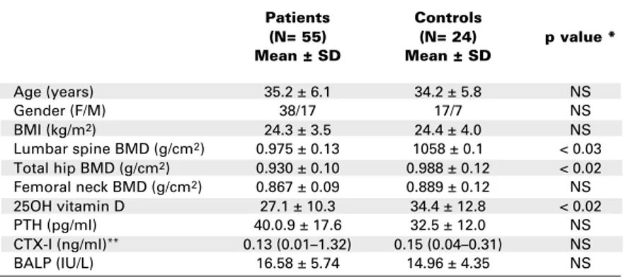
Table 1 summarizes the main characteristics of the studied groups. The median duration of treatment with AEDs was 6 years and varied from 2 to 21 years. At the time of the study, 17 patients (31%) were receiving single antiseizure agents, while 38 (69%) were receiving multiple antiseizure agents. The most common AED used was carbamazepine (400–1500 mg/dl), taken by 44 patients, median duration time of use was 6 years (1-17). The second most common AED was phenobarbital (50–150 mg/day) used by 28 patients for 5 years (1 to 12). Phenytoin (100–400 mg/day) was taken by 23 patients for 2 years (1 to 14) and sodium valproate (500–2250 mg/day) was used by 20 patients during 3.5 years (1 to 10).
Biochemical indices of bone metabolism
Mean results of routine biochemical testing total calci-um, albumin, phosphorus, magnesicalci-um, creatinine, ALP, liver function tests were normal with no statistic difference between patients and controls. At the male group, no statistic difference of serum levels of testos-terone was found (4). Levels of serum 25OHD were lower in patients than controls (27.1 ± 10.3 ng/ml vs. 34.4 ± 12.8 ng/ml; p< 0.02). Serum PTH concentra-tions were similar between study patients and controls, although there was a trend towards higher levels in those taking AEDs (40.09 ± 12.6 pg/ml vs. 32.54 ± 11.9 pg/ml; p= 0.062). No specific AED was associ-ated with a higher level of serum PTH than any other AED.
Serum BALP and CTX-I levels did not differ significantly between the groups (table 1). The mean level of BALP was 16.58 ± 5.74 U/L in the group of patients and 14.96 ± 4.35 U/L in the control group
(p= 0.22). The median serum CTX-I value was 0.13 ng/ml (0.01–1.32) in the study group and 0.15 ng/ml (0.04–0.31) in the control group (p= 0.28). By multiple regression analyses, the variability of serum CTX-I was positively related to levels of serum ALP (p< 0.01), phosphorus (p< 0.001), and PTH (p< 0.01) (R= 0.66). Serum BALP was positively related to total ALP (p< 0.0001) (R= 0.62) but not to any other parameters.
Serum CTX-I levels were significantly higher in the 28 patients who were exposed to phenobarbital (P< 0.01) than the 27 who had not been exposed to this drug. No other biochemical abnormality was found in subjects who had been exposed to phenobar-bital in comparison to those who had not (table 2). Serum BALP was not significantly different among the AED groups, although there was a tendency to higher values in patients exposed to phenobarbital versus those never exposed (p= 0.06).
Bone mineral density
Lumbar spine BMD in AED users was significantly lower than the control group (0.975 ± 0.13 g/cm2vs.
1.058 ± 0.1 g/cm2; p< 0.03). Similarly, total hip
BMD (0.930 ± 0.1 g/cm2vs.0.988 ± 0.12 g/cm2; p<
0.02) (table 1). No densitometric differences were found between studied subjects and controls at the femoral neck. With regard to exposure to specific AEDs, BMD of the total hip (0.890 ± 0.10 g/cm2vs.
0.970 ± 0.08 g/cm2; p< 0.003) and femoral neck
(0.830 ± 0.09 g/cm2 vs. 0.890 ± 0.09 g/cm2; p<
0.03) were significantly lower in patients who had been exposed to phenobarbital, in comparison to those who had not been exposed (figure 1). No other significant difference was found at any BMD site when other AEDs were analyzed separately. In addition, when combinations of AEDs were evaluated, only
Table 1.Baseline characteristics of the studied subjects.
Patients Controls
(N= 55) (N= 24) p value *
Mean ± SD Mean ± SD
Age (years) 35.2 ± 6.1 34.2 ± 5.8 NS
Gender (F/M) 38/17 17/7 NS
BMI (kg/m2) 24.3 ± 3.5 24.4 ± 4.0 NS
Lumbar spine BMD (g/cm2) 0.975 ± 0.13 1058 ± 0.1 < 0.03
Total hip BMD (g/cm2) 0.930 ± 0.10 0.988 ± 0.12 < 0.02
Femoral neck BMD (g/cm2) 0.867 ± 0.09 0.889 ± 0.12 NS
25OH vitamin D 27.1 ± 10.3 34.4 ± 12.8 < 0.02
PTH (pg/ml) 40.0.9 ± 17.6 32.5 ± 12.0 NS
CTX-I (ng/ml)** 0.13 (0.01–1.32) 0.15 (0.04–0.31) NS
BALP (IU/L) 16.58 ± 5.74 14.96 ± 4.35 NS
* p< 0.05: significative
therapeutic regimens that included phenobarbital showed a significant decrease in BMD.
By multiple regression analyses, exposure to phenobarbital accounted for variability both at the femoral neck (= 0.27, R= 0.37, p< 0.001) and at the total hip BMD (= 0.37, R= 0.50, p< 0.001). The relationship between phenobarbital use and BMD was also a function of time on drug (= -0.32) and the association with other AED, such as carbamazepine (= -0.25) and phenytoin (= -0.21), which could explain 43% of BMD variability (R= 0.43; p< 0.001). Regarding the time on drug, greater period of exposi-tion on phenobarbital was related to lower BMD.
DISCUSSION
This study defines further abnormalities in mineral metabolism among subjects treated with AEDs. Among users of AEDs, BMD was lower at the lumbar spine and total hip. Although there were no differ-ences in bone turnover markers between a control
group and AED users, further insight was gained when those taking phenobarbital were specifically analyzed. In subjects taking phenobarbital, serum CTX-I levels were higher along with a trend towards higher levels of serum BALP. In addition, exposure to phenobarbi-tal was specifically associated with lower BMD at the hip. In contrast, exposure to phenytoin, carba-mazepine or valproate, as mono- or polytherapy with-out phenobarbital, was not associated with abnormal-ities in bone turnover markers or BMD.
Phenobarbital is the most-widely used AED in the developing world, and because of its lower price, remains a popular choice in many industrialized coun-tries (20). This drug is known to impair bone metab-olism through several mechanisms. First, it stimulates hepatic mixed function oxidase activity, accelerating the breakdown of vitamin D metabolites (11). In addi-tion, both phenobarbital and phenytoin interfere with active transport of calcium in the small intestine (21). These two agents have also been shown to interfere with PTH-mediated bone resorption. Both the accel-erated metabolism of vitamin D and interference with
Figure 1.BMD of lumbar spine, femur neck, and total femur in controls and patients non-phenobarbital users and users.
Table 2.Biochemical data of patients exposed and never exposed to phenobarbital.
Patients exposed Patients never exposed
to phenobarbital to phenobarbital * p value
(N= 28) (N= 27)
BALP (U/L) 17.9 ± 6.6 15.1 ± 4.3 NS
CTX-I (ng/ml)** 0.15 (0.03–1.32) 0.08 (0.01–0.30) < 0.01
PTH (pg/ml) 43.3 ± 18.7 36.3 ± 15.8 NS
25OHD (ng/ml) 26.4 ± 11.0 29.3 ± 9.8 NS
Total hip BMD (g/cm2) 0.890 ± 0.10 0.970 ± 0.08 < 0.003
Femoral neck (g/cm2) 0.830 ± 0.09 0.890 ± 0.09 < 0.03
* p< 0.05: significative
PTH action could lead to an increase in circulating PTH levels (22,23). Although the PTH levels were not significantly different between the group exposed to phenobarbital and those patients not exposed, there was a trend for PTH levels to be higher in the pheno-barbital users.
Although others and we have demonstrated a decrease in 25OHD levels among chronic users of AEDs (4-6), the histomorphometric studies have established antiepileptic bone disease as a disorder of high bone turnover, more than a mineralization defect (24,25). However, there is no agreement in the litera-ture regarding the type of the drug, as well as dose and time of exposure that might influence bone remodel-ing. Verrotti et al. (16) have evaluated markers of bone turnover in 60 epileptic patients in 3 different stages of pubertal growth before and after monotherapy with carbamazepine. In their study, an increase in markers of bone turnover was seen after 2 years, but vitamin D metabolism was normal in all pubertal stage groups. In contrast, 2 other studies have failed to find an associa-tion between carbamazepine and altered bone turnover (15,26). Pack et al. (15) evaluated 93 pre-menopausal epileptic women receiving carbamazepine, phenytoin, lamotrigine or valproate as monotherapy. Only in women treated with phenytoin were there sig-nificant elevations in serum BALP.
One of the limitations of the present study is the cross-sectional nature, where BMD was measure only once, turning it difficult to know if the low bone density observed in the patients group is specifically related to AEDs use. However, by the fact that other secondary causes were excluded and the patients pre-sented lower BMD than the control matched group, we do believe that the use of AEDs may play a role on this issue.
Our results call attention to reduced bone min-eral density among chronic users of AEDs, with a potential for major complications, such as fractures. It seems reasonable, therefore, to consider a preventative approach to maintaining skeletal health when AED therapy is initiated, especially in children and teenagers, in whom adequate calcium and vitamin D are fundamental to the establishment of bone peak mass. In addition, further studies are needed to deter-mine appropriate therapeutic protocols for patients who present initially with low bone mass and low lev-els of vitamin D.
In conclusion, we have shown that patients with epilepsy on long-term AEDs demonstrate reduced BMD and lower serum levels of 25OHD. When indi-vidual effects of drugs were considered separately,
phe-nobarbital seems to become the main mediator of low bone density and increases in serum CTX-I. Further and prospective studies with larger number of patients are needed to elucidate the influence of AED on BMD and on bone turnover.
REFERENCES
1. Engel JJ. Epileptic syndromes. In:Seizures and Epilepsy. Philadelphia: Davis Company,1989. pp. 195-201.
2. Guerreiro CAM, Guerreiro MM, Cendes F, Lopes-Cendes I. Con-siderações gerais. In: Guerreiro CAM, Guerreiro MM, Cendes F, Lopes-Cendes I (eds).Epilepsia. 3ªed. São Paulo: Lemos,2000. pp. 1-10.
3. Mulder JE, Kulak CAM, Shane E. Secondary osteoporosis. In: Seibel MJ, Robins SP, Bilezikian JP (eds).Dynamics of Bone and Cartilage Metabolism. San Diego: Academic Press,1999. pp. 527-45.
4. Kulak CAM, Borba VZC, Bilezikian JP, Silvado CE, Paola L, Boguszewski CL. Bone mineral density and serum levels of 25OHvitamin D in chronic users of antiepileptic drugs.Arq Neu-ropsiquiatr 2004;62(4)940-8.
5. Baer TM, Kozlowski BW, Blyler EM, Trahms CM, Taylor ML, Hogan MP. Vitamin D, calcium and bone status in children with developmental delay in relation to anticonvulsant use and ambu-latory satus.Am J Clin Nutr 1997;65:1042-51.
6. Hahn TJ, Hendin BA, Scharp CB, Boisseai VC, Haddad JG. Serum 25-hydroxycalciferol levels and bone mass in children on chronic anticonvulsivant therapy.N Engl J Med 1975;292:550-4. 7. Andress DL, Ozuna J, Tirschwell D, Grande L, Meshell J,
Jacob-son AF, et al. Antiepileptic drug-induced bone loss in young male patients who have seizures.Arch Neurol 2002;59(5):781-6. 8. Farhat G, Yamout B, Mikati MA, Demirjian S, Sawaya R, El-Hajj
Fuleihan G. Effect of antiepileptic drugs on bone density in ambu-latory patients.Neurology 2002;58:1348-53.
9. Pack AM, Olarte LS, Morrel MM, Flasher E, Stanley RR, Shane E. Bone mineral density in an outpatient population receiving
enzyme-inducing antiepileptic drugs. Epilepsy Behav
2003;4:169-74.
10. Pack AM. The association between antiepileptic drugs and bone disease.Epilepsy Curr 2003;3(3):91-5.
11. Hahn TJ, Bridge SJ, Scarp CR, Avioli LV. Phenobarbital-induced alterations in vitamin D metabolism.J Clin Invest 1972 ;51:741-8.
12. Sheth RD, Wesolowski CA, Jacob JC. Effects of carbamazepine and valproate on bone mineral density.J Pediatr 1995 ;127:256-62.
13. Sarto Y, Kondo I, Ishida S, Motooka H, Takayama K, Tomita Y, et al. Decreased bone mass and increased bone turnover with val-proate therapy in adults with epilepsy.Neurology 2001 ;57:445-9.
14. Valimaki MJ, Tikhonen M, Laitinen K. Bone mineral density mea-sured by dual-energy absorptiometry and novel markers of bone formation and resorption in patients on antiepileptic drugs.J Bone Min Res 1994;9:631-7.
15. Pack AM, Morrell MJ, Marcus R, Holloway L, Flaster E, Done S, et al. Bone mass and turnover in women with epilepsy on antiepileptic drug monotherapy.Ann Neurol 2005;57:252-7. 16. Verrotti A, Greco R, Latini G, Morgese G, Chiarelli F. Increased
bone turnover in prepubertal, pubertal, and postpubertal patients receiving carbamazepine.Epilepsia 2002;43(12):1488-92. 17. Van Daele PL, Seibel MJ, Burger H, Hofman A, Grobbee DE, van
Leeuwen JP, et al. Case control analysis of bone resorption mark-ers, disability and hip fracture risk: the Rotterdam study.Br Med J 1996;312:482-3.
18. Gomez B, Jr, Ardakani S, Ju J, Jenkins D, Cerelli MJ, Daniloff GY, et al. Monoclonal antibody assay for measuring bone-specific
alkaline phosphatase activity in serum. Clin Chem
19. Bonde M, Garnero P, Fledelius C, Qvist P, Delmas PD, Chris-tiansen C. Measurement of bone degradation products in serum using antibodies reactive with an isomerized form of an 8 amino acid sequence of the C-telopeptide type I collagen. J Bone Miner Res 1997;12(7):1028-34.
20. Kwan P, Brodie MJ. Phenobarbital for the treatment of epilepsy in the 21st century: a critic review.Epilepsy 2004;45(9):1141-9. 21. Foss MC, Meneghelli UG, Tabosa Verissimo JM. The effect of the
anticonvulsants phenobarbital and diphenylhydantoin on intesti-nal absorption of calcium.Acta Physiol Lat Am 1979 ;29(4-5):223-8.
22. Jekins MV, Harris M, Willis MR. The effect of phenytoin on parathyroid extract and 25-hydroxycholecolecalciferol induced bone resorption: Adenosine 3’ 5’-cyclic monophosphatase pro-duction.Calcified Tissue Res 1974;16:163-7.
23. Hahn TJ, Scharp CR, Richardson CA, Halstead LR, Kahn AJ, Teit-elbaum ST. Intercation of dyphenylhydantoin and phenobarbital with hormonal mediation of fetal rat bone resorption in vivo.J Clin Invest 1978;62:406-14.
24. Mokesilde L, Melsen F. Dynamic differences in trabecular bone remodeling between patients after jejuno ileal by pass for obesi-ty and epileptic patients receiving anticonvulsant therapy. Metab Bone Dis 1980;2:77-82.
25. Weinstein RS, Bryce GF, Sappington LJ, King DW, Gallagher BB. Decreased serum ionized calcium and normal vitamin D metabo-lite levels with anticonvulsant drug treatment. J Clin Endocrinol Metab 1984;58:1003-9.
26. Bramswig S, Zittermann A, Berthold HK. Carbamazepine does not alter biochemical parameters of bone turnover in healthy male adults.Calcif Tissue Int 2003;73(4):356-60.
Endereço para correspondência:
Carolina Aguiar Moreira Kulak
Serviço de Endocrinologia e Metabologia Hospital de Clínicas da UFPR – SEMPR Avenida Agostinhio Leão Júnior 285 80030-110 Curitiba, PR