Case Report
Major Article
http://dx.doi.org/10.1590/0037-8682-0040-2014INTRODUCTION
Address to: Dr. Vagner Ricardo Lunge. Laboratório de Diagnóstico Molecular/ ULBRA. Av. Farroupilha 8001, Prédio 22/3º andar, Bairro São José, 92425-900 Canoas, RS, Brasil.
Phone: 55 51 3478-2777.
e-mail: [email protected]
Received 26 February 2014
Accepted 16 June 2014
A complete molecular biology assay for hepatitis C virus
detection, quantifi cation and genotyping
Yara Silva Casanova
[1],[2], Thais da Rocha Boeira
[2], Elisa Sisti
[1], Álvaro Celmer
[2],
André Salvador Kazantzi Fonseca
[2], Nilo Ikuta
[1],[2], Daniel Simon
[1]and Vagner Ricardo Lunge
[1],[2][1]. Laboratório de Diagnóstico Molecular, Programa de Pós-Graduação em Biologia Celular e Molecular Aplicada à Saúde, Universidade Luterana do Brasil, Canoas, RS. [2]. Simbios Biotecnologia, Cachoeirinha, RS.
ABSTRACT
Introduction: Molecular biology procedures to detect, genotype and quantify hepatitis C virus (HCV) RNA in clinical samples
have been extensively described. Routine commercial methods for each specifi c purpose (detection, quantifi cation and genotyping)
are also available, all of which are typically based on polymerase chain reaction (PCR) targeting the HCV 5’ untranslated region (5’UTR). This study was performed to develop and validate a complete serial laboratory assay that combines real-time nested reverse transcription-polymerase chain reaction (RT-PCR) and restriction fragment length polymorphism (RFLP) techniques for the complete molecular analysis of HCV (detection, genotyping and viral load) in clinical samples. Methods: Published HCV
sequences were compared to select specifi c primers, probe and restriction enzyme sites. An original real-time nested
RT-PCR-RFLP assay was then developed and validated to detect, genotype and quantify HCV in plasma samples. Results: The real-time nested RT-PCR data were linear and reproducible for HCV analysis in clinical samples. High correlations (> 0.97) were observed between samples with different viral loads and the corresponding read cycle (Ct - Cycle threshold), and this part of the assay had a wide dynamic range of analysis. Additionally, HCV genotypes 1, 2 and 3 were successfully distinguished using the RFLP method. Conclusions: A complete serial molecular assay was developed and validated for HCV detection, quantifi cation and genotyping.
Keywords: Hepatitis C virus. Polymerase chain reaction. Restriction fragment length polymorphism.
Hepatitis C virus (HCV) is recognized as one of the main
causes of chronic liver disease worldwide1. Hepatitis C virus
infection is usually asymptomatic during the acute phase, but more than 80% of patients progress to chronic hepatitis C (CHC). Approximately 130-170 million people worldwide are chronically infected with HCV, and these people are at risk of developing hepatic complications, such as cirrhosis and/or hepatocarcinoma2.
Hepatitis C virus is an enveloped, single-stranded, positive-sense ribonucleic acid (RNA) virus with a 50nm diameter viral particle and is a member of the Hepacivirus genus of the Flaviviridae family3. Its RNA genome encodes a unique
polyprotein of approximately 3,000 amino acids4,5. The HCV
genome is extremely heterogeneous. Published sequence data
indicate that the 5’ untranslated region (5´UTR) is highly conserved among various HCV isolates and is the target of most HCV molecular biology assays. This region, however, also contains genotypically variable sequences that allow the virus
to be classifi ed into six classical genotypes, which were recently
updated to seven main genotypes that differ by more than 30% in their nucleotide sequence6-8. Hepatitis C virus genotyping
is clinically relevant because improved treatment response rates have been observed with genotypes 2 and 3, compared to genotypes 1 and 42. Consequently, patients infected with
genotypes 1 or 4 are treated for longer times and with more potent combinations of antivirals than patients with genotypes 2 and 39,10.
Chronic hepatitis C is usually associated with mild symptoms. Elevation of aminotransferases, particularly alanine aminotransferase (ALT), is commonly observed, although up to one-third of asymptomatic patients have persistently
normal enzymes11. Liver histology in CHC generally consists
of infl ammatory infi ltrates and some degree of fi brosis, ranging from minimal expansion of portal tracts to cirrhosis. The fi brotic process, which is driven by liver infl ammation, can progress over
time, although patients differ greatly in this process depending on several viral and host factors12. Cirrhosis occurs in at least
20% of patients with CHC within 20 years13. In contrast to
other viral chronic infections, hepatitis B virus (HBV) and
human immunodefi ciency virus (HIV), HCV viral load is not
METHODS
it is an important prognostic factor for predicting the treatment response14. Additionally, monitoring viral load is essential during
treatment because this information is necessary for achieving an
early virological response (defi ned as negativity or a more than
2-log decrease in viral load by week 12) and allows clinicians to continuously monitor patient treatment15.
Several molecular biology assays have been described for hepatitis C virus-ribonucleic acid (HCV-RNA) detection and
quantifi cation. Most commercial methods are polymerase chain reaction (PCR)-based and target the 5’UTR, which is the most conserved region among various genotypes/subtypes16. The
introduction of real-time PCR-based assays improved both detection and viral load analysis17,18. These techniques have a
broad dynamic range of quantifi cation, which is well suited to the clinical needs (upper range of quantifi cation: 7-8 log10IU/mL).
Additionally, real-time PCR is more sensitive than classical PCR, with limits of detection of 10-15IU/mL. More recently, commercial real-time platforms have become available for the detection and
quantifi cation of HCV-RNA: the Cobas TaqMan platform, which
can be used together with automated sample preparation with the Cobas AmpliPrep_system (CAP-CTM; Roche Molecular System, Pleasanton, CA), and the Abbott platform (Abbott Diagnostic,
Chicago, IL), which uses the m2000RT amplifi cation platform
together with the m2000SP device for sample preparation19.
Molecular methods for genotyping HCV that target various HCV genomic regions have also been described20,21. Widely
used laboratory procedures include the line probe assay (LiPA) and 5’UTR sequencing22,23. Restriction fragment length
polymorphism (RFLP) analysis of the 5´UTR of the HCV
genome was one of the fi rst assays used in large genotyping studies. In this procedure, a PCR-amplifi ed hepatitis C
virus-deoxyribonucleic acid (HCV-DNA) fragment is digested into
fragments with restriction enzymes that recognize cleavage sites specifi c for each genotype24. Genotyping based on the
amplifi cation of this region has the advantage that it can be
performed on PCR amplification products obtained from HCV-RNA detection tests25. However, this procedure is not
widely used because it involves the use of 5 to 6 different
restriction enzymes and is hampered by partial digestions and
indeterminate results, making it laborious and time-consuming26.
This study aimed to develop and validate a complete analytical assay based on nested reverse transcription polymerase chain reaction (RT-PCR) and simpler RFLP methodology for the
detection quantifi cation and genotyping of HCV.
Sequences and comparative analysis
A total of 1,080 HCV 5´UTR nucleotide sequences (accession numbers EF558854-EF558890, EF564603-EF564609, EF571224-EF571247, AY306229-AY306686, AY309974-AY310119 and AY310921-AY311334) were retrieved using
Entrez from the National Center for Biotechnology Information
(NCBI, Bethesda, MD). These sequences were obtained from previous HCV genotype prevalence studies with 1,080 different HCV infected patients from different states of the 5 geographic
regions from Brazil27. Additional 26 reference sequences from
different genotypes (according to consensus proposals) were retrieved for comparative analysis6.
Sequences were edited and aligned with EditSeq and MegAlign (using the Clustal method) programs from the DNAstar package (LaserGene Inc., Madison, WI, USA). Primers and one probe were selected directly from the aligned sequences. The presence of the restriction sites for Hae III, Hinf I, BstN I, Rsa I, BfuC Iand BstU I was determined using Mapdraw program within the DNAstar package (LaserGene Inc., Madison, WI, USA).
Reference and clinical samples
A panel of 57 HCV-positive and HCV-negative plasma
samples was obtained from a Brazilian company (ControlLab, Rio de Janeiro - RJ, Brazil). Fifty anti-HCV-negative blood donor
samples were obtained from a hospital blood bank (Hospital das Clínicas de Porto Alegre - HCPA). A panel of 267 HCV-RNA-positive clinical samples (HCV genotypes 1, 2 and 3) was provided by a molecular diagnostic laboratory (Simbios Biotecnologia,
Cachoeirinha, RS, Brazil). All of the plasma samples were
collected with ethylenediaminetetraacetic acid (EDTA) in different clinical laboratories and were conserved at -20ºC until analysis.
RNA extraction
RNA was purifi ed from 100µL of plasma using a silica RNA
extraction method that has been previously described28. Briefl y,
100μL of plasma was added to 900μL of lysis buffer in a micro-tube and incubated at room temperature for 10 min. Then, 20μL
of the silica particle suspension was added to each micro-tube and centrifuged at 10,000rpm for 30 sec. The pellet was washed twice with washing solution, twice with 75% ethanol and once with acetone. After the last centrifugation step, supernatant was removed, and the pellet was dried at 56-60°C for 15 min.
RNA was eluted with 50μl of elution buffer, and the tube was
incubated at 65°C for 5 min.
RT-PCR and nested real-time PCR assay
Reverse transcription and the fi rst round of PCR amplifi cation was performed in a 20µL reaction volume using 14.5µL of a mastermix solution with a fi nal concentration of 75mM KCl,
50mM Tris-HCl, pH 8.3, 3mM MgCl2, 2.5mM DTT, 1mM
dNTPs, 2.0µM of the primers, 24U of MMLV-RT (Life
Technologies, Carlsbad, CA, USA), 4U RnaseOut (Life Technologies, Carlsbad, CA, USA), 1U Taq DNA polymerase
(Cenbiot Enzimas, Porto Alegre, RS, Brazil) and 5µL of
extracted RNA. Reverse transcription polymerase chain reaction
amplifi cation was performed for 30 min at 37°C followed by 15 cycles of the following temperatures and times: 94°C for
30 sec, 60°C for 30 sec and 72°C for 60 sec. A nested
real-time PCR (second amplifi cation stage) was performed in a 30µL reaction volume using 28.6µL of a mastermix solution with a fi nal concentration of 50mM KCl, 10mM Tris-HCl,
pH 8.3, 1.5mM MgCl2, 1mM dNTPs, 0.25µM of the primers,
and 0.125µM of the probe. The PCR amplification was
RESULTS
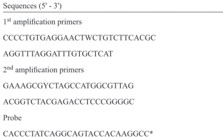
TABLE 1 - Primer and probe nucleotide sequences.
Sequences (5' - 3')
1st amplifi cation primers
CCCCTGTGAGGAACTWCTGTCTTCACGC
AGGTTTAGGATTTGTGCTCAT
2nd amplifi cation primers
GAAAGCGYCTAGCCATGGCGTTAG
ACGGTCTACGAGACCTCCCGGGGC
Probe
CACCCTATCAGGCAGTACCACAAGGCC*
*This probe was labeled with the fl uorophore FAM and the quencher TAMRA. FAM: fl uorescein amidite; TAMRA: tetramethylrhodamine. standard samples with pre-defi ned viral loads. Hepatitis C
virus concentrations were log10-transformed for analysis. The linear range was examined by plotting the data and comparing
them to a line of equality. Correlation coeffi cient calculations
and linear regression analyses were performed on scatter plots of log-transformed HCV RNA levels using Microsoft Excel (Microsoft Corp., Redmond, WA, USA).
5'UTR genotyping
Genotyping of all HCV-positive RNA samples was performed using an RFLP procedure adapted from the original method24. Briefl y, three restriction enzymes were used in two
separate digests to cleave each nested PCR product. First, the
HCV-amplifi ed fragment was digested with Hae III alone in the appropriate buffer and conditions (New England BioLabs, Ipswich, MA, USA). The fragment was also digested with Hinf I and BstN I simultaneously in appropriate buffer and conditions (New England BioLabs, Ipswich, MA, USA). Some samples were
digested with three other enzymes: Rsa I (Promega, Fitchburg, WI, USA), BfuC I and BstU I (New England BioLabs, Ipswich, MA, USA). Single digests were prepared with the respective
enzyme and the appropriate buffer and conditions: Rsa I (Promega, Fitchburg, WI, USA), BfuC I and BstU I (New England BioLabs, Ipswich, MA, USA). Digested products were separated
by electrophoresis on a 12.5% polyacrylamide gel and visualized
after rapid silver staining. Banding patterns of the various HCV genotypes were deduced from those previously established by analysis of the 5'UTR sequences obtained from gene databases.
Genotypes were then identifi ed according to previous fi ndings6. Confi rmation of RFLP analysis by sequencing PCR products were sequenced to confirm the results. Forward and reverse sequencing reactions were performed using template DNA, inner primers and BigDye Terminator v3.1 Cycle Sequencing reagent (Applied Biosystems Inc., Norwalk, CT, USA). Sequencing was performed using the thermocycler Veriti 96 (Applied Biosystems Inc., Norwalk, CT, USA) with an initial denaturation step at 95ºC for 3 min followed by 40 cycles of
95ºC for 10 sec and 60ºC for 240 sec. Samples were purifi ed using
an ethanol/EDTA/sodium acetate protocol, and DNA products were injected in the automated DNA sequencing ABI 3130 XL Genetic
Analyzer (Applied Biosystems Inc., Norwalk, CT, USA). Sequence
data were collected and quality analysis was performed using the Sequencing Analysis v.5.3.1 software by evaluating the main technical parameters as raw data, electropherograms and quality values of sequenced bases (Applied Biosystems Inc., Norwalk, CT, USA). The nucleotide sequences from the same amplicon (performed with sense and antisense primers) were edited and assembled using SeqMan software (DNAStar, Madison, WI, USA). Nucleotide sequences were aligned using the MegAlign program (DNAStar, Madison, WI, USA), and genotypes were deduced using the phylogenetic analysis protocols in this software. Additionally, data obtained were compared with sequences available in GenBank database (www.ncbi.nlm.nih.gov).
Ethical considerations
This project was approved by the Ethical Committee of the
Universidade Luterana do Brasil (Canoas, RS, Brazil).
Real-time nested RT-PCR primers and probe
All 5´UTR HCV sequences obtained from GenBank were aligned using PrimerSelect software (Lasergene Inc., Madison, WI, USA). Four primers and one probe were designed to accommodate all HCV types (Table 1). These oligonucleotides
were used to develop and standardize a molecular method to
detect, quantify and genotype HCV. The complete procedure consists of reverse transcription and a nested PCR. Reverse
transcription and the fi rst amplifi cation were performed in the same reaction tube. The second amplifi cation was performed
using TaqMan targeting all HCV genotypes.
RFLP patterns and HCV genotyping
A total of 1,080 NCBI sequences were evaluated for
genotype-specifi c RFLP patterns. The differentiation of the three main genotypes (1, 2, 3) present in Brazil could theoretically be accomplished using a double digest with restriction enzymes BstN I and Hinf I. The banding patterns of Genotype 3 samples consist of a 165bp fragment, while almost all genotype 1 samples (98.1%) contain a 117bp fragment and the majority of the genotype 2 samples (94.5%) have a 221bp fragment (Table 2). To further differentiate HCV genotypes 4 and 5 (which present the same patterns as genotypes 3 and 2, respectively) and to
differentiate a signifi cant proportion (5.5%) of the genotype 2 samples that have a 117bp-fragment, the restriction enzyme Hae III was selected because it was informative for all of these discriminations. Table 1 shows the predicted patterns using BstN I and Hinf I double digestion and Hae III single digestion for the
HCV Brazilian sequences. Based on these results, the following workfl ow for genotyping HCV was defi ned: BstN I + Hinf I double digestion and Hae III single digestion in a fi rst round for all samples. Samples with indeterminate patterns in the fi rst round
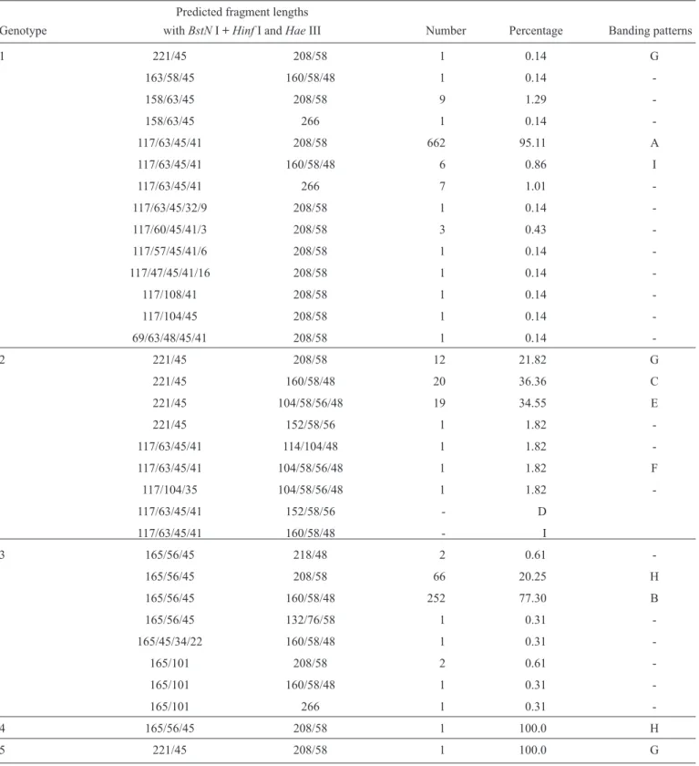
TABLE 2 - Restriction fragment sizes and frequency of HCV restriction patterns found in the samples of the different genotypes, originated from the NCBI sequences.
Predicted fragment lengths
Genotype withBstNI + HinfI andHaeIII Number Percentage Banding patterns
1 221/45 208/58 1 0.14 G
163/58/45 160/58/48 1 0.14
-158/63/45 208/58 9 1.29
-158/63/45 266 1 0.14
-117/63/45/41 208/58 662 95.11 A
117/63/45/41 160/58/48 6 0.86 I
117/63/45/41 266 7 1.01
-117/63/45/32/9 208/58 1 0.14
-117/60/45/41/3 208/58 3 0.43
-117/57/45/41/6 208/58 1 0.14
-117/47/45/41/16 208/58 1 0.14
-117/108/41 208/58 1 0.14
-117/104/45 208/58 1 0.14
-69/63/48/45/41 208/58 1 0.14
-2 221/45 208/58 12 21.82 G
221/45 160/58/48 20 36.36 C
221/45 104/58/56/48 19 34.55 E
221/45 152/58/56 1 1.82
-117/63/45/41 114/104/48 1 1.82
-117/63/45/41 104/58/56/48 1 1.82 F
117/104/35 104/58/56/48 1 1.82
-117/63/45/41 152/58/56 - D
117/63/45/41 160/58/48 - I
3 165/56/45 218/48 2 0.61
-165/56/45 208/58 66 20.25 H
165/56/45 160/58/48 252 77.30 B
165/56/45 132/76/58 1 0.31
-165/45/34/22 160/58/48 1 0.31
-165/101 208/58 2 0.61
-165/101 160/58/48 1 0.31
-165/101 266 1 0.31
-4 165/56/45 208/58 1 100.0 H
5 221/45 208/58 1 100.0 G
HCV: hepatitis C virus; NCBI: National Center for Biotechnology Information; BstN I + Hinf I and Hae III: restriction enzymes.
HCV detection and quantifi cation validation tests First, an HCV-positive (genotype 1) standard sample at a concentration of 10,000,000 IU/mL was diluted ten-fold to 1IU/mL, and the complete procedure (RNA extraction, RT-PCR
corresponding cycle threshold (Ct) had a wide dynamic range of analysis across the entire spectrum of clinically relevant viral loads (100 to 10,000,000IU/mL). Additionally, reproducibility was tested using four different HCV RNA genotype 1 samples (A = 6,000,000IU/mL, B = 30,000IU/mL, C = 3,000IU/mL, D = 1,500IU/mL) in 20 runs on different days. The total
coeffi cients of variation of the cycle threshold (Ct) were 8.77%
for sample A (mean Ct = 10.32 ± 0.90), 3.72% for sample B (mean Ct 18.36 ± 0.68), 3.96% for sample C (mean Ct = 21.72 ± 0.86) and 3.67% (mean Ct = 22.81 ±0.84).
The limit of detection was estimated using the Probit test by evaluating nine replicates of seven diluted HCV RNA samples ranging from 51 to 3,300IU/mL. The assay sensitivities were 1,500IU/mL for 95% positive repetitions and 500IU/mL for 50% positive repetitions. Specifi city was tested using hepatitis C-negative blood donors, and no false-positive results were observed in a total of 50 anti-HCV-negative samples.
The procedure was then used to analyze the 57 plasma samples
obtained from an inter-laboratorial program (ControlLab). In total, 36 samples tested positive and 21 negative, with 100% of agreement with the results reported by other laboratories. In the viral load analysis of the 36 HCV-positive samples, values ranging from 1,350 (log 3.13) to 5,250,000 (log 6.72) IU/mL were obtained. These data correlated well with the values reported by the ControlLab program. Linear regression of this comparison returned an intercept of -0.09 and a slope of 0.94.
The overall correlation coeffi cient was 0.91 (Figure 1).
HCV genotyping validation test
To validate the genotyping procedure, we performed a blind study comparing RFLP analysis and nucleotide sequencing for 20 random clinical samples. RFLP results with the Hinf I + BstN I
6.5
R = 0.9122
Nested real time RT PCR
Control lab
6.0
5.5
5.0
4.5
4.0
3.5
3.0
3.5 4 4.5 5 5.5 6 6.5 7
A
MW A B C D MW
1 2 1 2 1 2 1 2
B
MW E F G H I MW
1 2 1 2 1 2 1 2 1 2
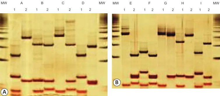
FIGURE 2 - Silver-stained polyacrylamide gels showing the different banding patterns obtained in the validation of the methodology (A) and in the analysis of the 804 clinical samples (B). The numbers indicate the digestion system (1 = Hinf I + BstN I and 2 = Hae III), and the letters indicate the banding patterns (A, B, C, D, E, F, G, H and I). The 50-bp molecular weight is also shown.
MW: molecular weight.
FIGURE 1 - The relationship between HCV RNA viral load levels given by ControlLab and obtained by nested real-time RT-PCR. HCV RNA: hepatitis C virus ribonucleic acid; RT-PCR: Reverse transcription polymerase chain reaction.
double digest and the Hae III digest revealed nine samples with the pattern predicted for genotype 1 (A), nine samples with the predicted pattern for genotype 3 (B), one sample with the primary predicted pattern for genotype 2 (C) and one sample with an unpredicted pattern (D) (Figure 2). All 19 samples from genotypes 1, 2 and 3 had exacting the same genotyping results with 5´UTR sequencing. The banding pattern of the remaining sample was not previously
Analysis of clinical samples
A total of 267 consecutive HCV-positive samples were tested using the nested real-time RT-PCR protocol in a routine laboratory. The mean viral load was 6,316,241 (log 6.32)IU/mL (standard deviation of log 0.80), ranging from 514 (log 2.71) to 93,850,240 (log 7.97)IU/mL. All samples were genotyped
using the RFLP method and were classifi ed according to the
banding pattern (Table 2). Patterns A, B, C, E, F, G, H and I
were identifi ed in 155 (58.1%), 96 (36.0%), 9 (3.4%), 1 (0.4%),
1 (0.4%), 1 (0.4%), 2 (0.7%) and 2 (0.7%) samples, respectively (Figure 2). Banding pattern D was not observed in any sample. A total of four randomly selected samples representing the three main banding patterns (A, B and C) and all samples presenting
patterns E, F, G, H and I were sequenced and confi rmed to be
of the expected genotypes (A and I genotype 1, C, E, F and G
Gen_1c
Gen_1a
Gen_1b
Gen_5a
Gen_2a Gen_2c
Gen_2b
Gen_4a
Gen_3b
Gen_3a RS02
RS18 RS03
RS12 58
49
46 42
98 88
77
90
90 RS17
RS13
RS16
RS11
RS19 RS05
RS07
RS08 RS04
RS20
RS06
RS10
0.005
RS15 RS14
RS01
RS09
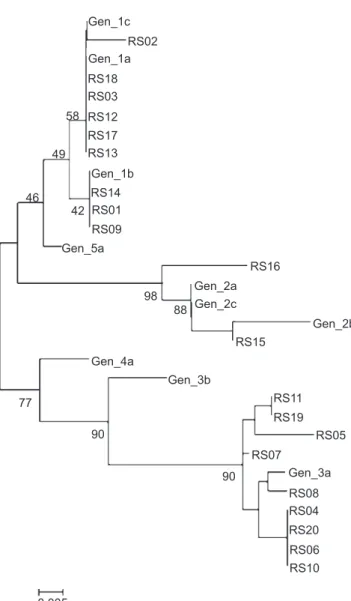
FIGURE 3 - Phylogenetic tree with the 5´UTR HCV sequences of the reference genotypes (1a, 1b, 1c, 2a, 2b, 2c, 3a, 3b, 4a and 5a) and 20 randomly selected HCV-positive plasma samples. RS: randomly selected; HCV: hepatitis C virus; 5' UTR HCV: HCV 5’ untranslated region HCV.
genotype 2, B and H genotype 3). In this group of samples, 157 (58.8%) were from genotype 1, 12 (4.5%) from genotype 2 and 98 (36.7%) from genotype 3. Genotypes 4, 5 and 6 were not found.
DISCUSSION
Hepatitis C virus detection, quantifi cation and genotyping are the crucial tests required to defi ne and monitor the treatment
of hepatitis C. Consequently, the introduction of assays with good analytical performance, high-throughput capacity and low
cost for these three analyses will benefi t both laboratories and
patients. Studies have described the development and validation of in-house and commercial real-time PCR assays for HCV
detection and quantifi cation17,18. We developed a new procedure
based on a nested PCR approach. Although this method is based
on two rounds of PCR amplifi cation, the results obtained by
real-time PCR were linear and reproducible, similar to other in-house or commercial real-time techniques previously described. The
use of RT-PCR amplifi cation with a limited number of cycles (in a fi rst round) followed by a real-time PCR with a higher
number of cycles (in a second round) allowed the precise
quantifi cation of clinical samples. Further, this real-time nested RT-PCR assay demonstrated similar sensitivity and specifi city
values compared to other methods29,30. According to the analyses
of 57 samples from an inter-laboratorial program, the correlation with other techniques was also very good. This assay could be useful in detecting and quantifying HCV in plasma samples.
Several methods have been used for assessing HCV genotypes in the clinical laboratories. The gold standard technique for genotyping HCV involves sequencing one or more genes in the HCV genome (mainly the 5´UTR, core, E1, NS3 and NS5) and comparing these sequences to the established genotypes by computer analysis. This approach is considered too expensive and time-consuming for large-scale diagnostics and has not been widely used in the majority of clinical laboratories26.
Commercial genotyping tests, such as the Invader assay, Trugene 5´NC and the INNO-LiPA HCV II test are available; however,
they all require an initial PCR amplifi cation step26.
Reverse transcription polymerase chain reaction analysis
of the 5´UTR of the HCV genome was the fi rst genotyping
method used for large-scale epidemiological studies20,24 and
became the preferred method of routine HCV genotyping in clinical laboratories for some years. However, its use declined
because of the diffi culty involved in performing the necessary
restriction analyses. The methodology was considered laborious,
time-consuming and diffi cult to evaluate (primarily when the banding patterns are visualized in ethidium bromide gels)26.
In the present study, we propose a simplifi ed procedure for HCV genotyping using only three restriction enzymes in two
digests. The analysis of the 1,080 available HCV sequences from
different Brazilian regions demonstrated that only four RFLP
banding patterns (A, B, C and E), characteristic of the three main genotypes (1, 2 and 3), would be obtained in the great majority
restriction enzymes or submitted to sequencing. This procedure
could also identify genotypes 4 and 5 (patterns H and G,
respectively), which are rarely found in Brazil27. Concordance
between this RFLP procedure and direct nucleotide sequencing was observed in the analysis of all HCV-RNA positive samples. Further analysis of clinical samples demonstrated a total of eight possible banding patterns (A, B, C, D, E, F, G H and I),
which are easily visualized in polyacrylamide gels. The most frequent banding patterns are characteristic of specifi c genotypes
(A - genotype 1; B - Genotype 3; C, D, E and F - genotype 2). Only a few exceptions (banding patterns G, H and I presented
in less than 3% of the positive samples) must be analyzed
using a more informative technique (e.g., sequencing). This RFLP procedure is certainly easier and less laborious than the original method20,24. The banding patterns are also clear, and the
identifi cation is easy to perform in a routine service (Figure 2). Finally, the complete molecular biology assay was successfully applied for the analysis of 267 HCV positive plasma samples in a clinical laboratory setting. The whole in-house procedure could be performed completely in less than 48 hours, including the determination of viral load and genotype in HCV-positive plasma samples. Moreover, this approach eliminates the
need for sequencing or hybridization with specifi c probes after PCR amplifi cation, eliminating the costs of expensive automated sequencers and/or hybridization platforms.
The authors declare that there is no confl ict of interest. CONFLICT OF INTEREST
FINANCIAL SUPPORT
This work was supported by the project titled ‘Development
of Molecular Test Kits to Detect and Quantify Viral Agents
– Hepatitis B and C and HIV – Based on the Real-Time PCR Technology, sponsored by FINEP and Simbios Biotecnologia (FINEP/FULBRA/LDM - Simbios Biotecnologia - 0.1.07.0102.00).
REFERENCES
1. Choo QL, Kuo G, Weiner AJ, Overby LR, Bradley DW, Hougthon M. Isolation of a cDNA clone derived from a blood-borne non-A, non-B viral
hepatitis genome. Science 1989; 244:359-362.
2. Hoofnagle JH. Course and outcome of hepatitis C. Hepatology 2002;
36:S21-S29.
3. Bostan N, Mahmood T. An overview about hepatitis C: A devastating virus. Crit Rev Microbiol 2010; 36:91-133.
4. Choo QL, Richman KH, Han JH, Berger K, Lee C, Dong C, et al. Genetic
organization and diversity of the hepatitis C virus. Proc Natl Acad Sci USA, 1991; 88:2451-2455 .
5. Choukhi A, Ung S, Wychowski C, Dubuisson J. Involvement of endoplasmic reticulum chaperones in the folding of hepatitis C virus
glycoproteins. J Virol 1998; 72:3851-3858 .
6. Simmonds P, Bukh J, Combet C, Deleage G, Enomoto N, Feinstone S,
et al. Consensus proposals for a unifi ed system of nomenclature of hepatitis C virus genotypes. Hepatology 2005; 42:962-973.
7. Smith DB, Bukh J, Kuiken C, Muerhoff AS, Rice CM, Stapleton JT, et al. Expanded classifi cation of hepatitis C virus into 7 genotypes and 67 subtypes: Updated criteria and genotype assignment web resource. Hepatology 2014; 59:318-327.
8. Ciotti M, Marcuccilli F, Guenci T, Babakir-Mina M, Chiodo F, Favarato M, et al. Perno CF. A multicenter evaluation of the Abbott RealTime HCV Genotype II assay. J Virol Methods 2010; 167:205-207.
9. Hadziyannis SJ, Sette Jr H, Morgan TR, Balan V, Diago M, Marcellin P, et al. Peginterferon-alpha2a and ribavirin combination therapy in chronic hepatitis C: a randomized study of treatment duration and ribavirin dose. Ann Intern Med 2004; 140:346-355.
10. Jacobson IM, Pawlotsky JM, Afdhal NH, Dusheiko GM, Forns X, Jensen DM, et al. A practical guide for the use of boceprevir and telaprevir for the treatment of hepatitis C. J Viral Hepat 2012; 19 (suppl II):1-26.
11. Conry-Cantilena C, Vanraden M, Gibble J, Melpolder J, Shakil AO, Viladomiu L, et al. Routes of infection, viremia, and liver disease in blood donors found to have hepatitis C virus infection. N Engl J Med 1996; 334:1691-1696.
12. Poynard T. Interferon alpha in hepatitis C: a cytokine for reducing fi brosis progression. Eur Cytokine Netw 1997; 8:310-320.
13. Seeff LB. Natural history of chronic hepatitis C. Hepatology 2002; 36:S35-46
14. Rotman Y, Liang TJ. Coinfection with hepatitis C virus and human immunodefi ciency virus: virological, immunological, and clinical outcomes. J Virol 2009; 83:7366-7374.
15. Hoofnagle JH, Seeff LB. Peginterferon and ribavirin for chronic hepatitis C. N Engl J Med 2006; 355:2444-2451.
16. Cantaloube JF, Laperche S, Gallian P, Bouchardeau F, Lamballerie X, Micco P. Analysis of the 5’ non-coding region versus the NS5b region in genotyping hepatitis C virus isolates from blood donors in France. J Clin Microbiol 2006; 44:2051-2056.
17. Halfon P, Bourliere M, Penaranda G, Khiri H, Ouzan D. Real-time PCR assays for hepatitis C virus (HCV) RNA quantitation are adequate for clinical management of patients with chronic HCV infection. J Clin Microbiol 2006; 44: 2507-2511.
18. Sizmann D, Boeck C, Boelter J, Fisher D, Miethke M, Nicolaus S, et al. Fully automated quantifi cation of hepatitis C virus (HCV) RNA in human plasma and human serum by the COBAS AmpliPrep/COBAS TaqMan system. J Clin Virol 2007; 38:326-333.
19. Chevaliez S. Virological tools to diagnose and monitor hepatitis C virus infection. Clin Microbiol Infect 2011; 17:116-121.
20. Davidson F, Simmonds P, Ferguson JC, Jarvis LM, Dow BC, Follet EA, et al. Survey of major genotypes and subtypes of hepatitis C virus using RFLP of sequences amplifi ed from the 5’ non-coding region. J Gen Virol 1995; 76:1197-1204.
21. Murphy DG, Willems B, Deschenes M, Hilzenrat N, Mousseau R, Sabbah S. Use of sequence analysis of the NS5B region for routine genotyping of hepatitis C virus with reference to C/E1 and 5’ untranslated region sequences. J Clin Microbiol 2007; 45:1102-1112.
22. Bouchardeau F, Cantaloube JF, Chevaliez S, Portal C, Razer A, Lefrère JJ, et al. Improvement of hepatitis C virus (HCV) genotype determination with the new version of the INNO-LiPA HCV assay. J Clin Microbiol 2007; 45:1140-1145.
23. Cook L, Sullivan K, Krantz EM, Bagabag A, Jerome KR. Multiplex real-time reverse transcription-PCR assay for determination of hepatitis C virus genotypes. J Clin Microbiol 2006; 44:4149-4156.
24. McOmish F, Yap PL, Dow BC, Follett EAC, Seed C, Keller AJ, et al. Geographical distribution of Hepatitis C virus genotypes in blood donors: an International collaborative survey. J Clin Microbiol 1994; 32:884-892.
25. Erensoy S. Diagnosis of hepatitis C virus (HCV) infection and laboratory monitoring of its therapy. J Clin Virol 2001; 21:271-281.
27. Campiotto S, Pinho JRR, Carrilho FJ, Silva LC, Souto FJD, Spinelli V, et al. Geographic distribution of hepatitis C virus genotypes in Brazil. Braz J Med Biol Res 2005; 38:41-48.
28. Boom R, Sol CJ, Salimans MM, Jansen CL, Wertheim-van Dillen PM, van der Noordaa J. Rapid and simple method for purifi cation of nucleic acids. J Clin Microbiol 1990; 28:495-503.
29. Vermehren J, Colucci G, Gohl P, Hamdi N, Abdelaziz AI, Karey U, et al. Development of a second version of the Cobas AmpliPrep/Cobas TaqMan
hepatitis C virus quantitative test with improved genotype inclusivity. J Clin Microbiol 2011; 49:3309-3315.