MASTICATION, DEGLUTITION AND ITS ADAPTATIONS
IN FACIAL PERIPHERAL PARALYSIS
Mastigação, deglutição e suas adaptações na paralisia facial periférica
Marion Renée Mory (1), Adriana Tessitore (2), LeopoldoNizam Pfeilsticker(3), Euro de Barros Couto Junior(4), Jorge Rizzato Paschoal(5)
(1) Speech therapist; volunteer at the Orofacial Rehabilitation Sector from the Facial Paralysis Ambulatory at Hospital das Clínicas from the Faculdade de Ciências Médicas at Uni-versidade Estadual de Campinas – UNICAMP; specialized in Oral Motricity by the Centro de Especialização em Fono-audiologia Clínica – CEFAC
(2) Speech therapist, Coordinator of the Orofacial Rehabilita-tion Sector from the Facial Paralysis Ambulatory at Hospital das Clínicas from the Faculdade de Ciências Médicas at Universidade Estadual de Campinas – UNICAMP; Doc-torate in Medical Sciences by the Faculdade de Ciências Médicas from the Universidade Estadual de Campinas – UNICAMP, São Paulo, Brazil.
(3) Otorhinolaryngologist Doctor; Cranio Maxillofacial surgeon from the Cranium Base and Facial Paralysis Ambulatory of the Otorinolaryngology subject of the Hospital das Clínicas from the Faculdade de Ciências Médicas of the Universi-dade Estadual de Campinas – UNICAMP – Campinas, São Paulo, Brazil.
(4) Statistician; Technical Assistant of the city of São Paulo City Hall. Phd in Sciences – FMUSP, São Paulo, Brazil. (5) Otorhinolaryngologist Doctor; Associate Professor at
Uni-versidade Estadual de Campinas – UNICAMP; Head of the Cranium Base and Facial Paralysis Ambulatory of the
Otorinolaryngology subject of the Hospital das Clínicas from the Faculdade de Ciências Médicas of the Universi-dade Estadual de Campinas – UNICAMP – Campinas, São Paulo, Brazil; Doctorate in Medical Sciences by the Univer-sidade Estadual de Campinas – UNICAMP, Campinas, São Paulo, Brazil.
Conlict of interest: non-existent INTRODUCTION
The partial or total interruption of impulses sent by the Facial Nerve to the muscles innvervated by it results in the abolishment of facial mimic 1-6.
The maxilla, mandible, temporomandibular joints, teeth, muscles, ligaments, tongue and lips form the Stomatognathic System (SS), which, controlled by the Central Nervous System is responsible by suction, mastication, deglution and speech7.
Mastication is the act of biting and crushing food, transforming it into smaller particles which, bonded by the mixing action of saliva, make up the bolus
ABSTRACT
Purpose: to describe mastication, swallowing oral phase and possible functional adaptations observed in Facial Peripheral Paralysis subjects. Method: there were 30 subjects with grade IV Facial Peripheral Paralysis, with at the most 30 days paralysis history and no etiology differentiation. They were separated in three groups, 1 to 10 days paralysis, 11 to 20 days and 21 to 30 days. Mastication and swallowing oral phase functions were assessed with both solid food and water. Participants
answered questions related to the dificulties right after the paralysis. Data were statistically analyzed
using the Likelihood Ratio Test and Fisher Exact Test. Results: changes were observed in mastication and swallowing oral phase due to the lowering of lips tonus, orbicular muscle and buccinator muscle that allows the escaping of food and liquid by decreasing intra-oral pressure. To the speech therapist’s
observation “spill liquid while drinking” presented statistically signiicant data (p=0,003) in the three observed groups. Variable “accumulate food between teeth and gums” was statistically signiicant in groups of 11 to 20 days (p= 0,002). Conclusion: sample subjects chew with dificulty in the paralyzed
side, showing slow and inconsistent mastication cycles. There is an increased tongue movements for cleaning the residues kept in oral vestibule in the paralyzed side. This is the most annoying symptom according to the patients. They develop adaptation strategies to compensate their functional
dificulties.
• At most 30 days history, and etiology differentiation
• Have all teeth in good conservation conditions. Exclusion criteria:
• Patients with neurological alterations related to deglution.
• Absence of dental elements.
30 individuals, who fulilled the criteria for inclu -sion, were selected, and divided into three groups, according to time of paralysis onset: from 1 to 10 days, 11 to 20 days, 21 to 30 days.
After that, they were examined by the medical
staff and classiied as to the degree of paralysis
according to the HB scale.
This research was made up by a clinical assess-ment and a questionnaire where the subjects could answer questions and where the examiner could
observe the functional dificulties (Picture 1).
The questionnaire, based in complaints which were previously reported by patients treated in the Facial Paralysis Ambulatory from the Hospital das Clínicas in the Universidade Estadual de Campinas – UNICAMP was only given to the subjects after our assessment, thus avoiding that
the patient be inluenced by its questions and inhibit
spontaneous behavior during the exam.
During the clinical assessment, the mastica-tion funcmastica-tion and the oral phase of deglumastica-tion were recorded and evaluated by the speech therapist, using solid food and natural water. The patients were instructed to chew normally, as they do at home. They were not oriented, neither received suggestions during the assessment, thus avoiding changes in their normal behavior.
The following protocol was used:
1. The patient was seated in a chair, with both feet fully touching the ground.
2. 1/5 of a cereal bar, weighing 25 grams was offered and the subject was oriented to eat normally.
3. The procedure was repeated.
4. After that, a glass of natural water was offered, to be drunk normally.
5. Assessment of the recorded material, focusing
in the dificulties found and in the compensatory
behaviors.
After that, the questionnaire was given, with
questions related to the dificulties met by the patient
to eat at home and at the moment of the clinical which is ready to be swallowed 7,8. The learned
func-tion depends of the neural pathways and synaptic connections established and commanded by the cerebral cortes9. Mastication contributes to the maintenance of the acquired tonus, and is consi-dered the most important function of the SS 10,11.
The bilateral pattern of mastication is the ideal condition for the functional harmony of the SS components, and is considered the model for normality 3,7,11.
From a functional point of view, the facial and mastication muscles act sinergically. During masti-cation, various muscle groups are coordinately contracted, with an obvious emphasis to the masti-cation muscles, followed by the tongue and facial muscles, specially the buccinator muscle and the orbicular muscle of the lips 3,12. The morphofunc-tional harmony of the SS presupposes the balance among its basic physiological components and the
performance of their functions with maximum efi -ciency. Any neuromuscular changes will lead to functional disruption of the system 9, 13, 14.
Among individuals with Peripheral Facial Paralysis (PFP), the paralyzed hemiface leads to unilateral mastication, in the non-affected side 13, 15. The containment of food between the dental arches
is jeopardized by the ineficiency of the buccinator
muscle and by the labial incompetence, which is a
consequence of the laccidity present in the affected
hemiface 3, 5, 12.
The lips, turned to the normal side and with little occlusion strength, determine the decrease of intra-oral pressure, altering the balance among the structures, lips, cheeks, palate and tongue 15-17.
With the orofacial muscle tonus jeopardized,
there will be extra-oral escape, dificulty in liquid
ingestion and impairment of the masticatory function 12-14, 17-20.
The patient develops individualized adaptations to feed 13,19.
In this research, the characteristics of the masti-cation, oral phase of deglution and possible func-tional adaptations used by patients with PFP to compensate for their limitations were studied.
METHOD
factor, since the alterations remain the same in relation to it.
The distribution of frequency with its respective percentiles of functional alterations as was observed by the speech therapist in subjects with PFP from the three groups while they were eating are shown in table 3. According to the Likelihood Ratio test, there
was a statistically signiicant alteration in variable
“spill liquid while drinking” among the groups of 11 to 20 days and 21 to 30 days in the comparison of the observations made by the speech therapist for the three groups studied, among which, the subjects of the last group were the ones who presented the
most dificulty in this item.
When comparing the answer of the subjects per groups and the observation of the speech
thera-pist, there was a statistically signiicant difference
in variable “accumulate food between teeth and gums on the paralyzed side” for the groups of 11 to 20 days, which was observed both by the speech therapist and the subjects.
In the comparison of functional alterations referred to by the subjects with PFP and the obser-vation of the speech therapist per group, shown in
table 4, there was no statistically signiicant relation
in the following variables:
For the 11 to 20 days group, “chew on the
paralyzed side”, “dificulty to apprehend the glass
between lips” and “ lips turn to the paralyzed side.” For the 21 to 30 days groups, “accumulate food between teeth and gums on the paralyzed side” and “no retention of saliva”.
291/2007, and was considered free of risks and a formal and informed consent was necessary.
In order to verify possible differences among the three groups when compared concomitantly as to the variables under study, the Likelihood Ration Test was applied:
The level of signiicance was 5% (0.050), for
the application of statistical tests, that is, when the
calculated signiicance (p) was less than 5% (0.050), a difference or a statistically signiicant relation was
observed. In order to check the level of association among the subjects and the speech therapist, for each of the variables under study, the Fisher Exact
Test was applied. A the calculated signiicance value (p) equal or higher than 5% (0.050) repre
-sents a difference or a “statistically non-signiicant”
relation. Thus, in this case, differences between the subject and the speech therapist will be found when p > 0.050.
RESULTS
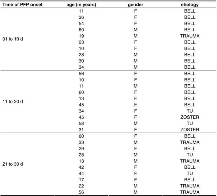
The characterization of the sample containing the PFP onset time, age, gender, and etiology of the subjects who were studied are presented in Table 1.
The frequency distribution with its respective percentiles of functional alterations as reported by the subjects with PFP from the three groups is presented in Table 2. When the results from the three groups are compared, it was possible to note the existence of alterations, but according to the Ratio Likelihood test, time is not a relevant
YES NO
Chews on the paralyzed side?
Slower mastication on the paralyzed side? Lack of strength to chew on the paralyzed side? Fatigue while chewing on the paralyzed side? Lips occluded during mastication?
Accumulation of food between the teeth and gums on the paralyzed side? Chews on cheeks or lips?
Difficulties to apprehend the glass between the lips? Spills liquid while drinking?
Does not retain saliva?
Other signs
Picture 1 – Protocol used with subjects with PFP related to the functinoal dificulties encountered to
Time of PFP onset age (in years) gender etiology
11 F BELL 36 F BELL 54 F BELL 60 M BELL
19 M TRAUMA
23 F BELL 10 F BELL 28 M BELL 30 M BELL 01 to 10 d
34 M BELL 56 F BELL 10 F BELL 11 M BELL 60 F BELL 13 F BELL 45 F BELL 34 F TU
45 F ZOSTER
58 M TU 11 to 20 d
31 F ZOSTER
60 F BELL
33 M TRAUMA
29 F BELL 28 M TU
13 M TRAUMA
42 F BELL 44 F TU 17 F BELL
22 M TRAUMA
21 to 30 d
56 M TRAUMA
Table 1 – characterization of the population under study according to gender, age and etiology
Legend: d: days F: female M: MALE
Time n=10 n=10 n=10
Alterations (%) 1 to 10d 11 to 20 d 21 to 30 d
Chews on the PS 40 50 70 0.384
Slower Mastication on the PS 80 80 80 0.999 Lack of strength to chew on the PS 20 60 40 0.178 Fatigue when chewing on PS 50 30 30 0.568 Lips are occluded during mastication 40 50 50 0.874 Lips are turned to the healthy side during mastication 50 50 60 0.874 Accumulation of food between the teeth and gums
in the PS 80 90 70 0.523
Bites cheeks or lips 50 60 40 0.669 Difficulties to apprehend the glass between the lips 80 70 60 0.617 Spills liquid while drinking 70 70 70 0.999 Does not retain saliva 20 40 30 0.617
Other signs 20 20 10 0.773
Table 2 – distribution of frequency (in %) with its respective percentiles of the functional alterations reported by the subjects with PFP in the three groups according to the categories analyzed
Legend: PFP: peripheral facial paralysis d: days
PS: paralyzed side
Statistical Test: Likelihood Ratio Test – Signiicance level of 5% (0.050). (p) < 5% (0,050) = statistically signiicant difference.
Time n=10 n=10 n=10
Alterations (%) 1 to 10d 11 to 20 d 21 to 30 d
Chews on the PS 60 70 40 0.384
Slower Mastication on the PS 80 40 40 0.104 Lack of strength to chew on the PS 30 10 10 0.404 Fatigue when chewing on PS 40 30 10 0.270 Lips are occluded during mastication 10 20 30 0.523 Lips are turned to the healthy side during mastication 70 70 60 0.862 Accumulation of food between the teeth and gums
in the PS
70 90 80 0.523
Bites cheeks or lips 10 10 10 0.999 Difficulties to apprehend the glass between the lips 90 60 60 0.199 Spills liquid while drinking 20 00 60 0.003 Does not retain saliva 20 00 20 0.170
Other signs 00 00 00 0.999
Table 3 – Distribution in frequency (in %) with its respective percentiles of the functional alterations as observed by the speech therapist in subjects with PFP in the three groups while they ate according to the categories analyzed
Legend: PFP: peripheral facial paralysis d: days
PS: paralyzed side
coordinated contraction of the mandible, cheeks and lips movement.
Different from what was reported by the subjects,
in the clinical assessment, 60% of them chewed
on the paralyzed side, needing manual support to remove the residues which were retained together with increased tongue movement to clean the oral vestibule of the paralyzed side.
In the initial stage of PFP, with laccid muscles
on the side of the paralysis, the subjects cannot chew or avoid chewing in this side, because of the
accumulation of food, due to a deiciency of the
buccinator muscle 13.
In the irst 10 days, some subjects do not report
or do not notice, during mastication, the lips slightly opened and turned to the non-paralyzed side, allo-wing the visualization of the bolus in the oral cavity, as was observed in the clinical assessment.
The duration and amplitude of the movements of
the facial muscles are inluenced by the mastication
cycles and by the contact of the upper and lower lips 3. The actions of the buccinator and orbicular muscles of the mouth are fundamental to the
masti-cation eficiency. Therefore, in the cases with PHP, the mastication eficiency is impaired by the func -tional inadequacy of these muscles.
Studies show that patients with PFP prefer to chew on the healthy side. Electromyographic data demonstrate that the conditions of the mouth
orbi-culars is not a factor that inluences the mastication preference. What does inluence is the lack of acti -vity in the buccinator muscle 3.
The patients reported a dificulty in apprehending
the glass between the lips, spilling liquid while they drank. The reduction of the labial occlusion strength implicates in the decrease of the intra-oral pres-sure, and, due to this, retention of liquids in the oral cavity 2,16,17.
DISCUSSION
People diagnosed with PFP are referred to Speech Therapy with the objective of maintaining the tonus and facilitating the function of the orofacial muscles.
Among the complaints presented, the dificul -ties related to feeding are constant and vary from patient to patient. In the literature consulted for this research, descriptions of the signs and symptoms of PFP and the therapeutic proposals are found,
but few studies emphasize the dificulties to eat and
drink.
In this research, the mastication dificulties
during the oral phase of deglution, and the adapta-tions developed to eat were studied.
Regardless of the severity of the paralysis, indi-viduals with PFP present many functional problems immediately after the onset of the paralysis 19.
It was possible to observe that in the irst 10 days,
subjects with PFP, still under the impact caused by the PFP in the impairment of facial expressions, do not notice or cannot be precise about the alterations related to feeding. The loss of voluntary movements
and orofacial tonus causes dificulties in the feeding
process 12,17.
The majority of the subjects in this study (60%)
does not spontaneously report chewing on the paralyzed side, saying that mastication is slower, with food accumulating in the lateral vestibule in the paralyzed side. The maintenance of food between
the dental arches is jeopardized due to the lacci -dity and to the non-participation of the buccinator
muscle and as a consequence, it is dificult to chew
on the paralyzed side 11.
During the functional clinic assessment, in the same period, the same signs reported above by the subjects were observed.
The neuromuscular functional synergy of the
1 to 10d 11 to 20 d 21 to 30 d Chews on the Paralyzed test --- 0.083 --- Accumulation of food between teeth --- --- 0.067 and gum on the paralyzed side --- 0.067 --- Difficulty to apprehend the glass --- --- 0.067
between the lips --- 0.083 ---
Table 4 – Comparison among the functional alterations reported by the subjects with PFP and the
speech therapist’s observation – group study – p’s signiicance
these patients and the patients with Bell’s PFP may have determined stronger signs.
When comparing the three groups in relation to time, it was possible to notice that the signs and symptoms did not alter. Therefore, time is not a rele-vant factor. Initially bothered, the subjects progressi-vely develop adaptations in order to be able to eat.
Even with dificulties, the subjects with PFP chew
on the paralyzed side, and the sign which mostly bothers them is the retention of food in the vesti-bule, with or without biting the cheeks. The subjects are aware that they eat and drink slower. They develop adaptations such as decrease the quantity of food and ingest smaller portions, prefer foods of a creamy consistency and use manual support to remove residues. They drink supporting the glass on the non-affected side, drinking little by little or using a straw.
As in most cases of Bell’s Paralysis a sponta-neous recuperation occurs, it would be interesting to study the alterations in the orofacial muscles in the paralysis occurring form other etiologies. Since
they remain a longer period in the laccid stage, and
due to a incomplete recovery in many cases, the best developed adaptations while eating could be observed.
In this research, the subjective examination of the speech therapist and the individual report of each subject with PFP were used. Maybe it could be interesting to go after objective examinations, such as the surface electromyography to characterize the oral motor disturbances in the alterations of the mastication, deglution and speech in patients with facial paralysis 3,17.
CONCLUSION
The subjects chew on the paralyzed side more
dificultly, presenting slow and inconsistent masti -cation cycles, associated with movements of the tongue to remove residues in the oral vestibule.
They ind it dificult to drink in a continuous manner
and search for individualized adaptations, aware or not that they try to compensate for their functional
dificulties.
This would already be an adaptation of the patient in relation to liquid ingestion.
The subjects with PFP from 11 to 20 days reported the same signs as the previous group, plus bites on the internal part of the cheeks during masti-cation and a lack of strength to chew. It is believed that trying to chew at the same pace as before the PFP increases the likelihood of biting the internal part of the cheeks, due to the absence of movement or little participation of the buccinator muscle during mastication.
Electromyographic studies during mastication, carried out in subjects with PFP, show that there
is no statistically signiicant difference between the
masseter muscles in the healthy side and in the side with PFP 3. This way, the lack of strength seems
to be related to the dificulty in maintaining the
synergism between the mastication muscles and the facial muscles, which determines synchronized movements of the mandible and the maintenance of the food on the teeth for crushing.
This group with PFP from 11 to 20 days did
not present statistically signiicant differences for
the variables “chew on the paralyzed side”, “lip
deviation”, “apprehension dificulty”, coinciding the
perception of the subjects with that of the speech therapist. It is understood that, because they have the face paralyzed for more than 10 days, they
could identify more precisely the dificulties found
for eating.
The subjects which presented PFP from 21 to 30 days, besides the symptoms reported by the previous groups, also reported chewing on the paralyzed side, but with slow mastication cycles. They notice the lips turning to the non-paralyzed side
(60%), and that they are not sealed during mastica
-tion (50%). They report dificulties to apprehend the
glass between the lips and reported having spilled liquid while they were drinking. This last item was
a statistically signiicant datum in the comparison
among the three groups in the clinical assessment. The accumulation of food in the lateral vestibule and the non-retention of saliva did not present a
statistically signiicant relation, and this was reported
8. Pereira LJ, Duarte Gavião MB, Van Der Bilt A.
Inluence of oral characteristics and food products
on masticatory function. Acta Odontol Scand. 2006; 64(4):193-201.
9. Whitaker ME, Trindade Jr AS, Genaro KF. Proposta de avaliação clinicada função mastigatória. Rev. CEFAC. 2009;11(3):311-23.
10. Bento RF, Brito Neto RV. Tratamento das paralisias faciais – presente e futuro. In: Lavinsky L (Org.). Tratamento em otologia. Rio de Janeiro (RJ): Revinter; 2005. p.709-15.
11. oPastana SG; Costa MS, Chiappetta MLA. Análise da mastigação em indivíduos que apresentam mordida cruzada unilateral na faixa-etária de 07 a 12 anos. Rev CEFAC. 2007;9(3):339-50.
12. Tessitore A, Paschoal JR, Pfeilsticker LN.Avaliação de um protocolo da reabilitação orofacial na Paralisia Facial Periférica. Rev. CEFAC. 2009;11(3):432-40.
13. Rahal A, Gofi-Gomez MVS. Avaliação eletromiográica do músculo masseter em pessoas
com paralisia facial periférica de longa duração. Rev CEFAC. 2007;9(2):207-12.
14. Bernardes DFF; Gomez,MVSG, Bento,RF.
Eletromiograia de superfície em pacientes
portadores de paralisia facial periférica. Rev
REFERENCES
1. Macedo FJM, Konig BJr, Chadi G. – Estudo do desenvolvimento do tubérculo articular da articulação temporomandibular em embriões humanos. Ortop Rev Int Ortop Func. 2004;1(1) 7-24. 2. Dib GC; Kosugi EM, Antunes ML. Paralisia facial periférica Como Diagnosticar e tratar. Rev.Bras. Méd. 2004;61(3):110-7.
3. Carvalho ARR. Preferência mastigatória em
pacientes com paralisia facial periférica lácida de
duração igual ou superior a seis meses: estudo
clínico e eletromiográico [tese]. São Paulo (SP):
Faculdade de Medicina, Universidade de São Paulo; 2008.
4. Bento RF, Tsuji RK. Paralisia facial. In: R Figueiredo (Org). Urgências em otorrinolaringologia. Rio de Janeiro (RJ): Revinter; 2006.p.129-44. 5. Magalhães Junior, Hipólito Virgílio. Fonoterapia na Paralisia Facial Periférica – Uma abordagem Miofuncional- Orofacial Rev. Bras. em Promoção
da Saúde. out-dez 2009;22(4):259-63.
6. Veillon F, Taboada LR, Eid MA; Riehm S, Derby C, Charpiot A. Pathology of the facial nerve.
RESUMO
Objetivo: caracterizar mastigação, fase oral da deglutição e possíveis adaptações funcionais obser-vadas nos portadores de Paralisia Facial Periférica. Método: participaram desta pesquisa 30 indiví-duos com Paralisia Facial Periférica grau IV, com história de até 30 dias, sem distinção de etiologia e divididos em três grupos, os que apresentavam a paralisia em até 10 dias, de 11 a 20 e de 20 a 30 dias. As funções mastigação e fase oral da deglutição foram avaliadas tanto comalimento sólido
e como com água natural. Os indivíduos responderam questões relacionadas às diiculdades ime -diatamente após a instalação da paralisia. Os dados foram analisados estatisticamente pelo Teste da Razão de Verossimilhança e pelo Teste Exato de Fisher. Resultados: foram constatadas
alte-rações nas funções de mastigação e fase oral da deglutição pela diminuição do tônus no músculo orbicular dos lábios e do músculo bucinador, que diminuindo a pressão intra-oral, favorece o escape
de alimento e líquido. À observação da Fonoaudióloga a variável “derrama líquido enquanto bebe”
apresentou dados estatisticamente signiicante (p=0,003) nos três grupos estudados. A variável “acú
-mulo de alimento entre os dentes e a gengiva no lado paralisado” foi estatisticamente signiicante nos grupos de 11 a 20 dias (p= 0,002). Conclusão: os indivíduos da amostra mastigam no lado paralisado
com diiculdade, mediante ciclos mastigatórios lentos e inconsistentes. Ocorre um incremento nos
movimentos de língua para limpeza de resíduos retidos no vestíbulo oral no lado paralisado. Este
é o sintoma que mais incomoda o paciente. Apresentam diiculdade no beber de forma contínua. Desenvolvem adaptações para compensar suas diiculdades funcionais.
http://dx.doi.org/10.1590/S1516-18462012005000076
Received on: September 09, 2011 Accepted on: December 07, 2011
Mailing Address: Marion Renée Mory
Rua Nilce Cottini Lombello nº 60 casa 9 Jardim das Palmeiras
Campinas – SP CEP: 13092-596
E-mail: marionfono@terra.com.br
periférica – relato de caso. ACTA ORL Técnicas em Otorrinolaringologia. 2007;25(4):312-7.
16. Tessitore A, Pfeilsticker LN, Paschoal JR.
Aspectos neuroisiológicos da musculatura facial
visando a reabilitação na paralisia facial. Rev. CEFAC.2008;10(1):68-75.
17. Tessitore A. Medida angular para determinação
do tônus muscular na paralisia facial. [tese].
Campinas: Faculdade de Ciências Médica, Universidade Estadual de Campinas; 2010.
18. Quintal M, Tessitore A, Pfeilsticker LN,Paschoal
JR. Quantiicação da Paralisia Facial com
paquímetro digital. Rev CEFAC. 2004;6(2):170-6. 19. de Swart BJ, Verheij JC, Beurskens CH. Problems with eating and drinking in patients with unilateral peripheral facial paralysis. Dysphagia. 2003;18(4):267-73.