ISSN 0100-879X
CLINICAL INVESTIGATION
www.bjournal.com.br
www.bjournal.com.br
Volume 43 (6) 522-599 June 2010
Institutional Sponsors
The Brazilian Journal of Medical and Biological Research is partially financed by
Hotsite of proteomics metabolomics developped by:
Braz J Med Biol Res, June 2010, Volume 43(6) 585-592
doi: 10.1590/S0100-879X2010007500041
Effect of aerobic training on EEG alpha asymmetry and
depressive symptoms in the elderly: a 1-year follow-up study
Effect of aerobic training on EEG alpha
asymmetry and depressive symptoms in
the elderly: a 1-year follow-up study
A.C. Deslandes
1,3, H. Moraes
1,2, H. Alves
2, F.A.M.S. Pompeu
4,
H. Silveira
1, R. Mouta
1, C. Arcoverde
1, P. Ribeiro
2,5, M. Cagy
6,
R.A.M. Piedade
2, J. Laks
1and E.S.F. Coutinho
31Centro de Doença de Alzheimer e Outros Transtornos da Velhice, 2Laboratório de Mapeamento Cerebral e Integração Sensório Motora, Instituto de Psiquiatria,
Universidade Federal do Rio de Janeiro, Rio de Janeiro, RJ, Brasil
3Escola Nacional de Saúde Pública, FIOCRUZ, Rio de Janeiro, RJ, Brasil 4Departamento de Biociências e Atividade Física, Escola de Educação Física e Desportos,
Universidade Federal do Rio de Janeiro, Rio de Janeiro, RJ, Brasil
5Instituto Brasileiro de Biociências Neurais, Rio de Janeiro, RJ, Brasil 6Departamento de Epidemiologia e Bioestatística, Universidade Federal Fluminense,
Rio de Janeiro, RJ, Brasil
Abstract
The effect of physical exercise on the treatment of depressive elderly adults has not been investigated thus far in terms of changes in cortical hemispheric activity. The objective of the present study was to identify changes in depressive symptoms, quality of life, and cortical asymmetry produced by aerobic activity. Elderly subjects with a diagnosis of major depressive disorder (DSM-IV) were included. Twenty patients (70% females, 71 ± 3 years) were divided into an exercise group (pharmacological treatment plus aerobic training) and a control group (undergoing pharmacological treatment) in a quasi-experimental design. Pharmacological treatment was maintained stable throughout the study (antidepressants and anxiolytics). Subjects were evalu-ated by depression scales (Beck Depression Inventory, Hamilton Depression Rating Scale, Montgomery-Asberg Depression Rating Scale) and the Short Form Health Survey-36, and electroencephalographic measurements (frontal and parietal alpha asymmetry) before and after 1 year of treatment. After 1 year, the control group showed a decrease in cortical activity on the right hemisphere (increase of alpha power), which was not observed in the exercise group. The exercise group showed a sig-nificant decrease of depressive symptoms, which was not observed in the control group. This result was also accompanied by improved treatment response and remission rate after 1 year of aerobic exercise associated with treatment. This study provides support for the effect of aerobic training on alpha activity and on depressive symptoms in elderly patients. Exercise facilitates the treatment of depressive elderly adults, leading to clinical and physical improvement and protecting against a decrease in cortical activity.
Key words: Alpha asymmetry; Quality of life; Depressive symptoms; Physical activity; Aerobic training; Major depression
Introduction
Correspondence: A.C. Deslandes, CDA, Instituto de Psiquiatria, UFRJ, Av. Venceslau Brás, 71, Fundos, 22290-140 Rio de Janeiro, RJ, Brasil. E-mail: [email protected]
Received August 30, 2009. Accepted April 23, 2010. Available online May 14, 2010. Published June 11, 2010.
As life expectancy increases, diseases related to the aging process have become more prevalent. Depression is acknowledged as the psychiatric disease with the high-est prevalence in old age (1), being related to functional, molecular, and structural changes in the brain (2). In this respect, interventions that improve the quality of life, decrease the incidence of disease, and contribute to the treatment of late life disorders are of increasing relevance.
586 A.C. Deslandes et al.
results following specific criteria (remission, response, effect
size) (7,8). In the few existing studies, both aerobic training (9-13) and strength training (14-16) showed a reduction of depressive symptoms, but the authors did not associate this
finding with any neurophysiological marker. The improve -ment was observed in different aspects, such as mood, quality of life, and positive and negative affects.
Studies on the antidepressant effects of physical ex-ercise based on the neurophysiological responses to this activity are also scarce. Electroencephalography (EEG) is a valuable tool used to assess electrophysiological changes associated with mental illness, and has shown increasing utility in assessing brain function in clinical studies of de-pression (17,18). The effects of exercise on the reduction of mood symptoms are observed by analyzing cortical hemispherical activity (19-23), especially in the alpha band (8-13 Hz). Based on Davidson’s hypothesis (24), cortical asymmetry observed through the EEG is associated with emotion, with positive affects being related to greater left frontal activity and negative affects to greater right frontal activity. Despite the vast literature on the relationship between alpha asymmetry and emotion (25-28), few stud-ies have been conducted with clinical samples and have assessed this behavior in elderly adults. To the authors’ knowledge, no study investigated the effects of exercise on the treatment of depression through EEG alpha asym-metry in elderly adults. The present study compares the
findings of cortical asymmetry, depressive symptoms, and
quality of life in depressive elderly adults with and without aerobic training.
Subjects and Methods
Subjects
Subjects included in this study were above 60 years of age, with a diagnosis of major depressive disorder (MDD) according to the DSM-IV (29). In case of diagnostic uncer-tainty, a second psychiatrist reassessed the patient in order
to confirm the diagnosis. Patients had been undergoing pharmacological treatment with antidepressants (fluox -etine or sertraline) or anxiolytics (diazepam, lorazepam, or clonazepam) for at least 6 months at the time of the evaluation. Subjects who were left-handed, were illiterate, or had any psychiatric comorbidity were excluded from the study. The initial sample consisted of 62 eligible patients, but only 20 patients agreed to participate in the trial. They were fully informed about the experimental procedure and were invited to participate in the exercise program. All sub-jects provided written informed consent and the protocol was approved by the Ethics Committee of the Institute of Psychiatry/UFRJ (#022/01).
Study design and intervention
A quasi-experimental design was adopted with partici-pants choosing to be assigned to the control or the exercise
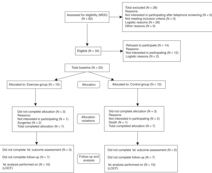
group. The present study is part of a research project on the effect of exercise on mental health (Major Depression, Alzheimer and Parkinson), and is a follow-up of another study that investigated the effect of 6 months of exercise as a non-pharmacological treatment (30). Figure 1 shows the
flow of participants according to the CONSORT statement
(31). The sample consisted of 20 Caucasian elderly subjects with MDD, 70% females, mean age 71 ± 3 years. Ten patients underwent pharmacological treatment [antidepressants (N = 5), anxiolytics + antidepressants (N = 5) plus aerobic training] (exercise group) and the other 10 received only pharmaco-logical treatment [anxiolytic (N = 1), antidepressants (N = 4), anxiolytics + antidepressants (N = 5)] (control group). Patients were evaluated with a thorough clinical examination and with a resting EEG. The functional capacity was measured by the Time to Up and Go (TUG) test (32) and the Multiple-Sit-to-Stand (MSTS) test (33). Each aerobic session began with a 10-min warm-up period (40% VO2max), followed by 20 min of continuous treadmill walking at an intensity correspond-ing to 60% VO2max. The exercise session was concluded with a 5-min cool down period. Heart rate (Polar® Sport Tester, Finland) and perceived exertion (Borg Scale) were monitored and recorded every 5 min during each exercise session by physical education instructors. Subjects attended two supervised exercise sessions per week for 12 months, and all subjects attended at least 75% of the total number of sessions (at least 6 days/month).
Assessment of the outcome
EEG recording, three depression scales and a quality of life questionnaire were used to evaluate the subjects at baseline before and after 1 year of exercise. The experi-mental procedure was carried out at the same time and none lasted more than 1 h. All scales were administered immediately after EEG recording. Handedness was as-sessed by the Edinburgh Handedness Inventory. Severity of cognitive impairment was evaluated by the Mini Mental State Examination (MMSE) (34). Treatment response was
defined as a 50% decrease in Depression Scale scores compared to baseline values. Remission was defined as the absence of significant signs or symptoms of depression,
whereby patients scoring below the cut-off was indicative of an asymptomatic state (Hamilton Depression Rating
Scale (HDRS) ≤7; Beck Depression Inventory (BDI) ≤10;
Montgomery-Asberg Depression Rating Scale (MADRS)
≤9). The validated Portuguese version of the Short Form
Health Survey-36 (SF-36) was used (35). SF-36 is a self-rating questionnaire that assesses eight domains, namely physical functioning, role-physical, bodily pain, social functioning, mental health, role-emotional, vitality, and general health.
Electroencephalogram recording and analyses
Cz, Pz, Oz, Fp1, Fp2, F3, F4, F7, F8, C3, C4, T3, T4, T5, T6, P3, P4, O1, and O2). The International 10/20 System (referred to linked earlobes) for electrode placement was used with a Braintech-3000 instrument (EMSA-Medical Instruments, Brazil). Eye-movement (EOG) artifact was monitored with a bipolar electrode montage using two 9-mm diameter electrodes attached aboveand on the external canthus of the right eye. Impedances for EEG and EOG
electrodes were below 5 and 20 kΩ, respectively. Artifacts
were detected and eliminated by visual inspection and independent component analysis was applied to remove other possible sources of artifacts using the EEGLAB tool
(36). Amplifier band-pass was 0.5-100 Hz (3 dB points), and a 60-Hz notch filter was employed. Data were digitized at
240 Hz with 12-bit resolution.
EEG reference remains one of the most controversial
issues in EEG asymmetry research. In our study, we used the linked ears reference. A classic power spectral density estimator was used (i.e., based on the squared absolute value of the Fourier Transform) for artifact-free 4-s EEG epochs (spectral resolution: 0.25 Hz) with Hamming window-ing. An overlapping factor of 50% (2 s) was used for con-secutive epochs. For each of the 20 monopolar derivations, absolute power (µV2) was computed for the alpha (8-13 Hz) frequency band. EEG measures were log-transformed (i.e., X’ = lognX) to provide a normal distribution. A metric value of asymmetry (ln [Right]) - ln [Left]) was computed for the alpha band (Fp2-Fp1, F4-F3, F8-F7, P4-P3, O2-O1). This difference provides a simple unidimensional scale repre-senting the relative activity of the right and left hemispheres, with higher scores putatively indicating relative greater left frontal activity (assuming that alpha is inversely related to
588 A.C. Deslandes et al.
activity) (26). The EEG data analysis was described in detail in a previous study from our laboratory (18).
Statistical analysis
Baseline differences between groups (control and exercise) were analyzed by an independent sample t-test or a Mann-Whitney U-test for all cognitive, emotional and clinical parameters. All endpoint analyses used a last ob-servation carried forward approach. Analysis of covariance (ANCOVA) models adjusting for baseline score and time of disease were used to analyze absolute changes over time (post-pre) in outcome measures between groups (control and exercise). Effect sizes were calculated by the method of Cohen (mean change/pooled standard deviation). For the EEG variable (log absolute power), four-way ANOVA was computed for the alpha band. Treatment (control x exercise) was established as a between-subject factor and time (pre x post), hemisphere (right x left) and area [anterior (F3, F4) x lateral frontal (F7, F8) x posterior (P3, P4)] as within-subject factors. ANOVA (area x hemisphere) is widely employed because it depicts the contribution of activity in each hemisphere. However, the asymmetry metric value (ln [Right]) - ln [Left]) conveniently summarizes the relative activity at homologous electrodes, providing a simple unidi-mensional scale and simplifying analyses such as correla-tions (26). Furthermore, two-way ANOVA (treatment x time) was performed for alpha asymmetry scores. We computed the correlation (Spearman) between alpha asymmetry and
depression scale scores. The levels of significance were
set at P ≤ 0.05 for all statistical analyses.
Results
Subject characteristics
There was no significant difference in cognition (MMSE
exercise: 26.5 ± 2.9; MMSE control: 27.8 ± 2.0) or educa-tional level (exercise: 9.5 ± 2.5; control: 8.7 ± 3.1) between
groups. Also, there were no significant differences in age,
duration of disease, depression scores, or EEG asymmetry between groups at baseline.
Adverse events and dropout
There were 6 dropouts from the study (3 from the exer-cise and 3 from the control group). The dropouts were due to factors unrelated to physical activity: lack of interest in continuing the program (1 in the exercise group and 2 in the control group), surgeries (2 in the exercise group) and death (1 in the control group). No adverse event took place during the training sessions.
Depressive symptoms
The exercise group (EG) showed a statistically signifi -cant decrease in depressive symptoms on the three scales employed (BDI, HDRS and MADRS) compared to the control group (CG; Table 1). Remission was more frequent in the exercise group, and this result was observed on the three scales: BDI (EG = 60%; CG = 10%), HDRS (EG = 90%; CG = 30%), MADRS (EG = 100%; CG = 70%). The same positive results were observed in the treatment response of the exercise group (BDI: EG = 30%; CG = 10%; HDRS: EG = 30%; CG = 20%; MADRS: EG = 40%; CG = 20%). The effect size observed in the three scales was greater in the group that associated physical activity with treatment (BDI: 0.12, HDRS: 0.5, MADRS: 0.74) compared to the group receiving conventional treatment (BDI: -0.27, HDRS: -0.25, MADRS: 0.23).
Quality of life and functional capacity
Although the exercise group improved in all aspects of quality of life (assessed by the SF-36) after 1 year, the ANCOVA results showed that the difference between exercise and
control groups was significant only in
role-physical (Table 1).
Functional capacity (assessed by physical tests) was investigated only
in the exercise group to confirm the
results of physical training. There was improvement in performance after 1
Table 1. Effect of aerobic training on depressive symptoms and quality of life (after-before treatment values).
Control (N = 10) Exercise (N = 10) F (P) value
After-before treatment After-before treatment
Depressive scale
HDRS 0.87 ± 8.59 -3.55 ± 7.12 11.16 (<0.001)*
BDI 2.11 ± 12.6 -1.44 ± 6.76 11.60 (<0.001)*
MADRS -3.14 ± 12.10 -6.88 ± 8.92 6.48 (0.02)*
SF-36 aspects
Role-physical -31.25 ± 47.32 10.71 ± 45.31 25.49 (<0.001)* Physical functioning -12.50 ± 6.45 4.28 ± 11.70 4.32 (0.07)
Bodily pain -0.50 ± 11.61 -2.42 ± 25.50 0.89 (0.37)
Social functioning -6.25 ± 52.53 26.78 ± 39.15 3.53 (0.09)
Mental health -7.00 ± 28.54 16.57 ± 18.68 3.02 (0.12)
Role-emotional -8.3 ± 41.94 38.09 ± 48.79 4.21 (0.07)
Vitality 13.75 ± 29.54 11.42 ± 18.41 0.55 (0.44)
General health 9.25 ± 32.18 1.85 ± 11.85 0.14 (0.71)
year. Both TUG and MSTS showed better results regarding functional capacity (TUG pre = 8.3 ± 2.1 s; post = 6.4 ± 0.9 s; T = 2.47; P = 0.038; MSTS pre = 9.7 ± 3.5; post = 14.0 ± 1.9; T = -3.47; P = 0.008). These results are noteworthy, since the baseline results of both tests were close to the scores related to risk for loss of functional mobility and they returned to normal range values after 1 year.
EEG results
Absolute alpha power (Ln). The results of four-way ANOVA indicated an expected main effect of area (F = 46.59; P < 0.001), demonstrating that frontal areas have less power than posterior areas. In addition, hemisphere x moment interaction (F = 18.40; P < 0.001) pointed to an increase in alpha power differences between hemispheres after 1 year. An area x moment interaction (F = 5.63; P < 0.001) indicated that alpha power in medial frontal (F4F3) and parietal (P4P3) areas increased after 1 year. Moreover, hemisphere x area x moment interaction (F = 3.35; P = 0.04) pointed to an increase in alpha power differences between hemispheres in the lateral frontal area (F8F7) after 1 year. However, the main result was observed in the hemisphere x moment x treatment (F = 4.54; P = 0.04) interaction, indicat-ing that, after 1 year, alpha power increased in the control group in the right hemisphere, whereas no changes were observed in the exercise group. There is some evidence in favor of a strong negative correlation between cortical activity and alpha power, i.e., increased alpha power is related to decreased cortical activity. Alpha power differ-ences among areas, hemispheres, and moments in the two groups are presented in Figure 2.
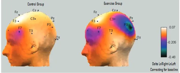
Alpha asymmetry (LnRight-LnLeft). There was no
sig-nificant difference in the metric value of asymmetry between
groups and moments. Figure 3 illustrates asymmetry values (LnRight-LnLeft) represented by the cortical half-map of the difference of frontal (F8-F7, F4-F3) and parietal (P4-P3) electrodes in the control and exercise groups, after 1 year of treatment (correcting for baseline differences). Although the exercise group presented a decrease in asymmetry in parietal areas (increased right parietal activity), this
differ-ence was not statistically significant.
Asymmetry and depressive symptoms. There was no correlation between frontal asymmetry and depressive
Figure 2. Absolute alpha power (Ln) of control and exercising depressed elderly subjects. A lower alpha power is indicative of greater activity. *Hemisphere x moment x treatment interaction (F = 4.54; P = 0.04).
590 A.C. Deslandes et al.
symptoms at any of the moments analyzed. The only cor-relation observed was in the parietal area (P4P3) at baseline in the control group: the greater the relatively left parietal activity (greater posterior alpha asymmetry indices Right-Left), the greater the depressive symptoms (Rs = 0.76, P = 0.01). However, after 1 year of treatment, the correlation
was no longer significant.
Discussion
The severity of depressive symptoms decreased signifi -cantly in the group of elderly adults who combined exercise and clinical treatment, whereas those not submitted to the physical training intervention showed an aggravation of depressive symptoms after 1 year. Treatment response and symptom remission also improved in the exercise group. Our results corroborate those of other studies, which indicate that exercise is effective in the treatment of depression (10,16). Despite the difference in effect size observed in the depression scales, all instruments showed an effect size agreeing with the literature. The improvement observed in the exercise group can be related to the several changes known to be produced by exercise: increased activity and production of neurotransmitters (serotonin, norepinephrine, dopamine), increased production of brain neurotrophic fac-tors (IGF1, BDNF, VEGF), neurogenesis, and hemodynamic changes (4,37).
Quality of life, as assessed by the SF-36, improved in the exercise group (all physical and mental parameters).
How-ever, a significant difference between groups was observed
only on the role-physical aspect after 1 year of treatment; the exercise group improved after training, whereas there was a worsening of this aspect in the control group. In ad-dition, the exercise group showed a trend to improvement in the physical functioning aspect and in two other mental aspects, i.e., role-emotional and social functioning. For all other aspects, improvements were observed in both groups. Despite the small difference between groups, the improvement seen in quality of life of the elderly subjects with exercise training was extremely relevant from a practical point of view. In addition, in the TUG and MSTS physical tests, the subjects improved their functional capacity. These
tests are closely related to aerobic capacity and confirm
the physiological results of exercise.
Given that few studies have assessed the effects of exercise in elderly subjects diagnosed with MDD, our study provides a novel approach by evaluating changes in cortical asymmetry using EEG before and after exercise training
and pharmacological treatment. The most significant EEG
results were related to the effects of exercise on right
corti-cal activity. Specificorti-cally, right alpha power increased in the
control group (decrease of cortical activity) and no changes were observed in the exercise group. The lack of changes in the exercise group may be regarded as a positive result since it implies maintenance of cortical activity.
Exercise has been shown to counteract the mental de-cline accompanying senescence and disease. The increase in right alpha power observed in the control group could be related to a decrease in EEG activity, also observed in elderly individuals with cognitive impairment (38). Several studies have used EEG parameters to investigate treatment response (30,39), since they could be helpful to predict the outcome of antidepressant treatment (39) or for the evalu-ation of treatment (30). Although alpha power is the most frequently investigated EEG variable, other bands (e.g., theta) and other variables (e.g., EEG mean frequency) have also been utilized with this purpose. Recently, our labora-tory observed that depressed elderly individuals showed an increase in EEG mean frequency after 6 months of exercise training (30). Iosifescu et al. (39) found that frontal relative
theta power at baseline was significantly lower in responder
than in non-responder subjects. However, in our study we
did not find differences in alpha power between groups
at baseline. In this respect, increased alpha power may be associated with a worse response to pharmacological treatment, since only the control group showed this result, associated with increased depressive symptoms. Alpha power may contribute to the evaluation of the treatment response and of the effect of the pharmacological and non-pharmacological intervention.
Based on Davidson’s hypothesis (24), we expected a decrease in relatively right frontal activity and in rela-tively left posterior activity. However, our results showed
that the frontal asymmetry was not significantly different
after exercise and clinical treatment. Although the
differ-ence between groups was not significant, we observed
a decrease in relatively left posterior area in the exercise group and an increase in the control group. This result cor-roborates those of investigators who detected a relatively right posterior activity (lower alpha power) in subjects with major depression, but no relatively left frontal activity in this same group (28). We also expected a correlation between frontal and posterior asymmetry (right-left) with depressed symptoms (an inverse and a direct correlation, respectively).
Specifically, a greater frontal asymmetry index is related to
less severe depressive symptoms and a greater posterior asymmetry index is related to more severe depressive symptoms. However, we observed this correlation only in the control group at baseline, whereas greater posterior asymmetry was related to more severe symptoms. In a
previous study from our laboratory, we also did not find
any correlation in depressed elderly subjects (18). In one of the few studies on elderly women, Kline et al. (25) found
that right frontal asymmetry did not vary significantly when
valerian odor (an unpleasant stimulus) was used. More
studies are necessary in order to investigate the influence
of the aging process on EEG asymmetry and to correlate it with mental disease.
It is important to mention that, although our study found
of elderly subjects, its quasi-experimental design raises some problems regarding the interpretation of the results. We cannot rule out an alternative explanation, according to which the subjects who chose the exercising group could have been more prone to improvement in general.
The present study confirms the beneficial effect of
exercise as an add-on treatment of depression in elderly
subjects. Our findings suggest that walking on the treadmill
twice a week for 30 min at moderate intensity seems to be enough to improve treatment response. Moreover, greater
functional capacity and physical and mental health could be achieved with this relatively simple and low-cost procedure. Thus, exercise facilitates the treatment of depressive elderly adults since it provides clinical and physical improvement and protects against a decrease in cortical activity.
Acknowledgments
Research supported by FAPERJ/CAPES and CNPq.
References
1. Blay SL, Andreoli SB, Fillenbaum GG, Gastal FL. Depres-sion morbidity in later life: prevalence and correlates in a developing country. Am J Geriatr Psychiatry 2007; 15: 790-799.
2. Maletic V, Robinson M, Oakes T, Iyengar S, Ball SG, Russell J. Neurobiology of depression: an integrated view of key findings. Int J Clin Pract 2007; 61: 2030-2040.
3. Deslandes A, Moraes H, Ferreira C, Veiga H, Silveira H, Mouta R, et al. Exercise and mental health: many reasons to move. Neuropsychobiology 2009; 59: 191-198.
4. Moraes H, Deslandes A, Ferreira C, Pompeu FAMS, Ribeiro P, Laks J. Physical exercise in the treatment of depression in the elderly: a systematic review. Rev Psiquiatr R Gd Sul
2007; 29: 79.
5. Ng F, Dodd S, Berk M. The effects of physical activity in the acute treatment of bipolar disorder: a pilot study. J Affect Disord 2007; 101: 259-262.
6. Carta MG, Hardoy MC, Pilu A, Sorba M, Floris AL, Mannu FA, et al. Improving physical quality of life with group physi-cal activity in the adjunctive treatment of major depressive disorder. Clin Pract Epidemiol Ment Health 2008; 4: 1. 7. Dunn AL, Trivedi MH, O’Neal HA. Physical activity
dose-response effects on outcomes of depression and anxiety.
Med Sci Sports Exerc 2001; 33: S587-S597.
8. Arent S, Landers D, Etnier J. The effect of exercise on mood in older adults: a meta-analytic review. J Aging Phys Activity
2000; 8: 407-430.
9. Blumenthal JA, Babyak MA, Moore KA, Craighead WE, Her-man S, Khatri P, et al. Effects of exercise training on older patients with major depression. Arch Intern Med 1999; 159: 2349-2356.
10. Blumenthal JA, Babyak MA, Doraiswamy PM, Watkins L, Hoffman BM, Barbour KA, et al. Exercise and pharma-cotherapy in the treatment of major depressive disorder.
Psychosom Med 2007; 69: 587-596.
11. Babyak M, Blumenthal JA, Herman S, Khatri P, Doraiswamy M, Moore K, et al. Exercise treatment for major depression: maintenance of therapeutic benefit at 10 months. Psycho-som Med 2000; 62: 633-638.
12. Mather AS, Rodriguez C, Guthrie MF, McHarg AM, Reid IC, McMurdo ME. Effects of exercise on depressive symptoms in older adults with poorly responsive depressive disorder: randomised controlled trial. Br J Psychiatry 2002; 180: 411-415.
13. Herman S, Blumenthal JA, Babyak M, Khatri P, Craighead WE, Krishnan KR, et al. Exercise therapy for depression in
middle-aged and older adults: predictors of early dropout and treatment failure. Health Psychol 2002; 21: 553-563. 14. Singh NA, Clements KM, Fiatarone MA. A randomized
con-trolled trial of progressive resistance training in depressed elders. J Gerontol A Biol Sci Med Sci 1997; 52: M27-M35. 15. Singh NA, Clements KM, Singh MA. The efficacy of exercise
as a long-term antidepressant in elderly subjects: a random-ized, controlled trial. J Gerontol A Biol Sci Med Sci 2001; 56: M497-M504.
16. Singh NA, Stavrinos TM, Scarbek Y, Galambos G, Liber C, Fiatarone Singh MA. A randomized controlled trial of high versus low intensity weight training versus general practi-tioner care for clinical depression in older adults. J Gerontol A Biol Sci Med Sci 2005; 60: 768-776.
17. Deslandes A, Veiga H, Cagy M, Fiszman A, Piedade R, Ribeiro P. Quantitative electroencephalography (qEEG) to discriminate primary degenerative dementia from major depressive disorder (depression). Arq Neuropsiquiatr 2004; 62: 44-50.
18. Deslandes AC, de Moraes H, Pompeu FA, Ribeiro P, Cagy M, Capitao C, et al. Electroencephalographic frontal asym-metry and depressive symptoms in the elderly. Biol Psychol
2008; 79: 317-322.
19. Petruzzello SJ, Landers DM. State anxiety reduction and exercise: does hemispheric activation reflect such changes?
Med Sci Sports Exerc 1994; 26: 1028-1035.
20. Petruzzello SJ, Tate AK. Brain activation, affect, and aerobic exercise: an examination of both state-independent and state-dependent relationships. Psychophysiology 1997; 34: 527-533.
21. Petruzzello SJ, Hall EE, Ekkekakis P. Regional brain activa-tion as a biological marker of affective responsivity to acute exercise: influence of fitness. Psychophysiology 2001; 38: 99-106.
22. Hall EE, Petruzzello SJ. Frontal asymmetry, dispositional affect and physical activity in older adults. J Aging Phys Activ
1999; 7: 76-90.
23. Hall EE, Ekkekakis P, Petruzzello SJ. Regional brain activity and strenuous exercise: predicting affective responses using EEG asymmetry. Biol Psychol 2007; 75: 194-200.
24. Davidson RJ. Anterior cerebral asymmetry and the nature of emotion. Brain Cogn 1992; 20: 125-151.
592 A.C. Deslandes et al.
26. Allen JJ, Coan JA, Nazarian M. Issues and assumptions on the road from raw signals to metrics of frontal EEG asym-metry in emotion. Biol Psychol 2004; 67: 183-218.
27. Thibodeau R, Jorgensen RS, Kim S. Depression, anxiety, and resting frontal EEG asymmetry: a meta-analytic review.
J Abnorm Psychol 2006; 115: 715-729.
28. Bruder GE, Fong R, Tenke CE, Leite P, Towey JP, Stewart JE, et al. Regional brain asymmetries in major depression with or without an anxiety disorder: a quantitative electroen-cephalographic study. Biol Psychiatry 1997; 41: 939-948. 29. American Psychiatric Association. Diagnostic and statistical
manual of mental disorders (DSM-IV). 4th edn. Washington: APA; 1994.
30. Silveira H, Deslandes AC, de Moraes H, Mouta R, Ribeiro P, Piedade R, et al. Effects of exercise on electroencepha-lographic mean frequency in depressed elderly subjects.
Neuropsychobiology 2010; 61: 141-147.
31. Toerien M, Brookes ST, Metcalfe C, de Salis I, Tomlin Z, Pe-ters TJ, et al. A review of reporting of participant recruitment and retention in RCTs in six major journals. Trials 2009; 10: 52.
32. Podsiadlo D, Richardson S. The timed “Up & Go”: a test of basic functional mobility for frail elderly persons. J Am Geriatr Soc 1991; 39: 142-148.
33. Jones CJ, Rikli RE, Beam WC. A 30-s chair-stand test as a measure of lower body strength in community-residing older adults. Res Q Exerc Sport 1999; 70: 113-119.
34. Folstein MF, Folstein SE, McHugh PR. “Mini-mental state”. A practical method for grading the cognitive state of patients for the clinician. J Psychiatr Res 1975; 12: 189-198. 35. da Mota Falcao D, Ciconelli RM, Ferraz MB. Translation and
cultural adaptation of quality of life questionnaires: an evalu-ation of methodology. J Rheumatol 2003; 30: 379-385. 36. Delorme A, Makeig S. EEGLAB: an open source toolbox for
analysis of single-trial EEG dynamics including independent component analysis. J Neurosci Methods 2004; 134: 9-21. 37. Dishman RK, Berthoud HR, Booth FW, Cotman CW,
Edgerton VR, Fleshner MR, et al. Neurobiology of exercise.
Obesity 2006; 14: 345-356.
38. Prichep LS, John ER, Ferris SH, Rausch L, Fang Z, Cancro R, et al. Prediction of longitudinal cognitive decline in normal elderly with subjective complaints using electrophysiological imaging. Neurobiol Aging 2006; 27: 471-481.