ASYMMETRY-IN:
THE ROLE OF
DMRT2
R AQUEL DE AMARO LOURENÇO
Dissertation presented to obtain the PhD degree in Biology at the Instituto de Tecnologia Química e Biológica
da Universidade Nova de Lisboa
ASYMMETRY-IN:
THE ROLE OF
DMRT2
R AQUEL DE AMARO LOURENÇO
SUPERVISOR: LEONOR SAÚDE, PhD
Dissertation presented to obtain the PhD degree in Biology at the Instituto de Tecnologia Química e Biológica
da Universidade Nova de Lisboa
Durante estes últimos anos tive a sorte e o privilégio de trabalhar com pessoas que em muito contribuíram para a minha formação. Em primeiro lugar quero agradecer à minha orientadora, obrigada Leonor por me ter aceite no grupo e supervisionar o meu PhD. Por tudo o que me ensinou e por todo o apoio e incentivo que me deu ao longo dos anos. Não podia ter tido melhor chefe!
Agradeço ao Instituto Gulbenkian de Ciência e ao Instituto de Medicina Mo-lecular por me terem acolhido durante a realização do meu doutoramento. À Fundação para a Ciência e Tecnologia agradeço o apoio financeiro essen-cial à realização deste trabalho (Apoio financeiro da FCT e do FSE no âmbito do Quadro Comunitário de Apoio, SFRH/BD/24861/2005).
A todo o grupo que fez com que o ambiente de trabalho fosse simples-mente óptimo, desde o tempo em que nos chamávamos SD até agora em que somos UDEV. O vosso apoio foi fundamental: Rita, a tua inteligência é inspiradora, obrigada por todos os ensinamentos, pela ajuda preciosa na construção do transgénico e por todas as boleias até casa; Susana L., gostei de poder trabalhar contigo num mesmo projecto, obrigada pelas discussões e por todo o incentivo para enfrentar a próxima etapa; Susana P., a minha companheira de secretária, já viste o espaço que vais ganhar?! Obrigada pelo apoio em momentos mais difíceis; Raquel M. e Sofia, foi bom ter-vos como colegas de doutoramento, boa sorte para a vossa fase final! João, che-gaste em último mas ainda partilhaste a tua boa disposição e conhecimen-tos, boa sorte no Canadá! Andreia, Patrícia e Ana Margarida, a vossa ajuda no laboratório fez muitas vezes a diferença. Ainda um grande obrigado aos grupos UMO e UBD por todas as discussões científicas e por contribuírem também para um ambiente de trabalho estimulante.
Um grande obrigado ao Miguel G. por me ensinar a técnica de Western blot, pelos ensinamentos e incentivo.
A todos os serviços técnicos do IGC e do IMM que tanto contribuíram para a realização deste trabalho. Às Unidade de Imagiologia do IMM e IGC. Às equipes dos biotérios de ambos os institutos, em especial ao Yuri Weber e à Alina Costa por toda ajuda no biotério do IMM. À equipe da Zebrafish Facil-ity do IMM, obrigada à Lara e ao Fábio por todo o trabalho em manter as diversas linhas de peixes, pelo número infindável de caixas de cruzamentos que tiveram de organizar e por serem sempre prestáveis para tudo.
A todos os amigos pela companhia durante estes anos. Marta, obrigada pelos cafés e por me tornares “tia”, um dia o Lourenço ainda ouve as nossas histórias. Ana Raquel, as tuas gargalhadas continuam contagiantes, como gosto de ouvir as histórias que vais contando, qualquer dia vejo um filme sobre a tua vida! Obrigada pelas longas conversas. À Tânia, pelos desabafos sobre tudo e mais alguma coisa e pelas guaridas, tu és a próxima, boa sorte! À Susana e à Lena, sempre boas companhias. Fred e Susana, longas conver-sas que tão bem me fizeram, boa sorte para os dois. Ao Pedro e aos seus discursos tão loucos e hilariantes, tu vais longe!
obrigada pela confiança, pelo ombro amigo e bons conselhos que tens dado, és extraordinária! Catarina, a força do norte que tanto apoio também me deu, obrigada por tudo! Terei sempre um cantinho para quando as três me visitarem, prometo escolher bons restaurantes para os nossos jantares! Lara, desde os tempos do IGC que estás presente. Nem sei por onde começar a agradecer todo o apoio que me tens dado, por cada palavra de força e por de certa forma teres ajudado a mudar a minha vida, sabes bem disso. Obrigada também ao Nuno, pela boa disposição e obras de arte!
A toda a minha família. Ao meu irmão, por todo o incentivo, conhecimento e ajuda gráfica na tese. Aos meus pais, obrigado por todo o apoio incondi-cional, sem vocês não estava onde estou.
Summary 1
Resumo 5
Abreviations 9
Index of Figures 10
ChApTer I
Introduction 13
I.1. What needs to happen in order to produce a symmetric body plan? 15
I.1.1. The bilateral symmetry of somitogenesis 15
I.1.2. The segmentation clock sets the periodicity of somite formation 15
I.1.3. The wavefront sets the position of somite formation 20
I.1.4. Somites epithelialization 22
I.2. Is the body plan all about symmetry? 24
I.2.1. How can cilia break symmetry? 29
I.2.2. Cilia distribution in the laterality organ 31
I.2.3. More than cilia: other players in the scene 33
I.3. How are symmetric tissues protected from LR asymmetric signals? 35
I.3.1. Retinoic acid buffers the PSM from the influence of LR signals 35
I.3.2. Bridge between LR patterning and somitogenesis 39
I.4. Human developmental disorders related to the LR axis 41
I.5. Conclusions 44
ChApTer II
left-right function of dmrt2 genes is not conserved between zebrafish and mouse 47
Abstract 49
Introduction 49
Materials and methods 52
Results 54
II.1. The murine Dmrt2 is not required for bilateral synchronization of somite formation 54
II.2. Mouse left-right LPM patterning is independent of Dmrt2 function 57
II.3. dmrt2a/terra is expressed in the zebrafish Kupffer’s vesicle but its
homologous gene is absent from the mouse node 59
ChApTer III
searching for downstream targets of Dmrt2a/Terra: a ChIp-on-chip approach 67
Abstract 69
Introduction 69
Results 72
Discussion 74
Materials and methods 81
ChApTer IV
Microarray approach for the search of Dmrt2a/Terra targets 87
Abstract 89
Introduction 89
Results 92
IV.1. Dmrt2a/Terra overexpression and somite formation 92
IV.2. Dmrt2a/Terra overexpression and LPM left-right patterning 98
IV.3. Construction of an inducible transgenic zebrafish line 102
Discussion 105
Materials and methods 109
ChApTer V
notch signalling regulates left-right asymmetry through ciliary length control 115
Abstract 117
Introduction 117
Material and methods 119
Results 120
V.1. DeltaD is the Notch ligand defining laterality in zebrafish 120
V.2. DeltaD maintains spaw expression in the posterior left LPM 124
V.3. DeltaD controls gut laterality 124
V.4. DeltaD regulates cilia length in the KV 125
V.5. DeltaD maintains proper cilia length through modulation of foxj1a 128
V.6. Short cilia generate a weak fluid flow inside the KV 129
V.7. A strong KV fluid flow promotes asymmetric charon expression 131
ChApTer VI
Discussion 139
VI.1. Left and Right sides combine symmetry with asymmetry 141 VI.1.1. Protection of somite formation from asymmetric signals to assure symmetry 141
VI.1.1.1. Conserved role of Retinoic Acid 142
VI.1.1.2. Notch signalling inputs in establishing asymmetry and symmetry 143
VI.2. Symmetry vs. asymmetry – the role of Dmrt2 145
VI.2.1. How does Dmrt2 regulates two opposite processes? 146
VI.3. Evolutionary origins of asymmetry 149
VI.3.1. Initial breaking of symmetry 150
VI.4. Evolutionary origins of segmentation 151
References 153
Appendix I
lourenço and saúde, 2010 173
Appendix II
lopes et al., 2010 203
Appendix III
It may come as a surprise that vertebrates are not entirely bilaterally sym-metric. In fact, at the internal level they are asymmetric with the organs acquiring a biased disposition along the left-right (LR) axis. Problems in the establishment of both symmetric and asymmetric features may give rise to several human developmental disorders. Thus, it is very important to study in more detail how LR embryonic development is initiated and maintained. The built of the axial skeleton and its associated skeletal muscles is depen-dent on somite formation and differentiation. The somites are formed as pairs and are bilaterally symmetric between both sides of the LR axis. Inter-estingly, it has been suggested that asymmetric somite formation may ac-count for problems at the level of the axial skeleton. At the same time that the somites are being formed, asymmetric signals are being transferred only to the left side of the axis being responsible to set up the correct asymmet-ric internal organs distribution. In fact, an incorrect transfer of information leads to serious problems in organ situs. It has become clear that several mechanisms are necessary to initiate the LR asymmetry pathway however, something that only recently has been acknowledge is that symmetry is not a default state and there are in fact mechanisms responsible for promoting it. It is not completely understood how, but there are indications that there is a crosstalk between the mechanisms necessary to regulate symmetry vs. asymmetry. Surprisingly, a zebrafish transcription factor named Dmrt2a/ Terra, a gene that belongs to a family mainly associated with sex determi-nation, as been shown to play a role in regulating these two contradictory features. Not only is it necessary for bilateral symmetric somite formation but also for the correct establishment of asymmetry in the left side of the axis. Given this, it is extremely important to study the signalling cascade where it operates and to identify its downstream targets.
Terra however, since it was not successfully obtained we decided to undergo to a microarray approach. To be able to compare changes in the expression levels of Dmrt2a/Terra target genes we would have used two different con-ditions where Dmrt2a/Terra would be downregulated, through morpholino injection, and upregulation. To overexpress it in a specific developmental time period, we constructed an inducible transgenic zebrafish line where Dmrt2a/Terra is under the control of a heat-shock promoter. With this tool built, we are now able to proceed with a controlled microarray experiment that will allow us to identify Dmrt2a/Terra direct targets during zebrafish embryonic development.
Different developmental processes have been shown to share the same molecular player and the Notch signalling pathway is no exception. Not only it drives the expression of oscillating genes that constitute the segmentation clock, associated with vertebrate somite formation, but it is also necessary to regulate the LR asymmetry cascade. Given the fact that this role during LR patterning is less understood during zebrafish embry-onic development, in Chapter V we address at what level Notch is having an impact in the LR pathway. We show that it is through the ligand DeltaD that Notch is regulating cilia length and consequently the correct transfer of information only to the left side of the axis. Interestingly, Notch is the first signalling pathway shown to be able to decrease or increase cilia length, either when it is downregulated or upregulated. With the regulation of cil-ia length being assoccil-iated with several human ciliopathies, understanding how Notch signalling modulates this regulation is thus of great importance for the search for other regulatory players.
onde o dmrt2a/terra é expresso. Uma vez que não sabemos como o Dmrt2a/ Terra regula estes dois processos, nos Capítulos III e IV é descrita a iniciação de duas abordagens diferentes com o intuito de descobrir quais os genes que regula. Discutimos o potencial de desenvolver as técnicas de “ChIP-o-chip” e “microarray” e ainda um transgene indutível que desenvolvemos. A técnica do “ChIP-o-chip” combina imuno-precipitação de cromatina com a tecnologia de “microarray” e permitiria investigar interacções in vivo entre o Dmrt2a/Terra e as sequências de DNA genómico às quais se liga. O seu sucesso depende do uso de um anticorpo específico contra o Dmrt2a/Terra contudo, como este não foi obtido com sucesso decidimos avançar para o uso da tecnologia de microarray. De modo a podermos comparar alterações nos níveis de expressão dos genes que o Dmrt2a/Terra regula, usaríamos duas condições diferentes em que a expressão do Dmrt2a/Terra seria ini-bida, através da injecção de um morpholino específico, e também aumen-tada ectopicamente. De modo a ser sobre-expresso num período específico do desenvolvimento embrionário, construímos uma linha transgénica de peixe zebra em que a expressão do Dmrt2a/Terra se encontra sob o con-trole de um promotor indutível pelo calor. Com este transgénico construído, podemos agora avançar para um microarray bastante controlado que nos permitirá identificar os genes alvo directos do Dmrt2a/Terra durante o de-senvolvimento embrionário do peixe zebra.
es-querdo do eixo. Surpreendentemente, a via de sinalização Notch é a primeira a ser demonstrada capaz de diminuir ou aumentar o tamanho dos cílios, uma vez inibida ou sobre-expressa, respectivamente. Uma vez que a regulação do tamanho dos cílios se encontra associada a várias ciliopatias em humanos, compreender como a via de sinalização Notch modula esta regulação é de extrema importância na procura de outras vias/genes reguladores.
Ap bhlh cDnA Cfp ChD ChIp Cyc Cyp26a1 DDC DfC’s Dll3 DM DMrT Dsx DV Dvl e eGf-CfC fGf fsi GAGs GJC Grp GsT h+/K+ ATpase
hsp Iv
Anterior-posterior
Basic-helix-loop-helix
Complementary DNA
Cyan Fluorescent protein
Congenital heart disease
Chromatin immunoprecipitation
Cyclops
Cytochrome p450 26a1
Duplication-degeneration-complementation
Dorsal forerunner cells
Delta-like 3
Doublesex and Mab-3
Doublesex and Mab-3 Related
Transcription Factor
Doublesex
Dorso-ventral
Dishevelled
Embryonic day
Epidermal Growth Factor-like -
Cripto, Frl-1, and Cryptic
Fibroblast growth factor
Frequent situs inversus
Glycosaminoglycans
Gap junction communication
Gastrocoel roof plate
Glutathione-S-Transferase Hydrogen-potassium adenosine triphosphatase Heat-shock protein Inversus viscerum KV lfng lpM lr lrd Ml Mo Mrf mrnA ntl oep pApC pCD pCp pCr pnC psM rA raldh2 rAr rbp-jk rnA rXr shh su(h) sur TGf-β Vang WT Kupffer’s vesicle Lunatic fringe
Lateral plate mesoderm
Left-right
Left-right dynein
Medio-lateral
Morpholino
Myogenic regulatory factor
Messenger RNA
No tail
One-eye pinhead
Paraxial protocadherin
Primary cilia dyskinesia
Planar cell polarity
Polymerase chain reaction
Posterior notochord
Presomitic mesoderm
Retinoic acid
Retinaldehyde dehydrogenase 2
Retinoic acid receptor
Recombination signal-binding
protein1 for j-kappa
Ribonucleic acid
Retinoid X receptor
Sonic hedgehog
Suppressor of Hairless
Schmalspur
Transforming growth factor
beta
Van gogh
Index of figures
ChApTer I Introduction
figure 1.1. The formation of a new pair of somites is under the control
of a molecular clock and a wavefront of differentiation 16
figure 1.2. Synchronized oscillations between adjacent cells in zebrafish presomitic mesoderm 18
figure 1.3. Somite differentiation 24
figure 1.4. Internal organ asymmetric organization 25
figure 1.5. Set of events that might culminate with the establishment of
the left-right patterning in different vertebrates 26
figure 1.6. Cilia distribution, tilt and flow generation 32
figure 1.7. Protection of PSM segmentation from LR asymmetric patterning cues 36
ChApTer II
left-right function of dmrt2 genes is not conserved between zebrafish and mouse figure 2.1. Dmrt2 is not required for symmetric somite formation in the mouse embryo 56
figure 2.2. Dmrt2 is not required for the left-right LPM patterning in the mouse embryo 58
figure 2.3. dmrt2/terra is expressed in the zebrafish kupfer´s vesicle 60
ChApTer III
searching for downstream targets of Dmrt2a/Terra: a ChIp-on-chip approach
figure 3.1. Zebrafish and mouse Dmrt2 polyclonal antibodies specificity 73
ChApTer IV
Microarray approach for the search of Dmrt2a/Terra targets
figure 4.1. Dmrt2a/Terra overexpression leads to desynchronization of the somite clock 93
figure 4.2. Asymmetric somite development in Dmrt2a/Terra overexpression embryos 95
figure 4.3. Dmrt2a/Terra overexpression induces a desynchronization of the wavefront
of differentiation 97
figure 4.4. Randomization of left-sided gene expression and internal organ localization
in the context of Dmrt2a/Terra overexpression 99
figure 4.5. Midline structure is intact in the context of Dmrt2a/Terra overexpression 101
figure 4.6. Transgenic approach 103
figure 4.7. Dmrt2a/Terra heat-shock in transgenic embryos leads to desynchronization
figure 5.2. Analysis of the midline markers shh and ntl 123
figure 5.3. DeltaD regulates cilia length in the Kupffer´s vesicle 126
figure 5.4. DeltaD modulates foxj1a expression 129
figure 5.5. Short cilia reduce fluid flow inside the Kupffer´s vesicle and compromise charon
Introduction
The introduction was partially published in lourenço, r. and saúde, l. (2010). Symmetry OUT, Asymmetry IN.
I.1. What needs to happen in order to produce
a symmetric body plan?
I.1.1. The bilateral symmetry of somitogenesis
The bilateral symmetric appearance of the vertebrate body plan is largely due to the symmetric organization of the skeleton and its associated mus-cles. The origin of this symmetry can be traced back to early developmen-tal stages when the somites, embryonic structures that will differentiate into the axial skeleton (vertebrae, intervertebral discs and ribs) and skeletal muscles, are being formed. Each new pair of somites is progressively laid down from the rostral region of the presomitic mesoderm (PSM) in a bi-lateral symmetric way on both sides of the axial structures i.e. neural tube and notochord. Somite formation is coupled with the constant supply of new somite precursors cells that are added into the caudal region of the PSM coming from a progenitor zone located in the tailbud. The long lasting fascination about the somites comes from the observation that these em-bryonic structures are produced in species-specific regular intervals in space and time. Every 4-5 hours a new pair of somites is formed in the human embryo, every 120 minutes in the mouse, every 90 minutes in the chick and every 30 minutes in the zebrafish (Dequéant and Pourquié, 2008).
I.1.2. The segmentation clock sets the periodicity of somite formation
The molecular evidence for the existence of a segmentation clock came with the discovery of the first cyclic gene, the avian basic-helix-loop-helix (bHLH) transcription factor hairy1. The chick hairy1 gene shows a dynamic and reiterated expression pattern in the PSM with the exact same periodicity of somite formation (Palmeirim et al., 1997). These hairy1 messenger RNA (mRNA) oscillations occur autonomously in PSM cells and because they are synchronized with adjacent cells, describe a wave of expression starting at the posterior PSM and moving towards the anterior PSM, where it slows down and eventually stops, concomitant with somite formation (Figure 1.1). There-fore, PSM cells undergo several periodic oscillations of hairy1 gene expression before they incorporate into the next somite (Palmeirim et al., 1997).
S-I RA RA RA RA . . . . . . S-0
Fgf / Wnt
Fgf / Wnt Fgf / Wnt
Fgf / Wnt
Fgf / Wnt
Time that takes to form a new pair of somites S-I S-0 S-II S-I S-0 A P
figure 1.1. The formation of a new pair of somites is under the control of a molecular clock and a wavefront of differentiation. mRNA oscillations of cyclic genes are detected in PSM cells (blue dots) and since they are synchronized with
neighbor-ing cells, describe a wave of expression that starts at the posterior region of the PSM and
moves towards the anterior region of the PSM. Here, the wave of expression slows down
and stabilizes in the formed somite (S-I). Somite formation is also under the control of a
wavefront of differentiation defined by a Fgf/Wnt signalling gradient in the posterior region
of the PSM, which is counteracted by a RA gradient in the anterior region of the PSM.
Soon after this discovery, a number of Hairy-Enhancer of Split-related genes, named hes in mice and her in zebrafish, were identified and shown to produce the same type of oscillating behavior at the level of the PSM (Dequéant and Pourquié, 2008). These genes are primary targets of the Notch signaling pathway and encode transcriptional regulators of the bHLH class that mainly work as repressors (Kageyama et al., 2007). This suggested that the oscillatory behavior of the cyclic genes could be due to feedback inhibition. Indeed the first indication that this was the case came from a pioneering study performed in cell culture. It was shown that not only the mRNA but also the Hes1 protein levels oscillate with the same periodicity as somite formation. These oscillations are generated by a mechanism of negative feedback, where the Hes1 protein periodically represses its own transcription (Hirata et al., 2002). It has been shown that negative feedback loops also underlie the oscillatory expression of a number of cyclic genes in the mouse, zebrafish and chick PSM (Oates et al., 2002; Holley et al., 2002; Dale et al., 2003; Bessho et al., 2003; Hirata et al., 2004; Sieger et al., 2003; Brend and Holley, 2009 A). A mathematical model based essentially on experimental data of zebrafish her genes clearly shows that sustained oscillations can indeed be generated by a negative feedback loop, if tran-scriptional and translational time delays are taken into account and if the lifetimes of cyclic mRNAs and proteins are short (Lewis, 2003).
Furthermore, a closer look at the expression of the Notch ligand del-taC, one of the oscillatory genes in zebrafish, in Notch signaling mutants revealed that individual PSM cells still expressed deltaC in a cyclic manner but the levels varied quite a lot between neighboring cells. This led to the idea that the main function of Notch signaling is to coordinate oscillations between individual cells and to keep them synchronized and not so much to drive the oscillations (Figure 1.2). Therefore, it was proposed that in zebraf-ish Notch mutants, PSM cells begin oscillations in synchrony forming normal anterior somites but after a few cycles gradually loose their synchrony form-ing abnormal somite boundaries (Jiang et al., 2000).
DeltaC
Notch her1/7 Her1/7
deltaC
DeltaC
. .
Tm Tp
.
Tm
.
Tp
her1/7
Her1/7
deltaC DeltaC
figure 1.2. synchronized oscillations between adjacent cells in zebrafish pre-somitic mesoderm. Activation of Notch, upon interaction with DeltaC, activates the ex-pression her genes. her genes encode transcriptional repressors that have the ability to inhibit their own expression generating negative feedback loops that are then responsible for the PSM
intracellular oscillations. Importantly, the period of these oscillations are dependent on both
transcriptional (Tm) and translational delays (Tp). Her proteins will not only inhibit their own
expression, but also inhibit the expression of deltaC that for this reason is also expressed in an oscillatory manner. DeltaC in one cell will then activate Notch signalling in the
neighbour-ing cell and as a consequence generates intracellular oscillations. Thus, oscillations between
adjacent cells are synchronized with one another since they are coupled via Notch signalling.
neigh-boring cells and finally their own intracellular oscillations. This Delta/Notch mediated coupling mechanism results in synchronization between adjacent cells (Lewis, 2003; Oates et al., 2002; Giudicelli et al., 2007; Jülich et al., 2005; Ozbudak and Lewis, 2008).
In contrast to what happens in zebrafish, it was recently shown that elimination of all Notch activity abolishes cyclic gene expression and somite formation in mouse PSM (Ferjentsik et al., 2009). This finding is not in-compatible with the possibility presented by others of Notch being a clock synchronizer during mouse somitogenesis (Aulehla and Pourquié, 2008; Feller et al., 2008) since it could integrate both functions. Although a clear demonstration that this pathway synchronizes the oscillations of PSM cells in amniotes is still missing, this could still be a possibility since perturbation of cell-cell interactions upon PSM cell dissociation in chick and mouse leads to irregular desynchronized oscillations (Maroto et al., 2005; Masamizu et al., 2006).
The different roles that Notch signaling seems to play during mouse and zebrafish somitogenesis could be due to different degrees of complex-ity of the segmentation clock mechanism in these two species. A mouse transcriptome study has shown that a large number of genes are in fact oscillating in the mouse PSM. Many of these genes belong not only to the Notch pathway as expected but also to the Wnt and Fibroblast growth factors (FGF) pathways. In addition, this study also revealed that while the Notch and FGF pathway genes oscillate in synchrony, they are in anti-phase with the oscillations of the Wnt pathway genes. This points to a possible cross-talk between them in a way that Wnt pathway may be coupled to Notch and FGF through mutual inhibition, so that all are integrated in one molecular clock (Dequéant et al., 2006). In zebrafish there is no evidence for the existence of Wnt or FGF-based cyclic genes (Dequéant and Pour-quié, 2008).
I.1.3. The wavefront sets the position of somite formation
2003) (Figure 1.1). While under the influence of FGF/Wnt signaling, the PSM cells are maintained in an immature state and are prevented from starting the genetic program of somite formation.
The posterior-to-anterior gradient of fgf8 mRNA in the PSM of sev-eral vertebrate embryos was the first one to be described (Dubrulle et al., 2001; Sawada et al., 2001; Dubrulle and Pourquié, 2004). This gradient is generated via a decay mechanism from a pool of transcripts produced by progenitor PSM cells (Dubrulle and Pourquié, 2004). According to the clock and wavefront model (Cooke and Zeeman, 1976), displacing the position of the determination front by altering the extent of the fgf8 gradient results in the shift of the somite boundary position (Dubrulle et al., 2001). It is thought that this gradient is translated into a protein gra-dient, which is then converted into a MAPK/ERK activity gradient along the PSM (Sawada et al., 2001; Dubrulle and Pourquié, 2004; Delfini et al., 2005).
Parallel to the fgf gradient there is also a posterior-to-anterior gradi-ent of Wnt signaling along the PSM. The expression of wnt3a in the poste-rior PSM (Aulehla et al., 2003) could be responsible for the nuclear β-catenin gradient reaching from the tailbud to the determination front (Aulehla et al., 2008) and for the graded expression of the Wnt target axin2 (Aulehla et al., 2003).
I.1.4. somites epithelialization
The maturation of the anterior PSM involves changes in cellular organization that ultimately lead to tissue epithelialization. This process is dependent on extracellular matrix proteins, such as fibronectin, with their presence in the PSM being essential for morphological somite formation (Georges-Labouesse et al., 1996; Rifes et al., 2007). Cadherins, such as the paraxial protocadherin(PAPC), have also been shown to regulate this process. PAPC is a cell adhesion molecule that is restricted to their anterior region of the last formed somite pair and the three next to be formed, being important to maintain somite cohesivity (Yamamoto et al., 1998; Kim et al., 2000; Johnson et al., 2001). In addition, somites are compartmentalized structures and the determination of their anterior-posterior (AP) polarity is actually established in the anterior PSM prior to the appearance of segmental borders. mesp genes encode bHLH transcription factors and are involved in the initial definition of the segments. They act downstream of the Notch signalling and are expressed in the anterior halves of the presumptive and forming somites, having a role in the acquisition of their anterior identity (Saga et al., 1997; Sawada et al., 2000; Durbin et al., 2000; Buchberger et al., 1998).
figure 1.3. somite differentiation. (A-B) Somites first differentiate into the dermo-myotome, which will give rise to the dermis and skeletal muscle precursors, and to the
scle-rotome that will give rise to the vertebral column and ribs. (C) Cells from the dorsomedial
edge of the dermomyotome delaminate to form the epaxial myotome, which differentiates
into the musculature of the back and intercostals muscles, while cells from the
dermo-myotome ventrolateral edge delaminate to form the hypaxial dermo-myotome, which will give
rise to the abdominal muscles. (D) At the limb bud level, cells from the ventrolateral edge
of the dermomyotome migrate to the lateral plate mesodermand form the limb muscles.
Adapted from Brent and Tabin, 2002.
I.2. Is the body plan all about symmetry?
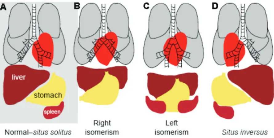
arise in single individuals and will result in laterality defects: situs inver-sus corresponds to a situation where the position of the internal organs is completely reverted as a mirror-image; left or right isomerism is a situation where bilateral symmetry is not broken and two left or two right sides will form; situs ambiguus or heterotaxia that corresponds to some organs being well oriented and others reversed (Fliegauf et al., 2007).
figure 1.4. Internal organ asymmetric organization. (A) Normal distribution of the internal organs referred to as situs solitus. Heart, stomach, spleen and the part of the lung with fewer lobes are found on the left side of the vertebrate body, while on the right
side we have the liver and the part of the lung with more lobes. Cases with laterality defects
may correspond to: (B) right isomerism, two right sides are formed; (C) left isomerism, two
left sides are formed; (D) situs inversus, the position of the internal organs is completely reverted as a mirror-image. Adapted from McGrath and Brueckner, 2003.
regula-tors, the lefty genes. Lefty1 in the midline prevents nodal activation on the right LPM, while lefty2 restricts the domain of nodal expression on the left LPM. Here, Nodal induces pitx2 expression. Pitx2 will promote LR asymme-try of the internal organs, through a mechanism not yet fully understood (Hamada, 2008) (Figure 1.5C).
midline lefty1 nodal lefty2 nodal pitx2 Left Right Ion transporters membrane voltage differences
asymmetric nodal
Left Right Left Right
Before gastrulation (Xenopus, zebrafish, chick)
Onset of gastrulation (zebrafish, chick, mouse)
During somitogenesis (Xenopus, zebrafish, chick, mouse)
A. B. C.
Left determinant
Gap-junction
Motile cilia
Mechanosensory cilia
Ca2+Ca2+ Ca2+ Ca2+ Ca2+ Ca2+ Ca2+ Ca2+ Leftward flow
figure 1.5. set of events that might culminate with the establishment of the left-right patterning in different vertebrates. A) Prior to gastrulationin Xenopus, zebrafish and chick, ion transporters asymmetrically distributed in the embryo generate differences in membrane voltage potential between the left and right side. It is
thought that this asymmetric membrane polarization promotes the accumulation of LR
determinants through directional transport involving gap-junction channels. B) In mouse, it
is though that mechanosensory cilia present in the node epithelia sense the leftward fluid
flow created by motile cilia and as a consequence trigger an asymmetric Ca2+ release which
will induce an asymmetric nodal expression around the node. This Ca2+ accumulation on the left side has also been described in zebrafish and chick although its relation with
cilia-driven flow has not been established. C) A conserved Nodal cascade is activated at the
onset of gastrulation in Xenopus, zebrafish, chick and mouse. nodal is asymmetrically transferred from the node to the left LPM. There, it induces its own expression through
Although the Nodal cascade is conserved among vertebrates, the mechanism that induces nodal in the node in the first place shows differences between vertebrates. In mouse and chick, Notch signaling activates nodal in the node region (Collignon et al., 1996; Raya et al., 2004). Mouse mutants for the notch1/notch2 receptors and for the recombination signal-binding protein1 for j-kappa (rbp-jk), the primary transcriptional mediator of Notch signaling, show absence of nodal expression around the node and LPM but do not interfere with the expression of its inhibitor cerl2 (Krebs et al., 2003; Raya et al., 2003;Takeuchi et al., 2007). In zebrafish, Notch signaling activates the Nodal negative regulator charon around the Kupffer’s vesicle (KV), a fluid-filled sac that lies between the tailbud and the yolk and is considered the node homologous LR organ. After blocking Notch signaling with DAPT, char-on expression is either reduced or absent while nodal expression in the KV region is not affected (Gourronc et al., 2007). Nevertheless, the involvement of Notch in establishing the LR axis is thus conserved among vertebrates.In addition to Notch signaling, Fgf8 also regulates nodal asymmetric expression in the mouse and chick node. In the mouse, Fgf8 acts as a positive nodal reg-ulator and therefore is a left determinant. In the chick, fgf8 is asymmetrically expressed on the right side of the node where it represses nodal, thereby acting as a right determinant (Meyers and Martin, 1999). The role of Fgf8 in controlling expression at the KV has not been determined, although fgf8 mutants show LR defects (Albertson and Yelick, 2005). Also Wnt signaling, namely Wnt3a, indirectly regulates nodal expression in the mouse node indi-rectly through Dll1 (Nakaya et al., 2005). In the chick, wnt8c is asymmetrically expressed on the right side of the node but functions as a left determinant controlling the expression of nodal (Rodríguez-Esteban et al., 2001).
LPM, Nodal will cross the PSM region. One question that arises is why is the PSM non-responsive to Nodal? An explanation comes from previous reports were it was shown that the zebrafish cryptic and one-eye pinhead (oep), membrane attached proteins members of the Epidermal Growth Factor-like - Cripto, Frl-1, and Cryptic (EGF-CFC) family, are necessary for the activation of Nodal in the left LPM. These proteins are extracellular cofactors for Nodal signalling and are expressed only in the LPM and not in the node and PSM (Shen et al., 1997;Zhang et al., 1998;Gritsman et al., 1999). Moreover, Cryp-ticis not required for the initiation of nodal expression in the node being only necessary for nodal expression in the left LPM (Gaio et al., 1999;Oki et al., 2007).Either in the absence of cryptic or oep, no asymmetric expression of nodal, lefty2/antivin and pitx2 is observed in the LPM (Gaio et al., 1999; Yan et al., 1999). These results indicate that Nodal travels from the node to the left LPM through GAGs, and that cryptic and oep are required for its activation in the LPM (Gaio et al., 1999;Yan et al., 1999; Oki et al., 2007).
Nodal was shown to initiate asymmetry in habenular neurogenesis that will indirectly bias parapineal migration toward the left, which will then main-tain the left-sided habenula identity (Concha et al., 2003; Roussigné et al., 2009). Interestingly, in zebrafish with situs inversus not only at the viscera level but also in the CNS, such as in the frequent situs inversus (fsi) mutant zebrafish, not all behaviors were reversed.This implies that behavior later-alization cannot be explained by a switch in the brain anatomical lateraliza-tion and that another level of regulalateraliza-tion exists (Barth et al., 2005).
I.2.1. how can cilia break symmetry?
that a directional fluid flow provides the asymmetry cue that determines laterality (Nonaka et al., 2002). Moreover, using the same experimental setup, it was possible to rescue the laterality phenotype of the iv mouse mutant, in which no flow is observed by simply exposing their nodes to a strong leftward fluid flow (Nonaka et al., 2002; Okada et al., 1999). In ad-dition, in mouse mutants for the cellular motor proteins kif3a-/- and kif3b
-/-no cilia are formed, resulting in an altered leftward flow and consequently perturbed LR asymmetry (Nonaka et al., 1998; Okada et al., 1999; Takeda et al., 1999). Given these results, nodal fluid flow was suggested to be a key factor in establishing LR asymmetry, with ciliary beating clearly being important in its generation.
How the flow is interpreted and then converted into LR asymmetric gene ex-pression is still unknown. Nevertheless, two models have been proposed to date. One model states that a morphogen might be transported as a conse-quence of the leftward fluid flow created by motile lrd-containing cilia. The resulting asymmetric morphogen distribution would initiate downstream molecular events that then establish LR asymmetries in the LPM (Nonaka et al., 1998). More recently, vesicular particles containing Shh and RA were shown to form in the node stimulated by FGF and are transported to the left edge of the node where they fragment and release their contents (Tanaka et al., 2005). An alternative model mentions the existence of two different subpopulations of cilia in the node. In addition to the motile lrd-containing cilia, there are also immotile mechanosensory cilia that lack dynein arms and containing the polycystin-2 (PKD) calcium activated channel (McGrath et al., 2003; Tabin and Vogan, 2003). It has been proposed that immotile mechanosensory cilia sense the fluid flow pressure on the left side of the node and trigger an asymmetric intracellular Ca2+ flux that then breaks LR
I.2.2. Cilia distribution in the laterality organ
PCP core proteins, were shown not to be able to place the basal bodies (from which cilia elongate) in the posterior region of the node cells and were un-able to generate a unidirectional leftward flow(Hashimoto and Hamada, 2010) (Figure 1.6B). Also, in mouse conditional mutants for both vangl1 and vangl2 (encode PCP transmembrane proteins homologues of the Drosophila gene van gogh (vang)), the basal bodies do not acquire a posterior localization within the cells and consequently cilia also do not acquire a posterior localization nor a posterior tilt orientation (Figure 1.6B). Further, the unidirectional leftward flow is compromised, left side specific genes become randomized and internal organs acquire laterality problems (Song et al., 2010). It was also shown in zebrafish and Xenopus that the PCP gene vangl2 is also regulating the posterior position and tilt of cilia in the KV and gastrocoel roof plate, respectively (Borovina et al., 2010;Antic et al., 2010). Given this, the PCP mediated polarity in ciliated cells is clearly a conserved approach in mouse, zebrafish and Xenopus.
figure 1.6. Cilia distribution, tilt and flow generation. (A) In a WT situation, cilia are localized in the posterior part of the node cells. This localization is important for
their posterior tilt orientation necessary for the induction of a leftward extracellular fluid
flow, required to regulate left-right asymmetry pathway. (B) PCP pathway is required for
the correct positioning and orientation of cilia in the cells. In PCP pathway mutants, the
basal bodies do not acquire a posterior localization within the cells and consequently cilia
also do not acquire a posterior localization nor a posterior tilt orientation. Given this, cilia
I.2.3. More than cilia: other players in the scene
Although it is clear that monocilia play a fundamental role in LR patterning, several lines of evidence suggest that earlier LR asymmetries already exist prior to the directional fluid flow generated by ciliary motion, at least in amphibians, chick and fish.
In Xenopus,an asymmetrically localized ion flux is set up through a hydrogen-potassium adenosine triphosphatase (H+/K+ ATPase) transporter
(pumps H+ out of the cell in exchange for K+). In fact, H+/K+ ATPase maternal
mRNA is already asymmetrically localized on the right-hand side of the em-bryo during the first two cell divisions. In addition, it was shown that inhibi-tion of this pump results in randomizainhibi-tion of left side specific genes and organ heterotaxia (Levin et al., 2002). It has been suggested that asymmet-ric ion flux might be responsible for directing the positioning of a LR deter-minant to the left side through gap junction communication channels (GJC) since inhibition of these channels induces heterotaxia in Xenopus (Levin et al., 2002; Levin and Mercola, 1998). Generation of LR voltage differences that control laterality also seem to be important in the chicken embryo. In fact, a differential H+/K+ ATPase activity across Hensen’s node results in left
side asymmetric ion flux, which creates a differential membrane potential between the left and right sides of the primitive streak. Asymmetric ion flux has also been suggested to direct LR determinants through GJC in chick, since GJC inhibition also leads to LR patterning problems (Levin et al., 2002; Levin and Mercola, 1998). H+/K+ ATPase activity results in extracellular Ca2+
accumulation on the left side of Hensen´s node, a possible candidate for being a LR determinant molecule to pass through the GJC. In fact, Ca2+
ac-cumulation was shown to induce an asymmetric activation of Notch on the left side of the node that then translates this differential activity into asym-metric nodal expression. Perturbing this early asymmetric ion flux, will lead to randomized gene expression and organ heterotaxia (Raya et al., 2004). In zebrafish it has been shown that the early activity of the H+/K+ ATPase pump
-V-ATPase, was shown to be important to establish LR asymmetries in Xeno-pus, fish and chick and in the case of zebrafish clearly impacts on cilia size/ number within the KV (Adams et al., 2006). In zebrafish another pump, the sodium-potassium ATPase (Na,K-ATPase) alpha2, modulates the levels of in-tracellular Ca2+ already in the cells that are going to give rise to the KV, the
dorsal forerunner cells (DFC’s). In turn, these Ca2+ levels regulate cilia motility
in the KV and consequently the cilia-driven leftward fluid flow (Shu et al., 2007). Propagation of the intracellular asymmetric Ca2+ flux is regulated by
inositol polyphosphates, which in turn are candidates for the LR determi-nant that passes through GJC and influence LR determination (Sarmah et al., 2005). Consistent with this idea, Connexin43.4 (Cx43.4), a protein of the GJC channel, is required for the LR patterning through the development of a functional KV with normal cilia (Hatler et al., 2009).
Another possible candidate for the LR determinant is the neurotrans-mitter serotonin, which has been demonstrated to regulate LR patterning in Xenopus and chick before the appearance of cilia. Maternal serotonin has a rightward gradient localization during cleavage stages and its localization requires the set up of an asymmetric voltage gradient created by the H+/K+
ATPase coupled with GJC channels (Fukumoto et al., 2005 A; Fukumoto et al., 2005 B).
laterality was not affected (Gardner, 2010). This finding sets the ground for the possibility that in mammals LR embryonic patterning may be set during the cleavage stages similarly to what has been described in snails, where manipulation of the first cleavages resulted in reverted shell coiling and visceral situs inversus (Kuroda et al., 2009). Thus, there is still specullation about the timing and regulatory mechanisms of the symmetry breaking between the various species.
I.3. how are symmetric tissues protected
from lr asymmetric signals?
The formation of a perfect vertebrate body plan involves the establishment of LR asymmetries in the LPM to position the internal organs. In addition, it is also crucial that bilateral symmetry is maintained in the PSM ensur-ing the symmetric formation of the somites and consequently of the axial skeleton and skeletal muscles. Besides sharing the same signaling pathways as discussed above, somitogenesis and LR patterning take place at around the same time during development in nearby embryonic regions. There-fore, the asymmetric signals that originate in the node have to be able to influence the LPM without affecting the bilateral symmetry of somite formation in the juxtaposed PSM. In fact, several lines of evidence show that bilateral symmetry is not a default state but instead has to be actively maintained through a mechanism that protects this territory from the LR asymmetric signals.
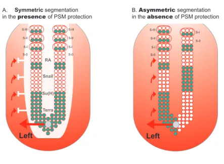
I.3.1. retinoic acid buffers the psM from the influence of lr signals
RA Snail Su(H) Terra S-I S-0
Symmetric segmentation in the presence of PSM protection
S-II S-III
S-I S-0
Asymmetric segmentation in the absence of PSM protection
Left Left S-I S-0 S-II S-III S-I S-0 S-II S-III A. B.
figure 1.7. protection of psM segmentation from lr asymmetric patterning cues. A) PSM is protected from LR signals that come from the node and are implicated in left-right patterning (red arrows). This protection consists of a “shield” (white) which so far
has been shown to be composed by RA, Snail, Su(H) and Terra. In its presence, cyclic gene
expression (blue) and somite formation are symmetric between the left and right sides. B)
In the absence of this protection, cyclic gene expression becomes desynchronised between
both sides. Consequently, somite formation proceeds in an asymmetric way, with the left
side exhibiting more somites than the right (this biased asymmetry towards the right side
is seen in mouse and fish embryos, while in chick asymmetries are biased to the left side).
node/KV to the LPM. This phenotype in not caused by a defect in LR pat-terning in general, since no laterality defects are observed in the absence of raldh2 in mouse and zebrafish (Kawakami et al., 2005; Vermot et al., 2005; Sirbu and Duester, 2006). In addition, epistatic experiments by cross-ing the raldh2 with lrd mouse mutants lead to randomized somite defects instead of the right biased defects seen in raldh2 single mutants (Vermot and Pourquié, 2005). This particular experiment shows that RA acts to coun-teract LR signals.
The right biased somite defects seen in the absence of raldh2 can be explained by the LR desynchronization of the segmentation clock (Kawakami et al., 2005; Vermot et al., 2005) (Figure 1.7B). In these embryos, the expres-sion of the cyclic genes hes7 and lfng (in mouse), deltaC, her1 and her7 (in zebrafish) are out of phase between the left and right sides. Also consistent with the somite phenotype is the anterior displacement of the wavefront seen by the anterior expansion of fgf8 on the right side of the PSM. Once again these LR desynchronization defects are only detected within a small time window that correlates with asymmetric somite formation (Kawakami et al., 2005; Vermot et al., 2005).
When raldh2 was inhibited with disulphiram in chick embryos, again a delay in somite formation was observed and this effect was restricted to a specific time window. But in contrast to mouse and zebrafish now fewer somites formed on the left side of the axis (Vermot and Pourquié, 2005). The segmentation clock is in different phases between the left and the right side of PSM, as seen by the asymmetric expression of lfng. However in contrast to mouse and zebrafish, fgf8 is not expanded anteriorly in an asymmetric manner in disulphiram-treated chick embryos (Vermot and Pourquié, 2005).
rea-sonable to predict that RA is performing its function in this mesodermal tis-sue. However, using a retinoic-acid response element (RARE)-LacZ-reporter transgene it was shown that RA signaling in mouse embryos is present not only in somites and anterior PSM, but it also extends to the adjacent neural plate and the node ectoderm (Sirbu and Duester, 2006). In the node ecto-derm, RA antagonizes fgf8 that is expressed nearby in the epiblast (primi-tive ectoderm). In raldh2 mutants already at early somite stages there is an expansion of fgf8 expression from the epiblast into the neural plate and node ectoderm. At later stages, these embryos show a right shift of fgf8 expression in the anterior PSM, suggesting that the expansion of fgf8 into the node ectoderm may be increasing its own levels in the adjacent PSM shifting it more anteriorly (Sirbu and Duester, 2006). These authors suggest that RA is ensuring bilateral somite formation not at the level of the PSM but at the level of the node ectoderm, where it controls the limits of fgf8.
Fgf8 does not seem to be the only LR cue corrected by RA signaling. In both chick and mouse embryos, snail1 is transiently asymmetrically expressed in the right LPM and plays a role in organ lateralization (Isaac et al., 1997; Sefton et al., 1998). Furthermore, it was shown that the period of asym-metric snail1 expression in the LPM coincides with the time window during which RA is necessary to protect the PSM from asymmetric signals (Morales et al., 2007). Indeed, in the chick it was shown that in the absence of RA signaling, snail1 expression is not affected in the LPM but starts to be asym-metrically expressed in the right anterior PSM. This asymmetric PSM expres-sion of snail1 results in asymmetric expression of the cyclic genes snail2 and lfng and later leads to asynchronous somitogenesis (Morales et al., 2007). Whether or not the asymmetric somite formation seen in the absence of RA signaling in the mouse (Vermot and Pourquié, 2005) is due to a misregu-lation of snail1 expression in the PSM is still unknown. However, in mice mutants for Rere, there is no sign of an asymmetric snail1 expression in the PSM (Vilhais-Neto et al., 2010).
RA signaling has emerged as a conserved keeper of bilateral somite formation. Perturbations of RA function lead to a biased somitogenesis de-fect in a specific time window that correlates with the timing of LR cues establishment (Kawakami et al., 2005; Vermot and Pourquié, 2005; Vilhais-Neto et al., 2010). In the same studies, it was shown that RA perturbation does not result in LR defects in the LPM since the expression of spaw and pitx2 in the LPM is normal. However, the somite laterality defects are linked to the LR pathway. In fact, the bias in somite defects is lost when RA signal-ing is perturbed along with randomization of the LR asymmetric cues upon lrd inactivation (Kawakami et al., 2005; Vermot and Pourquié, 2005; Vilhais-Neto et al., 2010).
I.3.2. bridge between lr patterning and somitogenesis
The downregulation of any of the two Su(H) paralog genes in zebrafish leads to randomization of LR markers in the LPM and to an unbiased asymmetric somite formation. In the su(H) morphants, the RA degrading enzyme cy-p26a1 is misregulated in the tailbud. Since cyp26a1 knockdown can also lead to asymmetric cycling gene expression, this suggests that Su(H) is required to regulate RA in the tailbud that will in turn regulate symmetric cycling gene expression in the PSM (Echeverri and Oates, 2007)(Figure 1.7A,B).
We have shown that Dmrt2a/Terra, a zinc finger-like transcription fac-tor belonging to the DMRT family, regulates the body plan along the LR axis in zebrafish. In Dmrt2a/Terra morphants, the LR asymmetry pathway is also affected, with the expression of left side LPM markers being randomized and consequently affecting the positioning of the heart. On the other hand, dynamic cyclic expression of deltaC, her1 and her7 becomes desynchronized between the left and right sides of the PSM in a specific time window, lead-ing to an unbiased somite number. Therefore, Dmrt2a/Terra has a dual role, it ensures the correct flow of LR asymmetry information to the LPM and in combination with RA signaling ensures the maintenance of symmetry in the PSM (Saúde et al., 2005) (Figure 1.7A,B). In the mouse, the knockout of dmrt2 strongly affected somite differentiation leading to severe rib and vertebral malformations (Seo et al., 2006). It would be interesting to know whether it also regulates synchronization of the clock genes and if it has an impact on heart laterality.
Even more striking is the observation that the simple disruption of the LR determination pathway results in asymmetric somite formation in zebraf-ish. Downregulation of the H+/K+-ATPase activity, with omeoprazole from
to the moment when LR information is being transferred from the KV to the LPM cascade (Kawakami et al., 2005).
At this moment, there is no evidence for the existence of a LR desyn-chronization phenotype in somite formation upon perturbation of early LR asymmetric information in mouse laterality mutants. Since the bilateral somite phenotype can only be detected in a specific time window, there is still the possibility that it was not noticed over the extensive organ laterality analysis.
I.4. human developmental disorders
related to the lr axis
The set up of the axial skeleton is dependent on somite formation and dif-ferentiation. After epithelialization from the anterior region of the PSM, each somite undergoes a dorsal-ventral compartmentalization so that the ventral region, enclosing the sclerotome, is different from the dorsal re-gion, the dermomyotome. This subdivision is important for later patterning events, with the sclerotome differentiating into the axial skeleton and ribs, and the dermomyotome giving rise to the dermis of the back and skeletal muscles (Andrade et al., 2007). A diverse number of human conditions as-sociated with vertebral malformations arise as a consequence of mutations in important somitogenesis-related genes. Mutations in Notch ligand delta-like 3 (dll3) (Bulman et al., 2000), mesp2 (Whittock et al., 2004) and lunatic
fringe (lfng) (Sparrow et al., 2006) are associated with the spondylocostal
which are “butterfly shaped” (McDaniell et al., 2006). Fgf signalling has also been implicated in disorders associated with skeletal development. A mutation in fgfr2 results in fused cervical vertebrae, known as the Apert syndrome (Kreiborg et al., 1992). A minor perturbation in segmentation can lead to severe clinical consequences. Thus the identification of molecules that can reduce vertebral patterning disorders will subsequently help in their prevention.
LR asymmetric cues are important to position the internal organs in a nor-mal configuration termed situs solitus. When LR patterning is disturbed by a series of events, as previously discussed, abnormal laterality phenotypes appear namely situs inversus and situs ambiguus. The morbidity and mor-tality associated with laterality defects is mainly due to congenital heart disease (CHD) (Ramsdell, 2005) Human patients with situs inversus have 3% incidence of CHD compared with normal situs solitus humans that show a 0.08% incidence (Lurie et al., 1995; Nugent et al., 1994; Sternick et al., 2004). In situs ambiguus patients the incidence of CHD is greater than 90% (Nugent et al., 1994). The cardiac defects in these patients include atrial and ventricular septal defects, transposition of great arteries, double outlet right ventricle, anomalous venous return and aortic arch anomalies (Bowers et al., 1996). The clinical observations together with a number of molecu-lar evidence from animal models are helping to understand the ethiology of CHD. It is becoming clear that heart diseases may result from abnormal looping and remodeling of the primitive heart tube into a multi-chambered organ as a consequence of LR patterning defects.
It should be noticed that laterality defects in humans are often associ-ated with abnormal vertebrae and scoliosis (Debrus et al., 1997). The genetic etiology of these conditions is unknown, however we speculate that they may relate to the recently uncovered molecular link between LR patterning and bilateral synchronization of the segmentation clock.
I.5. Conclusions
To design the vertebrate body plan it is fundamental to create asymmetry between the left and the right side of the lateral plate mesoderm, in or-der to correctly position the internal organs. Also, it is crucial to maintain symmetry between the left and the right side of the PSM to ensure the perfect allocation of symmetric body structures such as the axial skeleton, skeletal muscles and peripheral nerves. Although different strategies were shown to initiate the LR asymmetry in the vertebrate embryo (Levin et al., 2005), only recently the existence of mechanisms that promote symmetry have been described in several organisms (Vermot et al., 2005; Vermot and Pourquié, 2005; Kawakami et al., 2005; Saúde et al., 2005). Therefore, sym-metry is no longer perceived as a default embryonic state but rather as a developmental process involving an active molecular mechanism. Although the mechanism that bridges LR patterning and bilateral synchronization of the segmentation clock is not understood, the new studies here reviewed point to the idea that a correct flow of LR signals is necessary for bilateral somite formation.
The purpose of this work was to increase the knowledge regarding the role of Dmrt2 during embryonic development. The aim was:
– to analyze the degree of conservation of Dmrt2 function during vertebrate embryonic development. Since it has already been shown to be required to regulate both symmetric somite formation and LR asymmetry pathway dur-ing zebrafish development, we analyzed the phenotype of mice carrydur-ing the dmrt2 null mutation and compared it to the characterized phenotype in zebrafish;
– to identify the zebrafish Dmrt2a/Terra downstream targets and in this way better understand how it is regulating embryonic LR development. We initiated a ChIP-on-chip approach and also created the tools required to perform a microarray experiment;
left-right function of
dmrt2
genes is not
conserved between
zebrafish and mouse
The work presented here was published in
lourenço, r., lopes, s. s. and saúde, l.
(2010). Left-Right Function of dmrt2 Genes Is Not Conserved between Zebrafish and Mouse.
PLoS One5(12), e14438.
Abstract
Members of the Dmrt family, generally associated with sex determination, were shown to be involved in several other functions during embryonic development. Dmrt2 has been studied in the context of zebrafish devel-opment where, due to a duplication event, two paralog genes dmrt2a and dmrt2b are present. Both zebrafish dmrt2a/terra and dmrt2b are im-portant to regulate left-right patterning in the lateral plate mesoderm. In addition, dmrt2a/terra is necessary for symmetric somite formation while dmrt2b regulates somite differentiation impacting on slow muscle development. One dmrt2 gene is also expressed in the mouse embryo, where it is necessary for somite differentiation but with an impact on axial skeleton development. However, nothing was known about its role during left-right patterning in the lateral plate mesoderm or in the sym-metric synchronization of somite formation. Using a dmrt2 mutant mouse line, we show that this gene is not involved in symmetric somite forma-tion and does not regulate the laterality pathway that controls left-right asymmetric organ positioning. We reveal that dmrt2a/terra is present in the zebrafish laterality organ, the Kupffer’s vesicle, while its homologue is excluded from the mouse equivalent structure, the node. On the basis of evolutionary sub-functionalization and neo-functionalization theories we discuss this absence of functional conservation. Our results show that the role of dmrt2 gene is not conserved during zebrafish and mouse embry-onic development.
Introduction
The axial skeleton and skeletal muscles are derived from embryonic structures called the somites. The epithelialization of a new pair of somites occurs in a bilateral symmetric manner from the anterior-most region of the mesenchymal PSM (Dequéant and Pourquié, 2008). This process is tightly regulated in space and time, with a new pair of somites of approximately the same size being formed with a regular species-specific time period (De-quéant and Pourquié, 2008).
At the same time somites are being formed, LR asymmetric infor-mation is establishing laterality in the nearby LPM, culminating with the asymmetric positioning of internal organs. Before there are any signs of asymmetric organ localization in the vertebrate embryo, a conserved cas-cade of asymmetrically expressed genes is activated around the node in the mouse and around the KV, the functionally equivalent fish organ. An excess of Nodal activity on the left side of the node/KV is transferred to the left LPM and in this location Nodal exerts a positive feedback on itself. As a consequence, the expression of nodal is amplified in the left LPM. Nodal also activates its negative regulators, the lefty genes. Lefty1 in the midline prevents nodal activation on the right LPM, while Lefty2 restricts the do-main of nodal expression on the left LPM. The strong nodal expression on the left LPM induces pitx2 expression that in turn activates morphogenetic proteins required for LR asymmetry of the internal organs (Tabin, 2006). Even though this Nodal cascade is conserved, the mechanism that induces nodal in the node/KV is different between vertebrates. Notch signaling ac-tivates nodal in the murine node region, while in zebrafish it activates the Nodal negative regulator charon around the KV (Raya et al., 2003; Krebs et al., 2003; Takeuchi et al., 2007; Gourronc et al., 2007). In addition to Notch signaling, Fgf8 and Wnt3a regulate nodal expression in the mouse node (Meyers and Martin, 1999; Nakaya et al., 2005). The role of Fgf and Wnt sig-naling in controlling nodal expression at the KV has not been determined. Somitogenesis and LR patterning share the same signalling pathways
that occur at overlapping developmental time windows andin nearby
em-bryonic tissues. For this reason, the asymmetric signals from the node have to be able to reach the LPM without affecting the bilateral symmetry of so-mite formation in the adjacent PSM. In fact, several lines of evidence show that bilateral symmetry is not a default state but instead has to be actively maintained through a mechanism that protects this territory from the LR asymmetric signals (Brend and Holley, 2009 B).
Vermot et al., 2005; Vermot and Pourquié, 2005; Vilhais-Neto et al., 2010). Several lines of evidence show that Fgf8 and Snail1 are the LR cues that are being antagonized by RA signaling in the PSM (Vermot et al., 2005; Vilhais-Neto et al., 2010; Sirbu and Duester, 2006; Isaac et al., 1997; Morales et al., 2007). In zebrafish, another key player regulating development along the LR axis is the zinc-finger like transcription factor Dmrt2a/Terra, that belongs to the Dmrt family. We have previously shown that in zebrafish when Dmrt2a/ Terra function is blocked, the expression of the cycling genes becomes de-synchronized between the left and right sides and as a consequence somite formation is no longer symmetric. In addition, the positioning of the inter-nal organs is compromised as a result of a randomization of left side LPM markers (Saúde et al., 2005).
On the other hand, the mouse dmrt2 null mutants have severe somite differentiation defects but nothing was known regarding a possible role of Dmrt2 in regulating symmetric somite formation and establishing the LR asymmetry pathway (Seo et al., 2006).
Here we report that dmrt2homozygous mouse mutants do not show
LR desynchronization of somite formation and do not have LR defects re-garding internal organs positioning. We show that dmrt2a/terra is expressed in the zebrafish KV in agreement with its function in LR development. In contrast, we did not detect dmrt2 expression in the mouse node, consistent with its non-conserved function during the process of LR axis determination in this vertebrate.
Materials and methods
Zebrafish line
Mice breeding and genotyping
Mice carrying the dmrt2 null mutation where previously described (Seo et al., 2006) and obtained from David Zarkower´s laboratory. Dmrt2 heterozygous mice were maintained on a C57BL/6 genetic background. DNA was extracted from the tail tip of adult mice and genotyped by polymerase chain reaction (PCR) using Dmrt2 specific primers. Dmrt2 WT forward primer 5’-CTGGACCCGAGTACAGTTCC-3’, Dmrt2 WT reverse primer 5’-AATGGTGCGTTCAACTCAGG-3’, Dmrt2 mutant forward primer 5’- TGCGGAGGGCTGGATCTTAAGGAG-3’ and Dmrt2 mutant reverse primer 5’-AGGGGGTGGGGATTTGACACCATC-3’, which resulted in a 830 bp band and a 270 bp band for the WT and mutant alleles, respectively. Dmrt2 heterozygous mice were crossed and embryos collected at specific stages (8.0-9.0 dpc). Mutant embryos were identified by PCR on DNA isolated from the yolk sacs using the same primers as for the adult mice genotyping.
Cloning of mouse and zebrafish dmrt2a/terra and dmrt2b genes
Mouse dmrt2 complementary DNA (cDNA) (IMAGE: 1248080) was
used to synthesize an antisense RNA probe, linearized with EcoRI and transcribed with T3. Total RNA was extracted from appropriate staged zebrafish embryos using TRIzol (Invitrogen) and cDNA was synthesized with MMLV-Reverse Transcriptase kit(Promega). dmrt2a/terra full length sequence was amplified by PCR with the following primer set: dmrt2a/ terra forward primer 5’-ATGACGGATCTGTCCGGCACG-3’ and dmrt2a/ terra reverse primer 5’-AGCAAGAAGCCTTACTGAGATTTCCG-3’. dmrt2b was amplified by PCR with the following primer set: dmrt2b forward
primer 5’-TTTCTTCCCGCTGTCAGACC-3’ and dmrt2b reverse primer
5’-TTATCTCATGAGCAGTGCCTCG-3’. The dmrt2a/terra amplified DNA