Me tabo lic marke rs fo llo wing
be ta-adre no ce pto r ago nist infusio n
in fo o tsho ck-stre sse d rats
Departamento de Fisiologia e Biofísica, Instituto de Biologia, Universidade Estadual de Campinas, Campinas, SP, Brasil J.L. Verago,
D.M. Grassi-Kassisse and R.C. Spadari-Bratfisch
Abstract
Stress hormones can alter metabolic functions in adipose tissue and liver, as well as the sensitivity of rat white adipocytes and rat atrial responses to ß-adrenergic agonists. In this study, we examined the effects of three daily footshock stress sessions on the plasma corticos-terone, glucose, glycerol and triacylglycerol levels of fed, conscious male rats, and on the plasma glucose, glycerol and triacylglycerol levels of the same rats following iv infusions of ß-adrenergic agonists (isoproterenol: 0.4 nmol kg-1 min-1, noradrenaline: 5.0 µg kg-1 day-1, and BRL 37344 ([±]-[4-(2-[(2-[3-chlorophenyl]-2-hydroxyethyl) amino]propyl)phenoxy]acetic acid), a selective ß3-adrenoceptor ago-nist: 0.4 nmol kg-1 min-1). Plasma corticosterone levels increased significantly after each stress session, while triacylglycerol levels increased after the first session and glucose increased after the second and third sessions. Glycerol levels were unaltered after stress. These results suggest that repeated footshock stress may induce a metabolic shift from triacylglycerol biosynthesis to glucose release by hepatic tissue, with glycerol serving as one of the substrates in both pathways. Stressed rats were more sensitive to infusion of noradrenaline plus prazosin and to infusion of isoproterenol, with elevated plasma glu-cose, glycerol and triacylglycerol levels. The higher sensitivity of stressed rats to isoproterenol and noradrenaline was probably related to the permissive effect of plasma corticosterone. Only BRL 37344 increased plasma glycerol levels in stressed rats, probably because ß3 -adrenoceptors are not involved in hepatic triacylglycerol synthesis, thus allowing glycerol to accumulate in plasma.
Co rre spo nde nce R.C. Spadari-Bratfisch Departamento de Fisiologia e Biofísica
Instituto de Biologia, UNICAMP 13081-970 Campinas, SP Brasil
Fax: + 55-19-289-3124
E-mail: rspabrat@ obelix.unicamp.br
Publication supported by FAPESP. Part of a Master’s thesis presented by J.L. Verago to the Departamento de Fisiologia e Biofísica, Instituto de Biologia, UNICAMP, Campinas, SP, Brazil.
Received August 25, 2000 Accepted May 31, 2001
Ke y words ·Glucose ·Glycerol ·Triacylglycerol ·Triglycerides
·Beta-adrenergic agonist ·Corticosterone ·Infusion
Intro ductio n
The physiological responses to stressors include cardiovascular, renal, visceral, cuta-neous, and metabolic changes (1) in which catecholamines and peptides co-released from the sympathoadrenal system act as cru-cial mediators (1). Although the stress
affects glucose and lipid metabolism con-tinue to be of great interest, but no consensus has emerged. It is accepted that the central nervous system plays an important role in the regulation of hepatic glucose and lipid metabolism via the sympathetic nervous sys-tem and cytokines (2). However, the precise relationship between stress-induced incre-ments in metabolic hormones and in the sensitivity of tissues to catecholamines dur-ing repeated or prolonged stress has not been demonstrated in vivo.
Several experimental models have been used to elucidate stress mechanisms, includ-ing novelty, water immersion (3), immobili-zation (4,5), swimming (6), and footshock (7-9). Using a footshock stress paradigm (7), we demonstrated that ß-adrenergic receptor signaling was altered in cardiac and adipose tissues of stressed rats (6-10). Altered myo-cardial adrenergic receptor signaling has been demonstrated in several models of cardiac disease such as heart failure (11,12) and ischemic heart disease (12), and these pa-thologies had been associated with stress (13). Although ß1-, ß2-, and ß3
-adrenocep-tors use the same intracellular signaling path-way, it is now accepted that signaling through one or another subtype is fundamentally dif-ferent (12) and may involve unknown intra-cellular pathways.
Since these results were obtained for iso-lated tissues, the question of the physiologi-cal relevance of these stress-induced alter-ations in the organism in general remains to be answered. The effect of stress on glucose metabolism following electrical stimulation of the sympathetic nerves and the infusion of catecholamines, glucagon, or corticosterone in dogs has been examined (14). Yamada et al. (5) reported that in fed rats, glucagon and corticosterone as well as epinephrine act as synergistic factors to cause stress-induced hyperglycemia. Moreover, in stress situa-tions, lipolysis was greatly stimulated, with an accompanying increase in reesterification to remove the excess free fatty acids released
(15,16).
In the present study, we examined the effect of stress on the functional responses of conscious rats to ß-adrenoceptor agonist infusion. The plasma levels of corticoster-one before and after three sessions of foot-shock stress were used as indicators of stress intensity, whereas the plasma levels of glu-cose, triacylglycerol and glycerol were used as indicators of stress-induced substrate mobilization. Following the initial measure-ments, rats received an iv infusion of nor-adrenaline, isoproterenol or BRL 37344 ([±]-[4-(2-[(2-[3-chlorophenyl]-2-hydroxyethyl) amino]propyl)phenoxy]acetic acid), a selec-tive ß3-adrenoceptor agonist, and the above
parameters were again determined to evalu-ate whether the response was altered after stress.
Mate rial and Me thods
Anim als
Male Wistar rats (Rattus norvegicus) weighing 250 to 350 g at the beginning of the experiments were used. The animals were housed in individual cages (30 x 18 x 20 cm) in a temperature-controlled room (22oC), on
a 12-h light/dark cycle with lights on at 6:30 am. Standard laboratory chow and tap water were available ad libitum. During the experi-ments, the animals were cared for in accor-dance with the principles for the use of ani-mals in research and education, as specified in the Statement of Principles adopted by the Federation of the American Societies for Experimental Biology Board. The experi-mental protocols were also approved by the Institutional Committee for Ethics in Ani-mal Experimentation.
Blood ve sse l cathe te rization
PE20 tubing connected to PE50 tubing that was exteriorized in the dorsal interscapular region where it was fixed to the animals’ skin (17). This method permitted the collec-tion of blood samples from unanesthetized, undisturbed freely moving rats. The cath-eters were siliconized and filled with sodium citrate (5 mM) prior to insertion. In rats which received a drug infusion, the left jugu-lar vein was also cannulated.
Control procedure
On the first day of the experiment, each rat was placed in the footshock cage where it remained for 30 min before being returned to the animal care facilities. During this period, the power for the footshock cage was turned off (day 0). On the following three days, this procedure was repeated with each rat in the control group, whereas the rats in the stress group received shocks as described below. Some rats were cannulated to be used as blood donors.
Stress procedure
Each rat underwent three daily sessions of unsignaled, inescapable footshocks. The animals were placed in a Plexiglas chamber (26 x 21 x 26 cm) provided with a grid floor made of stainless-steel rods (0.3 cm in diam-eter and spaced 1.0 cm apart). During the 30-min sessions, which occurred between 7:30 and 11:00 am, the footshocks were delivered by a constant current source controlled by a microprocessor-based instrument con-structed at the University’s Biomedical En-gineering Center. Current intensity was 1.0 mA and duration was 1.0 s at random inter-vals of 5-25 s (mean interval of 15 s). The rats were returned to their cages at the end of each footshock session (10-13).
Expe rime nt 1
The rats remained in the animal care
facilities for at least one week before enter-ing the experimental protocol. Durenter-ing this period, the rats were handled for 15 min on five successive days. The left carotid artery was then cannulated and the animals were allowed to recover from surgery for 48 h before being used. On each of four consecu-tive days, the rats were brought to a silent room where they remained for 60 min. A 25-cm long piece of PE50 polyethylene tubing filled with 0.9% NaCl was then connected to the external tip of the cannula. Blood samples (500 µl) were withdrawn, and the volume was replaced with blood from a donor rat. Immediately after each blood sampling, the cannulas were filled with 5 mM sodium citrate solution and closed. Blood samples were collected each day immediately before and after the rats had been placed in the experimental cage. The rats were returned to the animal care facilities immediately after collection of the second blood sample.
Expe rime nt 2
In this group cannulation was performed after the second footshock session. On the next day, immediately after the third foot-shock or control session, a blood sample (500 µl) was withdrawn (-15 min) from the cannulated artery as described above, and the volume replaced with blood collected from a naive donor animal. Another sample was obtained after 15 min (0 min), when drug infusion was started. Blood samples were also obtained at 5, 15, 30, 45 and 60 min after starting the infusion which lasted for 30 min.
Using the cannula implanted into the left jugular vein, the rats received one of the following ß-adrenergic agonists: isoprote-renol (0.4 nmol kg-1 min-1), noradrenaline
(5.0 µg kg-1 min-1), or BRL 37344 (0.4 nmol
kg-1 min-1). Noradrenaline was also infused
kg). The infusion of noradrenaline (5.0 µg kg-1 min-1) plus prazosin (8.3 µg kg-1 min-1)
was then started 30 min later. The dose regime was based on Galitzky et al. (18).
Analytical de te rminations
All blood samples were collected into plastic vials in an ice-water bath and imme-diately centrifuged at 5000 rpm for 10 min at 4oC. Aliquots of plasma were removed and
stored at -20oC until assayed for plasma
corticosterone, glucose, glycerol and triacyl-glycerol levels. Plasma glucose was meas-ured by the glucose oxidase method (com-mercial kit, Laborlab S/A, São Paulo, SP, Brazil). Plasma corticosterone was deter-mined by radioimmunoassay (commercial kit, ICN Pharmaceuticals, Inc., Costa Mesa, CA, USA). For glycerol measurements, plasma was deproteinized prior to the enzy-matic assay (19). Plasma triacylglycerol lev-els were also measured by an enzymatic method (commercial kit, Laborlab).
Drugs and chemicals
ATP, bovine serum albumin (fraction V), glycerol phosphate dehydrogenase type I from rabbit muscle, glycerol kinase from
Candida mycoderma, (-)-isoproterenol, (-)-noradrenaline, prazosin HCl and sodium citrate were from Sigma (St. Louis, MO, USA); BRL 37344 was from Tockris Cookson (Ballwin, MO, USA), xylazine from Bayer S.A. (São Paulo, SP, Brazil), and ketamine from Konig S.A. (São Paulo, SP, Brazil).
Statistical analysis
The results are reported as means ± SEM. Differences between the plasma levels of each compound before and after the control or stress procedure during the four succes-sive days of experiment 1, and between the plasma levels at each time interval during or after drug infusion (experiment 2) were ana-lyzed by two-way ANOVA for repeated measures, followed by the Tukey test. Dif-ferences were considered significant for P<0.05.
Re sults
Effe ct of footshock stre ss on plasma
corticoste rone , glucose , glyce rol and
triacylglyce rol le ve ls
Figure 1 shows the plasma corticoster-one levels in fed conscious rats before and after the control procedure or footshock stress. There were no differences in the plasma corticosterone levels among any of the control groups. In stressed rats, there was a trend towards an increase in the levels of corticosterone after stress on days 1, 2 and 3. However, ANOVA for repeated measures followed by the Tukey test showed that only on days 2 and 3 were the plasma corticoster-one levels after footshock stress significant-ly higher than those before footshock. Plasma
Figure 1. Plasma corticosterone levels of fed conscious rats be-fore and after the control proce-dure (top) or 30 min of footshock stress (bottom). On day 0, ani-mals of both groups w ere placed in the footshock cage but did not receive footshocks (control pro-cedure). On days 1, 2 and 3, rats in the stressed group received 120 footshocks (1.0 mA, 1.0 s, int ervals of 5-25 s bet w een shocks) over a 30-min period. Rats from the control group re-mained in the footshock cage but did not receive footshocks. The colum ns represent t he m eans ± SEM f or 5 experi-ments. * P< 0.05 compared to the control group before and af-ter the session on days 0, 1, 2 and 3, and P<0.05 compared to the footshock group before the session on days 0, 1, and 2.
+P<0.05 compared to the
foot-shock group before the session on day 2 (ANOVA plus Tukey test).
C
o
rt
ic
o
s
te
ro
n
e
(
µ
g
/d
l)
60
40
20
0
0 1 2 3
Day Control
Stressed
+
*
Before After
0 1 2 3
Day
C
o
rt
ic
o
s
te
ro
n
e
(
µ
g
/d
l)
60
40
20
corticosterone levels on day 3 were also higher than the levels on days 0 and 1 (P<0.05).
Figure 2 shows that the plasma glucose, glycerol and triacylglycerol levels in the con-trol group were not significantly different before or after the control procedure during the four days of the experiment. In the footshock group, the plasma glucose levels on days 2 and 3 increased after footshock compared to the values before footshock (P<0.05). Furthermore, on day 3, the plasma glucose levels were higher than all values in the control group and in the footshock stress group, except on day 2 after footshock
(P<0.05). Plasma glycerol levels were not significantly different before or after the foot-shock stress on any of the three days. Plasma triacylglycerol levels increased after foot-shock stress only on day 1 (P<0.05; Figure 2).
Effe ct of isoprote re nol, noradre naline ,
noradre naline plus prazosin, or BRL 37344
infusion on plasma glucose , glyce rol and
triacylglyce rol le ve ls
After the third footshock session, the rats were kept in their home cages until the plasma glucose levels decreased to values which
Figure 2. Plasma glucose, glyc-erol and triacylglycglyc-erol levels of fed conscious rats before and after the control procedure (left panels) or 30 min of footshock stress (right panels). On day 0, animals of both groups w ere placed in the footshock cage but did not receive footshocks (con-trol procedure). On days 1, 2 and 3, rats in the stressed group received 120 footshocks (1.0 mA, 1.0 s, intervals of 5-25 s betw een shocks) over a 30-min period. Rats from the control group rem ained in the foot-shock cage but did not receive footshocks. The columns repre-sent the means ± SEM of 5 ani-mals in each group. +P< 0.05
compared to the control group before and after the session, and to the footshock group be-f ore session (ANOVA plus Tukey test). * P<0.05 compared to the control group after the session and to the footshock group bef ore t he session (ANOVA plus Tukey test).
G
lu
c
o
s
e
(
m
g
/d
l)
200
G
lu
c
o
s
e
(
m
g
/d
l)
200
150
100
50
0
Control
0 1 2 3
Day
150
100
50
0
0 1 2
Day 3
Stressed
0 1 2 3
Day
G
ly
c
e
ro
l
(µ
m
o
l/
d
l)
15
10
5
0
G
ly
c
e
ro
l
(µ
m
o
l/
d
l)
15
10
5
0
0 1 2 3
Day
0 1 2 3
Day
0 1 2 3
Day
Control Stressed
Control Stressed
T
ri
a
c
y
lg
ly
c
e
ro
l
(m
g
/d
l)
80
60
40
0 20
T
ri
a
c
y
lg
ly
c
e
ro
l
(m
g
/d
l)
75
50
25
0
Before After
+
+
were not significantly different from basal values (15 min). At this point, an iv infusion of saline solution (0.9% NaCl, w/v), isopro-terenol (0.4 nmol kg-1 min-1), noradrenaline
(5.0 µg kg-1 min-1), or BRL 37344 (0.4 nmol
kg-1 min-1) was started.
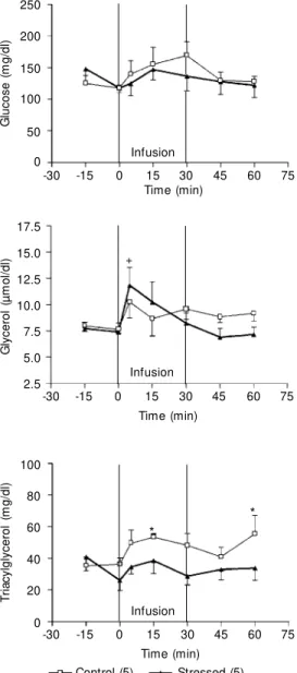
The infusion of saline solution in control rats did not significantly alter the plasma glucose, glycerol or triacylglycerol levels (Figure 3). Immediately after the third ses-sion, plasma glucose levels were higher in stressed rats than in control rats. During the subsequent 15 min, the plasma glucose lev-els of stressed rats decreased to the levlev-els
seen in the control rats. In the control group, isoproterenol (0.4 nmol kg-1 min-1) increased
the plasma glucose levels after 5 min fol-lowed by a return to basal values during infusion (P<0.05; Figure 3). In footshock-stressed rats, plasma glucose levels were higher than in the controls during infusion and 15 min, but not 30 min, after the end of infusion (P<0.05). Isoproterenol had no ef-fect on the plasma glycerol levels of control or footshocked rats (Figure 3). In control rats, isoproterenol increased the plasma tri-acylglycerol levels only 60 min after the start of the infusion (30 min after the end), whereas in footshock-stressed rats the increase in plasma triacylglycerol levels was significant at 30, 45 and 60 min after the onset of the infusion compared to the values obtained immediately before infusion (P<0.05; Fig-ure 3).
Noradrenaline (5.0 µg kg-1 min-1) had no
significant effect on plasma glucose or glyc-erol levels in either group (Figure 4). Nor-adrenaline produced a similar profile of varia-tion in the plasma triacylglycerol levels of control and footshock-stressed rats (Figure 4), with the levels of triacylglycerol being significantly lower in stressed rats than in control rats 15 min after the start of the infusion and 30 min after the end of infusion (Figure 4). Since noradrenaline has a potent vasoconstrictor effect that might affect the release and/or clearance of substrates in sev-eral tissues, we examined the effects of nor-adrenaline in rats which received prazosin before and during noradrenaline infusion.
An ip injection of prazosin did not affect the plasma glucose, glycerol or triacylglyc-erol levels in control rats (Figure 5). How-ever, in stressed rats which received prazo-sin, the plasma glucose levels remained high during the 30 min following ip injection of prazosin, so that when the infusion started the levels were higher in stressed rats than in control rats (Figure 5).
When the infusion of noradrenaline plus prazosin started, there was no significant
G
lu
c
o
s
e
(
m
g
/d
l)
250
75 200
150
100
50
0
-30 -15 0 15 30 45 60
Time (min)
G
ly
c
e
ro
l
(µ
m
o
l/
d
l)
17.5
75 15.0
12.5
10.0
7.5
2.5
-30 -15 0 15 30 45 60
Time (min) 5.0
T
ri
a
c
y
lg
ly
c
e
ro
l
(m
g
/d
l)
100
75 80
60
40
20
0
-30 -15 0 15 30 45 60
Time (min)
Saline (5) Control (8) Stressed (8) *
* Figure 3. Effect of iv infusion of
saline solution (0.9% NaCl, w /v) or isoproterenol (0.4 nmol kg-1
min-1) on plasma glucose,
glyc-erol and triacylglycglyc-erol levels in fed conscious rats after three daily sessions of confinement in footshock cages w ith no shocks (control) or w ith foot-shock stress for 30 min. The in-fusion (time 0) began 15 min af-ter the end of the third session. The points represent the means ± SEM of the number of rats indicated in parentheses. * P<0.05 compared to the control group;
+P<0.05 compared to the value
at 0 min; * * P<0.05 compared to the values at 5, 15 and 30 min;
#P<0.05 compared to the values
at -15, 0, 5, 15, and 30 min in the cont rol group (ANOVA plus Tukey test).
* * * *
+
Infusion
+ # +
* * Infusion
increase in plasma glucose levels in control or stressed rats. At the end of infusion, glu-cose levels tended to decrease towards pre-infusion levels, although the values in the stressed rats were higher than in control rats, even 30 min after the end of infusion (Figure 5). Similar trends were observed for plasma glycerol levels. However, there was a peak in the plasma glycerol levels of stressed rats 15 min after the end of infusion, when triac-ylglycerol levels were higher in stressed rats than in control rats (Figure 5).
BRL 37344 (0.4 nmol kg-1 min-1) had no
significant effect on plasma glucose or triac-ylglycerol levels in either group of rats (Fig-ure 6). BRL 37344 infusion also had no significant effect on plasma glycerol levels in control rats (Figure 6) but increased these levels in footshock-stressed rats towards the end of infusion (30 min) followed by a return to basal levels after the infusion had ended (Figure 6).
D iscussio n
An increase in plasma corticosterone in response to stress is directly related to the stressor intensity, so that corticosterone is a useful indicator (3,20). Plasma catechol-amines also increase during the stress reac-tion (3,21).
Figure 1 shows that 48 h after surgery, the plasma corticosterone level of rats was 26.1 ± 2.7 µg/dl. This value was significant-ly higher than the previoussignificant-ly reported level of 13.0 ± 0.3 µg/dl for male rats sedated with sodium pentobarbital (8) but was similar to that measured by De Boer et al. (3,20) in fed rats transferred to a novel cage and environ-ment, as done here. Thus, high plasma corti-costerone levels appear to be typical of con-scious rats brought to a different environ-ment and sampled for blood collection while freely moving in the cage. Confinement in a footshock cage for 30 min without receiving any footshock did not alter the plasma corti-costerone levels, indicating that remaining
in a novel room for 30 min was not an additional stressor stimulus. However, when rats received footshocks, plasma corticoster-one levels increased, as expected, and rep-etition of the stressor stimulus caused a more pronounced response, indicating that ani-mals had not adapted to the stressor. Many authors have shown that plasma corticoster-one responses are reduced in rats following repeated presentation of noise (21,22), han-dling (23), novelty (24,25) or restraint (26), whereas with the use of relative intense stres-sors such as footshock (27), cold exposure (27), forced running (27,28) or a
combina-Figure 4. Effect of iv infusion of noradrenaline (5.0 µg kg-1 min-1)
on plasma glucose, glycerol and triacylglycerol levels in fed con-scious rats after three daily ses-sions of confinement in foot-shock cages w ith no footfoot-shocks (cont rol) or w it h f oot shock stress for 30 min. The infusion (time 0) began 15 min after the end of the third session. The points represent the means ± SEM of the number of rats indi-cated in parentheses. +P<0.05
compared to the value at 45 min; * P<0.05 compared to the val-ues at 0 and 30 min for the foot-shock group (ANOVA plus Tukey test).
G
lu
c
o
s
e
(
m
g
/d
l)
250
75 200
150
100
50
0
-30 -15 0 15 30 45 60
Time (min)
G
ly
c
e
ro
l
(µ
m
o
l/
d
l)
17.5
75 15.0
12.5
10.0
7.5
2.5
-30 -15 0 15 30 45 60
Time (min) 5.0
T
ri
a
c
y
lg
ly
c
e
ro
l
(m
g
/d
l)
100
75 80
60
40
20
0
-30 -15 0 15 30 45 60
Time (min)
Control (5) Stressed (5)
+
* *
tion of restraint, light, noise and tailcutting (29), no such adaptation was detected. Our stressed rat model presented higher plasma corticosterone levels before the second and third footshock sessions compared to con-trol rats, indicating that with repetition of the stressful stimulus some anticipation rather than adaptation to the stressor may occur.
Plasma glucose levels followed a pattern similar to that of plasma corticosterone lev-els. In footshock-stressed rats, the glucose levels increased significantly after the sec-ond and third stress sessions (Figure 2). This probably reflects the permissive effect of corticosterone on the action of
catechol-amines plus the effect of glucagon in stimu-lating gluconeogenesis and glycogenolysis in hepatic cells (2,30) and the a-adrenergic effects of endogenous catecholamines in in-hibiting insulin secretion (31). In contrast to plasma glucose, which increased progres-sively with the repetition of stress, plasma triacylglycerol levels were not enhanced on day 2 or 3 but were significantly higher than control after the first footshock session (Fig-ure 2). Although we did not meas(Fig-ure any hepatic function indicators, this result sug-gests that, as the stress is repeated, glucose is released by the liver instead of triacylglyc-erol. The use of glycerol released by adipose tissue as a gluconeogenic substrate may help to maintain the plasma glycerol level un-changed after stress despite the adrenergic stimulation of lipolysis in white adipose tis-sue (32). Taken together, these findings indi-cate that repeated stress leads to a wide-spread or general metabolic response aimed at maintaining high plasma glucose levels. This post-stress hyperglycemia is enhanced by repetition of the stressful stimulus, sug-gesting a sensitization of this response.
Because we have previously shown that after this same stress protocol cardiac tissue and adipocytes are supersensitive to isopro-terenol and subsensitive to noradrenaline (7,8) and to BRL 37344 (8), we examined the effect of an infusion of these compounds on plasma glucose, glycerol and triacylglyc-erol levels in stressed rats.
In control rats, the infusion of isoprote-renol increased glycemia, which peaked by the fifth minute, followed by a slow decrease towards the basal value during infusion. This time course is probably a consequence of the effect of isoproterenol on hepatic glyco-genolysis and gluconeogenesis followed by insulin secretion (33). In footshock-stressed rats, the glycemia during isoproterenol infu-sion was higher than in control rats, indicat-ing that glycogenolysis and gluconeogenesis were also higher in these animals. Since De Boer et al. (3,20) have shown that in control
G
lu
c
o
s
e
(
m
g
/d
l)
250
75 200
150
100
50
0
-30 -15 0 15 30 45 60
Time (min)
G
ly
c
e
ro
l
(µ
m
o
l/
d
l)
17.5
75 15.0
12.5
10.0
7.5
2.5
-30 -15 0 15 30 45 60
Time (min) 5.0
T
ri
a
c
y
lg
ly
c
e
ro
l
(m
g
/d
l)
100
75 80
60
40
20
0
-30 -15 0 15 30 45 60
Time (min)
Control (5) Stressed (5)
Infusion Infusion
* *
*
* *
# +
Infusion Figure 5. Effect of iv infusion of
noradrenaline (5.0 µg kg-1 min-1)
plus prazosin (8.3 µg kg-1 min-1)
on plasma glucose, glycerol and triacylglycerol levels in fed con-scious rats after three daily ses-sions of confinement in foot-shock cages w ith no footfoot-shocks (control) or w ith footshock stress for 30 min. The rats received 0.2 mg of prazosin/kg, ip, immedi-ately after the third session. The infusion of noradrenaline began 30 min after the end of the third session. The points represent the means ± SEM of the num-ber of rats indicated in parenthe-ses. * P<0.05 compared to the value f or t he cont rol group; * * P<0.05 compared to the val-ues at -15 and 0 min for the con-trol group; +P<0.05 compared to
the value at 0 min; #P<0.05
com-pared to the values at all other time points, except for 45 min in the control group (ANOVA plus Tukey test).
rats brought to a novel room the plasma noradrenaline and adrenaline levels had al-ready returned to basal values by the end of a 30-min period, this effect observed in stressed rats seems to be a consequence of the high levels of endogenous catecholamine (34) or of stress-induced insulin resistance (35). Since isoproterenol stimulated lipoly-sis (36) as well as insulin release, plasma glycerol could be used by the liver for triac-ylglycerol synthesis, thereby maintaining the plasma glycerol levels unchanged, despite an increase in lipolysis. Furthermore, the late permissive effects of corticosterone on the action of endogenous catecholamine may have enhanced this metabolic pathway, since in stressed rats the increase in plasma triac-ylglycerol levels was faster and higher than in control rats.
Although noradrenaline has glycogeno-lytic and gluconeogenic effects in isolated hepatocytes and lipolytic effects in isolated adipocytes, when infused in rats its vasocon-strictor effect in vivo may mask its metabolic action (37). This may explain why there were no significant changes in plasma glu-cose or glycerol levels in the control and stressed rats infused with noradrenaline. In control rats, the plasma glucose levels were not modified by the a1-adrenoceptor antago-nist, prazosin, but increased during the ini-tial 5 min of noradrenaline plus prazosin infusion, slowly decreasing towards basal levels during and after infusion. However, when rats received prazosin (Figure 5) im-mediately after the third footshock session, the glycemia remained high before and throughout the period of noradrenaline plus prazosin infusion and up to 30 min after the end of the infusion. Thus, the ß-adrenoceptor-mediated effect of noradrenaline observed in the presence of prazosin was similar to the effect of isoproterenol (Figure 3), a selective ß-adrenoceptor agonist, and was more pro-nounced in stressed than in control rats.
The time course of changes in plasma triacylglycerol levels induced by the
infu-sion of noradrenaline was similar in control and stressed rats. The effect of noradrena-line in the presence of prazosin differed between stressed and control rats, with the former being more sensitive to this catechol-amine than to isoproterenol.
Tissues from stressed rats are supersensi-tive to adrenaline (2,7), which is elevated in stressed rats, and to isoproterenol (7). How-ever, subsensitivity to noradrenaline has been reported in cardiac tissue and adipocytes from stressed rats (7,8). In vivo conditions are much more complex and involve several factors which are difficult to control. Even if isolated tissues were subsensitive to
nor-G
lu
c
o
s
e
(
m
g
/d
l)
250
75 200
150
100
50
0
-30 -15 0 15 30 45 60
Time (min)
G
ly
c
e
ro
l
(µ
m
o
l/
d
l)
17.5
75 15.0
12.5
10.0
7.5
2.5
-30 -15 0 15 30 45 60
Time (min) 5.0
T
ri
a
c
y
lg
ly
c
e
ro
l
(m
g
/d
l) 100
75 80
60
40
20
0
-30 -15 0 15 30 45 60
Time (min)
Control (7) Stressed (7)
Infusion Infusion
Infusion
Figure 6. Effect of iv infusion of BRL 37344 (0.4 nmol kg-1 min-1)
on plasma glucose, glycerol and triacylglycerol levels in fed con-scious rats after three daily ses-sions of confinement in foot-shock cages w ith no footfoot-shocks (cont rol) or w it h f oot shock stress for 30 min. BRL 37344 infusion began 30 min after the end of the third session. The points represent the means ± SEM of the number of rats indi-cated in parentheses. +P<0.05
compared to the value at 0 min; * P<0.05 compared to the val-ues at 0, 5 and 15 min in the cont rol group (ANOVA plus Tukey test).
adrenaline, this stress-induced subsensitiv-ity seems not to be sufficient to overcome the combined effect of the high levels of endog-enous catecholamines plus the infused exog-enous noradrenaline.
BRL 37344 is a selective ß3-adrenoceptor
agonist (38,39) which stimulates lipolysis in rat white adipocytes (39) and insulin secre-tion by pancreatic islets (38), but has no effect on hepatic triacylglycerol synthesis (40). The infusion of BRL 37344 did not alter the plasma glucose levels of control or stressed rats. However, this compound in-duced a more pronounced increase of plasma glycerol levels in stressed than in control
rats. Again, the adipocyte subsensitivity to BRL 37344 demonstrated in vitro seems to be overcome by the high plasma corticoster-one and catecholamine levels of stressed rats.
Our results suggest that stress caused by repeated footshock may induce metabolic changes leading to glucose production in-stead of triacylglycerol biosynthesis. This pathway probably involves the use of glyc-erol as a substrate. The in vivo responses to the infusion of isoproterenol, noradrenaline and BRL 37344 were more pronounced in stressed rats than in control rats.
Re fe re nce s
1. Nankova B, Kvetnansky R, Hiremagalur B, Sabban B, Rusnak M & Sabban EL (1996). Immobilization stress elevates gene ex-pression for catecholamine biosynthetic enzymes and some neuropeptides in rat sympathetic ganglia: effects of adreno-corticotropin and glucocorticoids. Endo-crinology, 137: 5597-5604.
2. Nonogaki K (2000). New insights into sym-pathetic regulation of glucose and fat me-tabolism. Diabetologia, 43: 533-549. 3. De Boer SF, Koopmans SJ, Slanger JL &
Van Der Gugten J (1990). Plasma cate-cholamine, corticosterone and glucose responses to repeated stress in rats: ef-fect of interstressor interval length. Phys-iology and Behavior, 47: 1117-1124. 4. Kvetnansky R, Nankova B, Hiremagalur B,
Viskupic E, Vietor I, Rusnak M , M cM ahon A, Kopin IJ & Sabban EL (1996). Induction of adrenal tyrosine hydroxylase mRNA by single immobilization stress occurs even after splanchnic transection and in the presence of cholinergic antagonists. Jour-nal of Neurochemistry, 66: 138-146. 5. Yamada F, Inoue S, Saitoh T, Tanaka K,
Satoh S & Takamura Y (1993). Glucoregu-latory hormones in the immobilization stress-induced increase of plasma glu-cose in fasted and fed rats. Endocrinol-ogy, 132: 2199-2205.
6. M arcondes FK, Vanderlei LCM , Lanza LLB & Spadari-Bratfisch RC (1996). Stress-in-duced subsensitivity to catecholamines depends on the estrous cycle. Canadian Journal of Physiology and Pharmacology, 74: 663-669.
7. Vanderlei LCM , M arcondes FK, Lanza LLB & Spadari-Bratfisch RC (1996). Influence of the estrous cycle on the sensitivity to catecholamines in right atria from rats submitted to foot-shock stress. Canadian Journal of Physiology and Pharmacology, 74: 670-678.
8. Farias-Silva E, Grassi-Kassisse DM , Wolf-Nunes V & Spadari-Bratfisch RC (1999). Stress-induced alteration in the lipolytic response to beta-adrenoceptor agonists in rat w hite adipocytes. Journal of Lipid Research, 40: 1719-1727.
9. Spadari-Bratfisch RC, Santos IN, Vanderlei LCM & M arcondes FK (1999). Pharmaco-logical evidence for ß2-adrenoceptor in
right atria from stressed female rats. Ca-nadian Journal of Physiology and Pharma-cology, 77: 432-440.
10. Spadari RC & De M oraes S (1988). Re-peated sw imming stress and responsive-ness of the isolated rat pacemaker to the chronotropic effects of noradrenaline and isoproterenol: role of adrenal corticoste-roids. General Pharmacology, 19: 553-557.
11. Brodde OE (1993). Beta-adrenoceptors in cardiac disease. Pharmacology and Thera-peutics, 60: 405-443.
12. Koch WJ, Lefkow itz RJ & Rockman HA (2000). Functional consequences of alter-ing myocardial adrenergic receptor signal-ing. Annual Review of Physiology, 62: 237-260.
13. Goldstein DS (1995). Stress, Catechol-amines and Cardiovascular Diseases. Ox-ford University Press, Inc., New York.
14. Eigler N, Sacca L & Sherw in RS (1979). Synergistic interactions of physiologic in-crements of glucagon turnover in the dog.
American Journal of Physiology, 235: E287-E290.
15. Wolfe RR, Klein S, Herndon DN & Jahoor F (1990). Substrate cycling in thermogen-esis and amplification of net substrate flux in human volunteers and burned patients.
Journal of Trauma, 30: S6-S9.
16. Coppack SW, Jensen M D & M iles JM (1994). In vivo regulation of lipolysis in humans. Journal of Lipid Research, 35: 177-193.
17. Popovic V & Popovic P (1960). Permanent cannulation of aorta and vena cava in rats and ground squirrels. Journal of Applied Physiology, 15: 727-728.
18. Galitzky J, Reverte M , Carpéné C, Lafon-tan M & Berlan M (1993). ß3
-adrenocep-tors in dog adipose tissue: studies on their involvement in the lipomobilizing effect of catecholamines. Journal of Pharmacol-ogy and Experimental Therapeutics, 266: 358-366.
19. Wieland O (1957). Eine enzymatische M ethode zur Bestimmung von Glycerin.
Biochemische Zeitschrift, 329: 313-319. 20. De Boer SF, Koopmans SJ, Slanger JL &
Van Der Gugten J (1989). Effects of fast-ing on plasma catecholamine, corticoster-one and glucose concentrations under ba-sal and stress conditions in individual rats.
Physiology and Behavior, 45: 989-994. 21. Konarska M , Stew art RE & M cCarty R
expo-sure to chronic intermittent stress. Physi-ology and Behavior, 47: 647-652. 22. Armario A, Castellanos JM & Balasch J
(1984). Adaptation of anterior pituitary hor-mones to chronic noise stress in rats. Be-havioral and Neural Biology, 41: 71-76. 23. Dobrakovova M & Juricovicova J (1984).
Corticosterone and prolactin responses to repeated handling and transfer of male rats. Experimental and Clinical Endocri-nology and Diabetes, 83: 21-27. 24. Basset JR, Cairncross KD & King M G
(1973). Parameters of novelty, shock pre-dictability and response contingency in corticosterone release in the rat. Physiol-ogy and Behavior,10: 901-907.
25. Pfister HP (1979). The glucocorticoster-one response to novelty as a psychologi-cal stressor. Physiology and Behavior, 23: 649-652.
26. Pitman DL, Ottenw eller JE & Natelson BH (1988). Plasma corticosterone levels during presentation of tw o intensities of restraint stress: chronic stress and habitu-ation. Physiology and Behavior, 43: 47-55. 27. Kant GJ, Egglestone T, Landman-Roberts L, Kenion CC, Driver GC & M eyerhoff JL (1985). Habituation to repeated stress is stressor specific. Pharm acology, Bio-chemistry and Behavior, 22: 631-634. 28. Kant GJ, Bunnell BN, M ougey EH,
Pennington LL & M eyerhoff JL (1983). Effects of repeated stress on pituitary cyclic AM P, and plasma prolactin, corti-costerone and grow th hormone in male rats. Pharmacology, Biochemistry and
Behavior, 18: 967-971.
29. M urison R, Overmier JB & Skoglund EJ (1986). Serial stressors: prior exposure to a stressor modulates its later effective-ness on gastric ulceration and corticoster-one release. Behavioral and Neural Biol-ogy, 45: 185-195.
30. Exton JH, Friedman N, Hee-Aik Hong E, Brineaux P, Corbin JD & Park CR (1972). Interaction of glucocorticoids w ith gluca-gon and epinephrine at the control of glu-coneogenesis and glycogenolysis in liver and of lipolysis in adipose tissue. Journal of Biological Chemistry, 247: 3579-3588. 31. Porte D & Robertson RP (1973). Control of insulin secretion by catecholamines, stress and the sympathetic nervous sys-tem. Federation Proceedings, 32: 1792-1796.
32. Wolfe RR, Shaw JH & Durkot M J (1983). Energy metabolism in trauma and sepsis: the role of fat. Progress in Clinical and Biological Research, 111: 89-109. 33. John GW, Doxey JC, Walter DS & Reid JL
(1990). The role of alph and bet a-adrenoceptor subtypes in mediating the effects of catecholamines on fasting glu-cose and insulin concentrations in the rat.
British Journal of Pharmacology, 100: 699-704.
34. Pilkis SJ & El-M aghrabi M R (1988). Hor-monal regulation of hepatic gluconeogen-esis and glycolysis. Annual Review of Bio-chemistry, 57: 755-783.
35. Chaouloff F, Laude D, M erino D, Serrurier B & Elghozi JL (1989). Peripheral and
cen-t ral consequences of im m obilizacen-t ion stress in genetically obese Zucker rats.
American Journal of Physiology, 256: R435-R442.
36. Van Lief de I, Van W it zenburg A & Vauquelin G (1992). M ultiple beta adre-nergic receptor subclasses mediate the I -isoproterenol-induced lipolytic response in rat adipocytes. Journal of Pharmacolo-gy and Experimental Therapeutics, 262: 552-558.
37. Guimarães S (1986). Postsynaptic alpha-adrenoceptors in blood vessels: discrep-ancies betw een results obtained in vivo
and in vitro. In: Grobecker H, Philippou K & Starke K (Editors), New Aspects of the Role of Adrenoceptors in the Cardiovas-cular System. Springer-Verlag, Berlin, Heidelberg, M unich.
38. Yoshida T (1992). The antidiabetic beta-3-adrenoceptor agonist BRL 26830A w orks
by release of endogenous insulin. Ameri-can Journal of Clinical Nutrition, 55: 237S-241S.
39. Atef N, Lafontan M , Double A, Helary C, Ktorza A & Penicaud L (1996). A specific beta 3-adrenoceptor agonist induces in-creased pancreatic islet blood flow and insulin secretion in rats. European Journal of Pharmacology, 298: 287-292. 40. Evans BA, Papaioannou M , Bonazzi VR &