Transient outward potassium current
and Ca
2+
homeostasis in the heart:
beyond the action potential
Centro de Engenharia Biomédica, Universidade Estadual de Campinas, Campinas, SP, Brasil
R.A. Bassani
Abstract
The present review deals with Ca2+-independent, K+-carried transient
outward current (Ito), an important determinant of the early
repolariza-tion phase of the myocardial acrepolariza-tion potential. The density of total Ito
and of its fast and slow components (Ito,f and Ito,s, respectively), as well
as the expression of their molecular correlates (pore-forming protein isoforms Kv4.3/4.2 and Kv1.4, respectively), vary during postnatal development and aging across species and regions of the heart. Changes in Ito may also occur in disease conditions, which may affect
the profile of cardiac repolarization and vulnerability to arrhythmias, and also influence excitation-contraction coupling. Decreased Ito
den-sity, observed in immature and aging myocardium, as well as during several types of cardiomyopathy and heart failure, may be associated with action potential prolongation, which favors Ca2+ influx during
membrane depolarization and limits voltage-dependent Ca2+ efflux
via the Na+/Ca2+ exchanger. Both effects contribute to increasing
sarcoplasmic reticulum (SR) Ca2+ content (the main source of
contrac-tion-activating Ca2+ in mammalian myocardium), which, in addition
to the increased Ca2+ influx, should enhance the amount of Ca2+
released by the SR during systole. This change usually takes place under conditions in which SR function is depressed, and may be adaptive since it provides partial compensation for SR deficiency, although possibly at the cost of asynchronous SR Ca2+ release and
greater propensity to triggered arrhythmias. Thus, Ito modulation
appears to be an additional mechanism by which excitation-contrac-tion coupling in myocardial cells is indirectly regulated.
Correspondence R.A. Bassani
Centro de Engenharia Biomédica UNICAMP
Caixa Postal 6040 13084-971 Campinas, SP Brasil
Fax: +55-19-3289-3346 E-mail: [email protected] Publication supported by FAPESP.
Received May 18, 2005 Accepted November 24, 2005
Key words
•Action potential •Repolarization •Ca2+ current •K+ current
•Excitation-contraction
coupling
Introduction
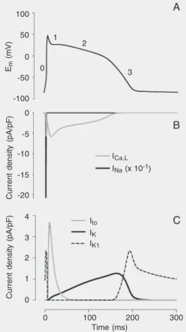
The action potential (AP) is the trigger-ing signal for contraction in striated muscle. Ion currents through sarcolemmal voltage-and ligvoltage-and-dependent channels, as well as electrogenic ion transporters, determine the AP waveform. In a typical mammalian
car-diac myocyte, 4 phases of the AP (Figure 1) can be identified (1,2):
Phase zero (upstroke): during this brief phase, the rapidly activating Na+ current
drives the membrane potential (Em) from its
the transsarcolemmal Na+ electrochemical
gradient.
Phase 1 (early repolarization): the rapid, transient outward current (Ito) is the
pre-dominant contributor to the partial mem-brane repolarization.
Phase 2 (plateau): this usually long phase may last up to a few hundred milliseconds, and is characterized by slow Em variation,
which is dependent on the delicate balance between depolarizing (mostly L-type Ca2+
current, ICa,L) and repolarizing (mainly
me-diated by delayed rectifier K+ channels)
cur-rents.
Phase 3 (late repolarization): this phase relies on both a decrease in ICa,L due to ICa,L
inactivation/deactivation, and activation of delayed rectifier K+ channels. As
repolariza-tion proceeds, K+ efflux through inwardly
rectifying K+ channels (I
K1) becomes greater
(Figure 1C) due to relief of channel
rectifi-cation and contributes to late restoration and stabilization of the diastolic Em.
Recent investigation has provided evi-dence that the early repolarization phase may considerably influence the AP wave-form. Ito, the main current responsible for
this phase, has been shown to be carried by distinct ionic components: in addition to the Ca2+-independent, voltage- and
time-depend-ent K+ current (I
to-1), a Ca2+-dependent Cl
-current (ICl(Ca) or Ito-2) was also identified. In
this review, I will refer to the 4-aminopyri-dine-sensitive, Ca2+-insensitive transient
outward K+ current (I
to-1) as Ito. More
infor-mation on ICl(Ca) can be found in a recent
review (4).
Ito, in turn, is the net result of K+ flux
through at least two different types of chan-nel associated with different isoforms of the pore-forming protein. The behavior of these channels differs especially in the time course of voltage-dependent inactivation and steady-state recovery from inactivation. Fast Ito (Ito,f)
is mediated by channels that recover from inactivation in less than 100 ms, whereas for channels that carry slow Ito (Ito,s) recovery
from inactivation takes a few seconds (5,6). Both experimental data and mathemati-cal models have shown that Ito magnitude
and composition (i.e., the relative contribu-tions of Ito,f and Ito,s to total Ito) may markedly
affect AP duration and shape (7-13). Ex-pression of Ito,f and Ito,s channel proteins is
highly regulated, and may vary with species, developmental stage, and region of the heart (6,11). For instance, Ito,f is more prominent
in ventricular epicardial myocytes than in endocardial myocytes. In the former, the AP waveform displays the so-called spike-and-dome configuration, with distinct phases 1 and 2, while in the latter AP duration tends to be greater and repolarization to the plateau level is slower and more gradual. Differ-ences in Ito,f density correlate with
differ-ences in the AP profile within and between ventricles (e.g., 5,11,14,15). The Ito,f and AP
duration gradients across and along the
ven-Figure 1. Action potential (AP) and ion currents in a rabbit
ven-tricular myocyte. A, AP
wave-form, in which phases zero (upstroke), 1 (early repolariza-tion), 2 (plateau), and 3 (late
repolarization) are indicated. B,
Voltage-dependent ion currents that contribute to membrane
de-polarization during the AP: Na+
current (INa) and L-type Ca2+
current (ICa,L). Note: the actual
INa density is 10-fold greater
than that shown in the figure. C,
Voltage-dependent K+ currents
that contribute to membrane re-polarization during the AP:
tran-sient outward current (Ito), total
delayed rectifier K+ current (IK)
and inwardly rectifying K+
cur-rent (IK1). For clarity, IK
compo-nents (IKr and IKs) were
com-bined, and currents carried by
ion transporters (e.g., Na+-Ca2+
exchanger, Na+-K+ ATPase), as
well as ICl(Ca), were omitted.
Traces were obtained by simu-lation with the LabHEART 4.9.5 program (Ref. 3).
Em
(mV)
100
50
0
-50
-100
0
-5
-10
-15
-20
4
3
2
1
0 0
1 2
3
ICa,L
INa (x 10-1)
Ito
IK
IK1
Current density (pA/pF)
Current density (pA/pF)
0 100 200 300
Time (ms)
A
B
tricular wall have been proposed to contri-bute to the dispersion of ventricular refracto-riness, which markedly influences ventricu-lar repoventricu-larization path, direction and time course. It has been proposed that region-dependent changes in Ito reported in certain
disease states (e.g., post-myocardial infarc-tion, ventricular hypertrophy, heart failure) may underlie increased susceptibility to re-polarization abnormalities and reentry ar-rhythmias (11,16,17). However, in this brief review, I will not deal with the influence of Ito on cardiac electric conduction (see Ref.
16 for further discussion), but its indirect effects, via the AP waveform, on Ca2+
ho-meostasis, which is of paramount impor-tance for the development of adequate ven-tricular mechanical function and blood pump-ing.
Myocardial Ca2+ cycling
Ca2+-induced Ca2+ release (CICR) is the
most accepted mechanism underlying exci-tation-contraction coupling (ECC) in the mammalian myocardium (18,19). Ca2+
in-flux through voltage-dependent, sarcolem-mal ICa,L during the AP is the main trigger for
the release of a greater amount of Ca2+ by the
sarcoplasmic reticulum (SR), which is the source of most Ca2+ that activates
contrac-tion. SR Ca2+ release causes an increase in
the cytosolic free Ca2+ concentration ([Ca2+] i),
which results in greater interaction of the ion with myofilament proteins and development of force and cell shortening. Thus, the AP is the signal for contraction development, and Ca2+ acts as the second messenger in the
electro-mechanical coupling process. Con-traction is limited by Ca2+ removal from the
cytosol by several transporters, which pro-mote [Ca2+]
i decline, Ca2+ dissociation from
myofilaments and cell relaxation. The domi-nant transporter that ultimately determines relaxation is the SR Ca2+-ATPase (SERCA),
which is responsible for 70-90% of cytosolic Ca2+ clearance and repletion of the SR Ca2+
store. Ca2+ efflux is mainly mediated by the
Na+-Ca2+ exchanger (NCX), a sarcolemmal
counter-transporter that is driven by the transsarcolemmal Na+ and Ca2+
electro-chemical gradients. NCX accounts for 7-30% of total cytosolic Ca2+ removal during
relaxation, which is approximately equiva-lent to the amount of Ca2+ entering the cell
via ICa,L during excitation (1,20,21).
The Ca2+ transient amplitude is an
impor-tant determinant of contraction amplitude and is greatly dependent on the amount of Ca2+ released by the SR during ECC. During
each AP, the SR releases a fraction of its content, which increases with increasing trig-ger (i.e., ICa,L) amplitude and/or SR Ca2+
content (12,22,23). Moreover, ECC effi-ciency may be modulated by additional regu-lation of the SR Ca2+ channel activity by ions
(e.g., Mg2+) and proteins (e.g., Ca2+
-calmod-ulin-dependent protein kinase II, FK506-binding protein, sorcin) (1).
It is important to note that Ca2+ transport
by the main influx (ICa,L) and efflux (NCX)
pathways during the cardiac cycle is strongly influenced by Em: the former because of the
voltage dependence of ICa,L activation and
inactivation (and also because of the driving force for Ca2+ flux), and the latter because
the direction and driving force for Ca2+
trans-port are determined by the difference be-tween Em and the exchanger reversal
poten-tial (ENCX = 3ENa - 2ECa, where ENa and ECa
are the Nernst equilibrium potentials for Na+
and Ca2+, respectively (24)). The
conse-quences of the voltage dependence of ICa,L
and NCX operation for ECC, [Ca2+] i and
contraction amplitude are many. The ampli-tude and time course of the trigger Ca2+
signal were shown to markedly affect its ability to induce SR Ca2+ release (19) and the
so-called fractional SR Ca2+ release, i.e., the
fraction of the SR Ca2+ content that is
re-leased at a twitch (22). On the other hand, Ca2+ efflux by the NCX is
Ca2+ content and vulnerability to triggered
arrhythmias (1). Thus, one can conclude that the AP does not represent simply an impulse to trigger ECC, but a complex input wave-form that can modulate directly or indirectly ECC efficiency. On the other hand, Ca2+
cycling may conversely modulate the AP waveform, because of the effects of SR-released Ca2+ on membrane currents, for
instance, inactivation of ICa,L, activation of
Ca2+-dependent Cl- channels and
NCX-me-diated current (25).
Transient outward K+ current (I
to)
Ito is characteristic of neurons and
car-diac muscle, and its voltage-dependent acti-vation and inactiacti-vation kinetics is much faster than that of other cardiac K+ currents. The
channel is a macromolecular protein com-plex composed of pore-forming subunits α
(which belong to the Kv gene subfamily), accessory ß subunits (several types of ß sub-unit have been identified) and other regula-tory proteins, such as minimal K+ channel
subunit homologues, K+ channel-associated
proteins (possibly chaperone proteins) and K+ channel-interacting proteins (KChIP,
which belong to a family of neuronal Ca2+
binding proteins). KChIP2 is present in the heart, and its co-expression with Kv4.2, but not with Kv1.4, increases current amplitude, changes the biophysical channel properties and allows the channel to be regulated by protein kinase A. The α subunit presents 6
transmembrane domains, a K+-selective pore
region and a highly charged S4 domain, which is considered to be the region where the voltage sensor is located. The functional channel consists of the assembly of 4 α
subunits. In rodent heart, it seems that the channel that mediates Ito,f consists of Kv4.2
and/or 4.3 subunits co-assembled with KChIP2. A detailed description of the mo-lecular aspects of the channels that mediate Ito can be found elsewhere (2,6,11,17).
Two types of α subunits may form
chan-nels with different kinetic properties. Kv4.2/ 4.3 expression correlates with Ito,f, which
shows fast inactivation and recovery from steady-state inactivation (milliseconds). Kv4.2 appears to be the pore-forming sub-unit in rodent atria, whereas Kv4.3 mediates Ito,f in canine and human ventricle. Kv1.4 is
thought to form the channels that mediate Ito,s, which displays a longer time course,
especially of recovery from steady-state in-activation (seconds) (5,6,11,17).
The expression of these rapidly activat-ing K+ channels is influenced by several
factors.
Species
Kv4.2/4.3 and Ito,f are strongly expressed
in the ventricular myocardium of adult ro-dents, ferrets, dogs,andhumans, with a lesser contribution of Kv1.4 and Ito,s (6,12). Ito,f has
been considered to be responsible for the typically brief rodent ventricular AP (6,26). In other species (e.g., rabbit), Ito,s and Kv1.4
are the dominant Ito component and channel
isoform, respectively (6,12). In the guinea pig ventricle, Ito is absent, a fact that may
contribute to the prolonged AP in this spe-cies (6).
Developmental stage
In several species, even those in which the heart presents large Ito expression during
adulthood, Ito density is considerably small
or absent during the fetal and neonatal pe-riod. In neonatal rodent ventricle, Ito,f and Ito,s
(whose density is paralleled by Kv4.2/4.3 and Kv1.4 expression, respectively) show similar contributions to total Ito, while in
adults the former contributes over 90% (2,11,27,28). In the neonatal myocardium, the AP is longer than in the adult, and AP shortening with maturation coincides with an increase in myocardial Ito density and
in Ito, but in this species the dominant
com-ponent shifts from Ito,f to Ito,s (29). On the
other hand, aging is associated with a de-crease in Ito density and AP prolongation
(30).
Region of the ventricle
Ito density (particularly Ito,f) is greater in
epicardium vs endocardium, in the apex vs
base, and in right vs left ventricle. This varia-tion is considered to underlie the regional differences in the AP profile, as well as in the dispersion of repolarization (5,11,14,15). While in rodents this regional variation may rely on a gradient of Kv4.3 expression only, it has not yet been established whether the origin of this variation in canine and human ventricle depends on the expression of Kv4.3, KChIP2, or both (17,31,32).
Disease
Ito,f down-regulation associated with AP
prolongation has been reported following myocardial infarction, in hypertension, dia-betic cardiomyopathy, ventricular hypertro-phy, and heart failure (7-9,32-35), although in some cases AP lengthening is not accom-panied by changes in Ito density (36). The
mechanisms responsible for these changes have not been ascertained, but it is possible that they involve increased activity of the sympathetic and renin-angiotensin-aldoste-rone systems, since norepinephrine (via α
-adrenoceptors), angiotensin II and aldoste-rone may negatively regulate Ito,f-mediating
channels (11,17,35,37,38). Although in most cases a decrease in Ito,f is accompanied by
Kv4.2/4.3 down-regulation, sometimes with an increase in Ito,s and Kv1.4 up-regulation
(9,32,33,38), the functional change may be also associated with direct modification of the biophysical properties of the current (e.g., by angiotensin II) (37). Because of the de-crease in Ito,f in the epicardium (where
cur-rent density is greater) during disease, the
transmural heterogeneity of AP duration is largely suppressed or even reversed, an event that may lead to repolarization abnormali-ties and possibly increase the predisposition to reentry arrhythmias (11,15).
How Ito can affect Ca2+ homeostasis
In the physiological context, Ito regional
variability within the heart is thought to partially underlie regional differences in Ca2+
transient amplitude (15,39). The role of Ito
magnitude and composition in cell Ca2+
ho-meostasis and contraction is mediated by Ito
influence on AP waveform, especially the rate at which a plateau is attained, as well as its amplitude and duration. These AP fea-tures affect voltage-dependent Ca2+
trans-port pathways involved in ECC, in relax-ation and in general regulrelax-ation of cell Ca2+
load, such as ICa,L and NCX (Figure 2).
Most of ICa,L develops during the AP
plateau, in the voltage range at which the current peak is nearly maximal (1). A de-crease in Ito density results in AP
prolonga-tion, especially of the plateau phase. It is expected that a longer AP is associated with greater total Ca2+ influx, and this has been
confirmed by experimental evidence (12, 21,40). Paucity of Ito and prolonged AP are
observed in some conditions in which the SR function is depressed, such as immatu-rity and senescence, ventricular hypertro-phy and heart failure. In these cases, in-creased Ca2+ influx may contribute not only
to the contraction-activating cytosolic Ca2+
pool, but also to facilitating ECC by en-hancement of the fractional SR Ca2+ release
(22,23). It is tempting to speculate whether there is a negative relationship between Ito
functional expression and the SR relative contribution to ECC. Ito density has been
found to be higher in cardiomyocytes in which Ca2+ cycling between the SR and
short AP would allow large and short-lived ICa,L, better suited for triggering
synchro-nized SR Ca2+ release than for providing
Ca2+ for contraction and SR loading (see
below).
During early postnatal development, SR contribution to Ca2+ cycling, although
im-portant, is smaller than in adult myocardium (1,41), probably due to structural SR under-development and paucity of sarcolemma-SR specialized junctions, as well as to di-minished sensitivity of the CICR mechan-ism (18). Because of the small volume and
reduced Ca2+ buffering capacity in
imma-ture myocytes (42), a greater Ca2+ influx
should have a considerable impact directly on cytosolic [Ca2+] and/or indirectly via the
induction of SR Ca2+ release, and thus on
contractile activity. It has been recently shown that immature human myocytes (in which AP is long) stimulated with a wave-form similar to the adult AP show depressed Ca2+ transients, which indicates that
pro-longed AP in developing myocytes is of paramount importance for proper Ca2+
cy-cling (43). Similar results were observed in myocytes from senescent animals (44), which present, as neonatal cells, reduced Ito density
and prolonged AP (30).
In adults, enhanced cardiac workload due to increased hemodynamic load and/or de-creased myocardial contractile function, such as in chronic ventricular hypertrophy duced by pressure overload, myocardial in-farction and heart failure, are commonly associated with diminished total Ito or
selec-tively Ito,f, and AP prolongation (7-9,35).
These changes are accompanied by greater total Ca2+ influx during the long AP
wave-form, which may result in maintenance of Ca2+ transient amplitude, just as observed in
immature and senescent ventricle. Interest-ingly, in these conditions the SR function is depressed, usually in association with SERCA down-regulation and diminished CICR sensitivity (1). One could thus inter-pret these findings from the viewpoint that Ito reduction may enable a presumably
adap-tive increase in Ca2+ influx during the long
AP, so as to maintain Ca2+ cycling and
con-tractile function compatible with survival. Kassiri et al. (45) reported that Ito
inhibi-tion in cultured cardiac myocytes was effec-tive in inducing myocyte hypertrophy by a Ca2+ influx-dependent mechanism.
Expres-sion and function of Kv4.2/4.3 channels are inhibited by signaling pathways normally activated during cardiac overload, possibly via phosphorylation by protein kinase C re-sulting from stimulation of angiotensin, α1
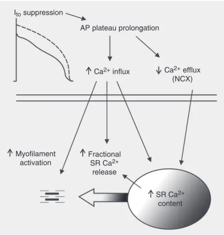
-Figure 2. How lengthening of the action potential (AP) plateau by transient outward current
(Ito) suppression may affect excitation-contraction coupling in mammalian cardiac
myo-cytes. Prolonged depolarization increases Ca2+ influx mainly by voltage-dependent L-type
Ca2+ channels, and decreases the driving force of Ca2+ extrusion via the sarcolemmal Na+
-Ca2+ exchanger (NCX). Both changes enhance Ca2+ availability for uptake by the
sarco-plasmic reticulum (SR) Ca2+-ATPase, leading to an increase in SR Ca2+ content. Both
increased Ca2+ influx and SR Ca2+ load synergistically increase the fractional SR Ca2+
release during systole, which causes greater myofilament activation and contraction.
In-creased Ca2+ influx can also permit a greater direct contribution to myofilament activation
by Ca2+ originating from the extracellular medium.
Ito suppression
AP plateau prolongation
Ca2+ influx Ca2+ efflux
(NCX)
Fractional SR Ca2+
release
SR Ca2+
content Myofilament
adrenergic and endothelin-1 receptors (11, 37,46). Additionally, mineralocorticoid re-ceptor activation has been implicated in early channel down-regulation following myocar-dial infarction (35). Thus, it has been specu-lated whether hypertrophy induction by ac-tivation of these pathways would partially involve enhanced Ca2+ influx resulting from
Ito depression (45). Greater cell Ca2+ cycling,
in addition to contributing to the preserva-tion of cardiac mechanical funcpreserva-tion, might also play a role in excitation-transcription coupling, for instance via Ca2+
-calmodulin-dependent kinases and phosphatases (calci-neurin), in the development of ventricular hypertrophy and remodeling, which may eventually deteriorate to heart failure (1,36,45). However, although there is strong evidence of the implication of Ca2+
-depend-ent biochemical pathways in hypertrophy development (see, e.g., 1,36,38), Ito,f
reduc-tion may not be the only way by which cell Ca2+ cycling can be increased. A few days
after aortic banding (before hypertrophy de-velopment), Ca2+ cycling is enhanced by
augmented SR function, without signs of increased Ca2+ influx (47), whereas a few
weeks later the observed AP prolongation may be associated with an increase in ICa,L
density, rather than a decrease in Ito (36).
Also, Bodi et al. (38) observed that Ito
sup-pression, reversal of Kv4.2/4.3 vs Kv1.4 dominance and AP prolongation in trans-genic mice overexpressing ICa,L occurred only
several months after hypertrophy develop-ment. Thus, Ito suppression may also be a
consequence, rather than a cause, of greater Ca2+ cycling. For instance, enhancement of
Ca2+ transient amplitude by SERCA
overex-pression has been shown to cause Kv4.2/4.3 and KChIP2 down-regulation, a decrease in Ito density and AP lengthening, even in the
absence of hypertrophy or heart failure (48). In summary, there is no compelling evi-dence supporting the hypothesis that Ito
sup-pression is necessarily involved in hypertro-phy signaling. Part of the discrepancies
among studies might be due to the multiplic-ity of experimental hypertrophy models and of the signaling pathways involved.
Although greater Ca2+ influx during a
long AP may help prevent a dramatic de-pression of Ca2+ cycling in disease
condi-tions, this compensation may be only partial in heart failure, because: a) a decrease in SR Ca2+ content in this condition (49) probably
limits the amount of released Ca2+, even
though influx is increased, and b) the slow AP phase 1 to phase 2 transition may slow down the ICa,L time course. To investigate
the latter aspect, Sah et al. (50) employed stimulating AP waveforms of similar total duration, but with different rates of phase 1 repolarization. They observed that when early repolarization is slowed, ICa,L shows a
de-creased peak and slower kinetics, in spite of similar or greater total Ca2+ influx compared
to that with fast early repolarization. Slow-developing ICa,L results in asynchronous SR
Ca2+ release, which is less efficient to
rap-idly increase [Ca2+]
i at systole (50,51). This
alteration may become more accentuated, with progression to heart failure, when in-trinsic reduction of ICa,L amplitude may
oc-cur (51). One of the expected consequences would be slower and weaker contraction activation. Thus, an increase in Ca2+ influx
due to Ito suppression and AP prolongation
may provide a partial adaptation that is even-tually offset by decreased ECC efficiency with maintenance and worsening of the dis-ease condition.
Thus, the kinetic aspect involves an addi-tional, subtler aspect of ICa,L modulation by
Ito, i.e., the timing and rate of early
repolar-ization. Linz and Meyer (52) showed that, during the respective AP waveforms, ICa,L is
briefer and attains greater amplitude in rat (strong Ito) than in guinea pig myocyctes
(where Ito and AP phase 1 are absent), even
though the plateau is much shorter in the former. This difference was attributed to the ability of the large rodent Ito to rapidly drive
ICa,L is greater, before the channels rapidly
inactivate and deactivate, causing ICa,L to
assume a quasi impulse-like waveform. Al-though total Ca2+ influx is lower, it develops
much faster in the rat than in the guinea pig, which is consistent with an optimal trigger-ing signal for SR Ca2+ release. On the other
hand, comparing epicardial vs endocardial canine ventricular myocytes (both of which present much longer APs than observed in rodents), the greater Ito density in the former
causes the AP to assume the spike-and-dome configuration, which apparently favors Ca2+
channel reopening during the plateau (see secondary ICa,L peak in Figure 1B), resulting
in greater Ca2+ influx (53). Thus, it appears
that the fine-tuning of Ca2+ influx by the
modulation of the AP waveform by Ito
strongly depends on the type, density and behavior of the ion current profile present in a specific cell type and/or animal species.
An important side effect of Ito
down-regulation and AP prolongation present in disease states may be the greater vulnerabil-ity to arrhythmias. In addition to reentry-predisposing changes in refractoriness dis-persion (16), triggered activity is often asso-ciated with increased AP duration (1). Pro-longed depolarization may result in dimin-ished Ca2+ efflux via NCX (due to a
de-creased driving force), which, in combina-tion with greater Ca2+ influx, leads to a greater
cell and SR Ca2+ load. Although this might
cause a further increase in Ca2+ transient and
contraction amplitude because SR Ca2+
con-tent greatly influences the fractional SR Ca2+
release (22,23), it may also facilitate ar-rhythmogenesis. SR Ca2+ overload is often
accompanied by enhanced diastolic SR Ca2+
release (1,54) that results in the generation of a depolarizing, inward membrane current by electrogenic efflux of the leaked Ca2+ via
NCX (stoichiometry of 1 Ca2+:3 Na+) (24). If
of sufficient magnitude, this current can drive Em to the excitation threshold and give rise
to triggered arrhythmias. In the particular case of heart failure, arrhythmogenesis by
this mechanism would be additionally fa-vored by NCX up-regulation and Em
insta-bility due to decreased IK1 density (1).
Finally, it is also worthwhile to consider the relationship between AP duration and cycle length. This relation seems to stem from multifactorial mechanisms: Ito may be
one of the underlying mechanisms in some species, in addition to modulation of ICa,L
and other Ca2+-dependent currents by
SR-released Ca2+, and a rate-dependent increase
in density of delayed-rectifier K+ currents
(e.g., 25,55). In most large mammals, in-cluding man, which present marked Kv4.2/ 4.3-dependent Ito, AP duration is decreased
or little affected by increasing rate, espe-cially in epicardial cells (16,56,57). This response might be important to allow ad-equate relaxation and ventricular filling dur-ing the shortened diastole. However, in rab-bit and hamster myocardium, in which total Ito density is lower and Ito,s is the dominant
component, the AP is lengthened with in-creasing rate (12,56,58). This might be due, at least in part, to the slow Ito,s recovery from
inactivation, which would decrease channel availability at short intervals (2,55,56). A positive relationship between AP duration and rate may limit ventricular function and predispose the heart to triggered arrhyth-mias at high rates. As pointed out earlier, Ito,f
down-regulation has been described in ca-nine and human hypertrophied and failing ventricle. However, this event per se does not seem to affect the AP duration-rate rela-tion, since the AP is prolonged at long, but not short cycle lengths (16,25). Recent re-sults from computer simulations predicted that the positive rate-AP duration relation-ship relies on the presence of Ito,s, rather than
on a decrease in Ito,f, since a negative relation
is still present if Ito is totally suppressed (12).
Interestingly, our prediction is in agreement with experimental data that show a negative AP duration-rate curve in the guinea pig ventricle (in which Ito is absent (55,59)) which
hyper-trophy induced by pressure overload (59). The finding that diabetic cardiomyopathy is associated with a frankly positive AP dura-tion-rate relationship in the rat is intriguing, in contrast with the weak rate dependence observed in controls (60). However, infor-mation on Ito,s or Kv1.4 expression in this
condition is lacking, and thus a possible link between Ito,s dominance and positive AP
du-ration-rate still requires experimental con-firmation.
Changes in Ito density and profile caused
by pathological cardiovascular conditions may be overall adaptive, as they contribute to increase Ca2+ influx and may confer some
protection against reentry due to prolonged refractoriness, although excessive Ca2+
load-ing, in conjunction with a decrease in mem-brane electrical stability, may favor the ap-pearance of triggered arrhythmias. However,
the fact that these changes are superimposed on the naturally occurring regional hetero-geneity of this current (and its components) in the adult ventricle makes it difficult to predict their net effect on vulnerability to arrhythmia. This also complicates the devel-opment of pharmacological and gene thera-py strategies targeted to Ito channels.
Hope-fully, this might be overcome with novel information to be gathered in the coming years.
Acknowledgments
I am grateful to Dr. José W.M. Bassani for helpful criticism and comments about this manuscript, and to Mr. Rafael A. Ricardo for assistance with the illustrations, both from Departamento de Engenharia Biomédica, FEEC, UNICAMP, Campinas, SP, Brazil.
References
1. Bers DM (2001). Excitation-Contraction Coupling and Cardiac
Con-tractile Force. 2nd edn. Kluwer Academic Press, Dordrecht, The Netherlands.
2. Nerbonne JM & Kass RS (2003). Physiology and molecular biology of ion channels contributing to ventricular repolarization. In: Gussak
I, Antzelevich C, Hammill SC et al. (Editors), Contemporary
Cardiol-ogy: Cardiac Repolarization: Bridging Base and Clinical Science. Humana, Totwa, NJ, USA, 25-62.
3. Puglisi JL & Bers DM (2001). LabHEART: an interactive computer model of rabbit ventricular myocyte ion channels and Ca transport. American Journal of Physiology, 281: 2049-2060.
4. Hartzell C, Putzier I & Arreola J (2005). Calcium-activated chloride
channels. Annual Review of Physiology,67: 719-758.
5. Xu H, Guo W & Nerbonne JM (1999). Four kinetically distinct
depo-larization-activated K+ currents in adult mouse ventricular
myo-cytes. Journal of General Physiology, 113: 661-677.
6. Nerbonne JM (2001). Molecular analysis of voltage-gated K+
chan-nel diversity and functioning in the mammalian heart. In:Page E,
Fozzard HA & Solaro RJ (Editors), Handbook of Physiology, Section
2: The Cardiovascular System. Vol. 1. The Heart. Oxford University Press, Oxford, UK, 568-594.
7. Beuckelmann DJ, Nabauer M & Erdmann E (1993). Alterations of K+
currents in isolated human ventricular myocytes from patients with
terminal heart failure. Circulation Research, 73: 379-385.
8. Käab S, Nuss HB, Chiamvimonvat N et al. (1996). Ionic mechanism of action potential prolongation in ventricular myocytes from dogs
with pacing-induced heart failure. Circulation Research, 78:
262-273.
9. Kaprielian R, Wickenden AD, Kassiri Z et al. (1999). Relationship
between K+ channel down-regulation and [Ca2+]i in rat ventricular
myocytes following myocardial infarction. Journal of Physiology,
517: 229-245.
10. Greenstein JL, Wu R, Po S et al. (2000). Role of
calcium-independ-ent transicalcium-independ-ent outward currcalcium-independ-ent Ito1 in shaping action potential
mor-phology and duration. Circulation Research,87: 1026-1033.
11. Oudit GY, Kassiri Z, Sah R et al. (2001). The molecular physiology
of the cardiac transient outward potassium current (Ito) in normal
and diseased myocardium. Journal of Molecular and Cellular
Cardi-ology,33: 851-872.
12. Bassani RA, Altamirano J, Puglisi JL et al. (2004). Action potential
duration determines sarcoplasmic reticulum Ca2+ reloading in
mam-malian ventricular myocytes. Journal of Physiology, 559: 591-607.
13. Bondarenko VE, Szigeti GP, Bett GCL et al. (2004). Computer
model of action potential of mouse ventricular myocytes. American
Journal of Physiology,287: H1378-H1403.
14. Liu DW, Gintant GA & Antzelevitch C (1993). Ionic basis for electro-physiological distinction among epicardial, midmyocardial, and en-docardial myocytes from the free wall of the canine left ventricle. Circulation Research, 72: 671-687.
15. Kaprielian R, Sah R, Nguyen T et al. (2002). Myocardial infarction in
rat eliminates regional heterogeneity of AP profiles, Ito K+ currents,
and [Ca2+]i transients. American Journal of Physiology,283:
H1157-H1168.
16. Wolk R, Cobbe SM, Hicks MN et al. (1999). Functional, structural and dynamic basis of electrical heterogeneity in healthy and dis-eased cardiac muscle: implications for arrhythmogenesis and
anti-arrhythmic drug therapy. Pharmacology and Therapeutics, 84:
17. Birnbaum SH, Varga AW, Yuan LL et al. (2004). Structure and
function of Kv4-family transient potassium channels. Physiological
Reviews,84: 803-833.
18. Fabiato A (1982). Calcium release in skinned cardiac cells:
varia-tions with species, tissues, and development. Federation
Proceed-ings, 41: 2238-2244.
19. Fabiato A (1985). Time and calcium dependence of activation and inactivation of calcium-induced calcium release from the
sarcoplas-mic reticulum of a skinned canine cardiac Purkinje cell. Journal of
General Physiology, 85: 247-290.
20. Bassani JWM, Bassani RA & Bers DM (1994). Relaxation in rabbit and rat cardiac cells: species-dependent differences in cellular
mechanisms. Journal of Physiology, 476: 279-293.
21. Yuan W, Ginsburg KS & Bers DM (1996). Comparison of sarcolem-mal calcium channel current in rabbit and rat ventricular myocytes. Journal of Physiology, 493: 733-746.
22. Bassani JWM, Yuan W & Bers DM (1995). Fractional SR Ca release is regulated by trigger Ca and SR Ca content in cardiac myocytes. American Journal of Physiology, 268: C1313-C1319.
23. Shannon TR, Ginsburg KS & Bers DM (2000). Potentiation of frac-tional SR Ca release by total and free intra-SR Ca concentration. Biophysical Journal, 78: 334-343.
24. Blaustein MP & Lederer WJ (1999). Sodium/calcium exchange: its
physiological implications. Physiological Reviews, 79: 763-854.
25. Carmeliet E (2004). Intracellular Ca2+ concentration and rate
adap-tation of the cardiac action potential. Cell Calcium, 35: 557-573.
26. Josephson IR, Sanchez-Chapula J & Brown AM (1984). Early
out-ward current in rat single ventricular cells. Circulation Research, 54:
157-162.
27. Kilborn MJ & Fedida D (1990). A study of the developmental change
in outward currents in rat ventricular myocytes. Journal of
Physiolo-gy, 430: 37-60.
28. Wickenden AD, Kaprielian R, Parker TG et al. (1997). Effects of
development and thyroid hormone on K+ currents and K+ channel
gene expression in rat ventricle. Journal of Physiology, 504:
271-286.
29. Sanchez-Chapula J, Elizalde A, Navarro-Polanco R et al. (1994). Differences in outward currents between neonatal and adult
ventric-ular cells. American Journal of Physiology, 266: H1184-H1194.
30. Walker KE, Lakatta EG & Houser SR (1993). Age-associated
changes in membrane currents in rat ventricular myocytes.
Cardio-vascular Research, 27: 1968-1977.
31. Rosati B, Pan Z, Lypen S et al. (2001). Regulation of KChIP2 potassium channel ß subunit gene expression underlies the gradi-ent of transigradi-ent outward currgradi-ent in canine and human vgradi-entricle. Journal of Physiology, 533: 119-125.
32. Zicha S, Xiao L, Stafford S et al. (2004). Transmural expression of transient outward current subunits in normal and failing canine and
human hearts. Journal of Physiology, 561: 735-748.
33. Takimoto K, Li D, Hershman KM et al. (1997). Decreased
expres-sion of Kv4.2 and novel Kv4.3 K+ channel subunit mRNAs in
ven-tricles of renovascular hypertensive rats. Circulation Research, 81:
533-539.
34. Qin D, Huang B, Deng L et al. (2001). Downregulation of K+ channel
genes expression in type I diabetic cardiomyopathy. Biochemical
and Biophysical Research Communications, 283: 549-553. 35. Perrier E, Kerfant BG, Bideaux P et al. (2004). Mineralocorticoid
receptor antagonism prevents the electrical remodeling that
pre-cedes cellular hypertrophy after myocardial infarction. Circulation,
110: 776-783.
36. Wang Z, Kutschke W, Richardson KE et al. (2001). Electrical
remod-eling in pressure-overload cardiac hypertrophy: role of calcineurin. Circulation, 104: 1657-1663.
37. Yu H, Gao J, Wang H et al. (2000). Effect of renin-angiotensin
system on the current Ito in epicardial and endocardial ventricular
myocytes from canine heart. Circulation Research, 86: 1062-1068.
38. Bodi I, Muth JN, Hahn HS et al. (2003). Electrical remodeling in hearts from a calcium-dependent mouse model of hypertrophy and
failure: complex nature of K+ current changes and action potential
duration. Journal of the American College of Cardiology, 41:
1611-1622.
39. Volk T, Nguyen THD, Schultz JH et al. (1999). Relationship between
transient outward K+ current and Ca2+ influx in rat cardiac myocytes
of endo- and epicardial origin. Journal of Physiology, 519: 841-850.
40. Sah R, Ramirez RJ, Kaprielian R et al. (2001). Alterations in action potential profile enhance excitation-contraction coupling in rat
car-diac myocytes. Journal of Physiology, 533: 201-214.
41. Bassani RA & Bassani JWM (2002). Contribution of Ca2+
transport-ers to relaxation in intact ventricular myocytes from developing rats. American Journal of Physiology, 282: H2406-H2413.
42. Bassani RA, Shannon TR & Bers DM (1998). Passive Ca2+ binding
in ventricular myocardium of neonatal and adult rats. Cell Calcium,
23: 433-442.
43. Wagner MB, Wang Y, Kumar R et al. (2005). Calcium transients in
human infant myocytes. Pediatric Research, 57: 28-34.
44. Janczewski AM, Spurgeon HÁ & Lakatta EG (2002). Action potential prolongation in cardiac myocytes of old rats is an adaptation to
sustain youthful intracellular Ca2+ regulation. Journal of Molecular
and Cellular Cardiology, 34: 641-648.
45. Kassiri Z, Zobel C, Nguyen TT et al. (2002). Reduction of Ito causes
hypertrophy in neonatal ventricular myocytes. Circulation Research,
90: 578-585.
46. Nakamura TY, Coetzee WA, Miera EVS et al. (1997). Modulation of Kv4 channels, key components of rat ventricular transient outward
K+ current, by PKC. American Journal of Physiology, 273:
H1775-H1786.
47. Bassani RA, Carvalho BMR, Franchini KG et al. (2005). Greater
sarcoplasmic reticulum (SR) Ca2+ release in early ventricular
hyper-trophy induced by pressure overload. Biophysical Journal, 88 (Suppl
1): 1556 (Abstract).
48. Xu Y, Zhang Z, Timofeyev V et al. (2005). The effects of intracellular
Ca2+ on cardiac K+ channel expression and activity: novel insights
from genetically altered mice. Journal of Physiology, 562: 745-758.
49. Piacentino III V, Weber CR, Chen X et al. (2003). Cellular basis of abnormal calcium transients of failing human ventricular myocytes. Circulation Research, 92: 651-658.
50. Sah R, Ramirez RJ & Backx PH (2002). Modulation of Ca2+-release
in cardiac myocytes by changes in repolarization rate: role of phase-1 action potential repolarization in excitation-contraction coupling. Circulation Research, 90: 165-173.
51. Harris DM, Mills GD, Chen X et al. (2005). Alterations in early action potential repolarization causes localized failure of sarcoplasmic
reticulum Ca2+ release. Circulation Research, 96: 543-550.
52. Linz KW & Meyer R (2000). Profile and kinetics of L-type calcium current during the cardiac ventricular action potential compared in
guinea-pigs, rats and rabbits. Pflügers Archives, 439: 588-599.
53. Bányász T, Fulop L, Magyar J et al. (2003). Endocardial versus epicardial differences in L-type calcium current in canine ventricular
myocytes studied by action potential voltage clamp. Cardiovascular
Research, 58: 66-75.
54. Bassani RA, Bassani JWM, Lipsius SL et al. (1997). Diastolic Ca
Ameri-can Journal of Physiology, 273: H886-H892.
55. Ravens U & Wettwer E (1998). Electrophysiological aspects of
changes in heart rate. Basic Research in Cardiology, 93 (Suppl 1):
60-65.
56. Fermini B, Wang Z, Duan D et al. (1992). Differences in rate
depend-ence of transient outward current in rabbit and human atrium.
Ameri-can Journal of Physiology, 263: H1747-H1754.
57. Pucelik P, Kralicek P, Holicka M et al. (1987). Influence of a period of inactivity on the duration of the post-rest action potentials of the mammalian working ventricular myocardium in correlation to the
preceding stimulation frequency. Physiologia Bohemoslovaca, 36:
394-402.
58. Kocic I, Hirano Y & Hiraoka M (2002). Rate-dependent changes in action potential duration and membrane currents in hamster
ventric-ular myocytes. Pflügers Archives, 443: 353-361.
59. Davey P, Bryant S & Hart G (2001). Rate-dependent electrical, contractile and restitution properties of isolated left ventricular
myo-cytes in guinea-pig hypertrophy. Acta Physiologica Scandinavica,
171: 17-28.
60. Pacher P, Ungvári P, Nánási PP et al. (1999). Electrophysiological changes in rat ventricular and atrial myocardium at different stages
of experimental diabetes. Acta Physiologica Scandinavica,166: