maintain intrinsic calcium cycling. The IKCablocker (apamin 100 nM) was used to examine the role of the current in atrial and
ventricular myocytes. A canine tachypacing induced model of HF (1 and 4 months, n = 5 per group) was used, and compared to a group of 4 month HF with 6 weeks of superimposed atrial fibrillation (n = 7). A group of age-matched canine controls were used (n = 8). Human atrial and ventricular myocytes were isolated from explanted end-stage failing hearts which were obtained from transplant recipients, and studied in parallel. Atrial myocyte action potentials were unchanged by IKCablockade in all of the groups studied. IKCablockade did not affect ventricular myocyte repolarization in controls. HF
caused prolongation of ventricular myocyte action potential repolarization. IKCa blockade caused further prolongation of
ventricular repolarization in HF and also caused repolarization instability and early afterdepolarizations. SK2 and SK3 expression in the atria and SK3 in the ventricle were increased in canine heart failure. We conclude that during HF, IKCa
blockade in ventricular myocytes results in cellular arrhythmias. Furthermore, our data suggest an important role for IKCain
the maintenance of ventricular repolarization stability during chronic heart failure. Our findings suggest that novel antiarrhythmic therapies should have safety and efficacy evaluated in both atria and ventricles.
Citation:Bonilla IM, Long VP III, Vargas-Pinto P, Wright P, Belevych A, et al. (2014) Calcium-Activated Potassium Current Modulates Ventricular Repolarization in Chronic Heart Failure. PLoS ONE 9(10): e108824. doi:10.1371/journal.pone.0108824
Editor:Blanca Rodriguez, University of Oxford, United Kingdom
ReceivedMarch 27, 2014;AcceptedAugust 26, 2014;PublishedOctober 1, 2014
Copyright:ß2014 Bonilla et al. This is an open-access article distributed under the terms of the Creative Commons Attribution License, which permits unrestricted use, distribution, and reproduction in any medium, provided the original author and source are credited.
Data Availability:The authors confirm that all data underlying the findings are fully available without restriction. All relevant data are within the paper. Funding:Pacemakers and leads provided by St. Jude Medical, Sylmar, CA as an in-kind donation (Carnes). Research supported in part by NIH grants (R01-HL115580 to CAC and VVF; R01-HL089836 to CAC; HL074045 to SG; HL084583, HL083422, HL114383 to PJM) and the American Heart Association (PJM). The funders had no role in study design, data collection and analysis, decision to publish or preparation of the manuscript.
Competing Interests:The authors have declared that no competing interests exist. * Email: [email protected]
¤ Current address: University of LaSalle, Bogota, Columbia
Introduction
Heart failure (HF) is a chronic disease that develops over months to years, and is defined by insufficient cardiac output to meet the physiologic and metabolic needs of the body. Atrial fibrillation (AF) and HF are common coexisting disease states, and HF results in a 4.5 to 5.9 fold increase in the risk of developing AF. [1] Moreover, in patients with HF, the development of AF significantly increases the risk of death. [2] Thus, identifying and elucidating pharmacological targets to treat AF may significantly reduce mortality and morbidity in HF.
Small-conductance Ca2+
- activated K+
(SK) channels are expressed in multiple tissues such as skeletal and smooth muscle, the central and peripheral nervous system and the heart.[3–5] Cardiac myocytes express SK1, SK2 and SK3 gene products. [6] SK- encoded current is voltage-independent and activated by intracellular calcium. [7] All three members of the SK family have similar calcium sensitivity for activation (0.6–0.7mM) [8].
SK-encoded current is blocked by apamin, a constituent of bee venom, which appears to be highly selective for IKCa. [7,9,10].
IKCa, the potassium current conducted by SK channels, contributes to repolarization, [3,11] but the importance of IKCa in repolarization remains poorly elucidated. For example, ventricular IKCashortens repolarization and promotes peri-infarct arrhythmias in rats. [12] Conversely, blockade of IKCapromotes ventricular arrhythmias in human HF and a non-ischemic rabbit HF model, suggesting a protective role for IKCa. [13,14] The contribution of IKCatoatrialrepolarization is also unclear as some reports demonstrate that IKCa is proarrhythmic while others suggest it is protective. [15,16].
We measured the impact of IKCablock on action potentials in intact myocytes using perforated patch recordings to maintain intrinsic Ca2+cycling. We utilized a well-validated canine model
handling, increased predisposition to AF and increased myocar-dial fibrosis. [17,18] Complementary experiments were conduct-ed in end-stage human HF. The role of IKCa in AF was evaluated in atrial myocytes from a canine model of chronic HF with sustained AF.
Methods
Heart failure and atrial fibrillation animal models
All animal procedures conformed to the Guide for the Care and Use of Laboratory Animals of the National Institutes of Health, and were approved by the Ohio State University Institutional Animal Care and Use Committee (Protocol: 2010A00000103-R1). Canine heart failure was induced by right ventricular (RV) tachypacing as previously described. [19] Animals were assigned to one or four months of RV tachypacing to induce heart failure. Atrial fibrillation was induced in dogs with HF using a customized pacemaker (St Jude Medical, Sylmar, CA). One pacing lead was implanted in the right atria (RA) and the second lead was implanted in the RV, with HF induced as previously described. [20] After 10 weeks of RV tachypacing, RA tachypa-cing was initiated, with the RA stimulated at 10 Hz for 60 seconds, followed by a 10 second pause for automated interrogation of atrial rhythm. This cycle of RA tachypacing was repeated every 70 seconds until AF was detected. Subsequent detection of normal atrial rhythm resulted in resumption of the atrial tachypacing. The total HF duration in the HF+AF group was 4 months. Ventricular pacing was stopped during atrial stimulation, and the ventricular rate was 150–200 BPM during atrial pacing or AF. Serial
echocardiograms and ECGs were performed as previously reported. [17,21] Serial pacemaker interrogations were used to monitor cardiac rhythm.
Myocyte Isolation and Tissue Collection
On the day of the terminal procedure, the dogs were anesthetized with pentobarbital sodium (50 mg/kg intravenously; Nembutal, Abbott Laboratories). The heart was rapidly removed and perfused with cold cardioplegia solution containing the following (mM): NaCl 110, CaCl2 1.2, KCl 16, MgCl2 16 and NaHCO310. Cannulation of the left circumflex artery was used to perfuse left atria and ventricle following removal of the right atrium and right ventricle, as previously described. [22] Adjacent tissue samples were collected and snap frozen for protein analyses. Tyrode’s solution (mM) containing NaCl 130, KCl 5.4, MgCl2 3.5, NaH2PO4 0.5, Glucose 10, HEPES 5 and taurine 20, was used as the initial perfusate. During the cell isolation process the heart was perfused with three different solutions (36uC). The heart was initially perfused for 10 minutes with Tyrode’s solution with 0.1 mM EGTA; this was followed by perfusion with Tyrode’s solution containing 0.3 mM Calcium, 0.12 mg/ml of Trypsin Inhibitor (NIBCO) and 1.33 mg/ml of collagenase (Type II, Worthington) for a maximum of 45 minutes. Then following enzymatic digestion, the heart was perfused with normal Tyrode’s solution for five minutes to remove residual enzyme. Subsequently, left ventricular mid-myocardial and left atrial myocytes were obtained through secondary digestion, as previously described. [22] After secondary digestion the cells were re-suspended in Figure 1. In vivo data from 1 month (1 Mo), 4 month (4 Mo), and 4 month HF with sustained AF (4 Mo HF+AF) canine groups. A. Representative ECG recording from a 4 month HF+AF dog showing the absence of P waves and irregularly irregular QRS complexes characteristic of
AF.B.LVFS was similarly decreased in the 1 month HF, 4 month HF and the 4 month HF+AF groups compared to baseline. (p,0.05 vs baseline).C. Atrial contractility was decreased in 4 month HF and 4 month HF+AF groups compared to baseline. (p,0.05, N = 5–7 per group).
doi:10.1371/journal.pone.0108824.g001
incubation buffer. [23] This isolation procedure typically yields 70–90% and 40–60% rod shaped ventricular and atrial myocytes, respectively. All myocyte electrophysiology experiments were conducted within 10 hours of isolation.
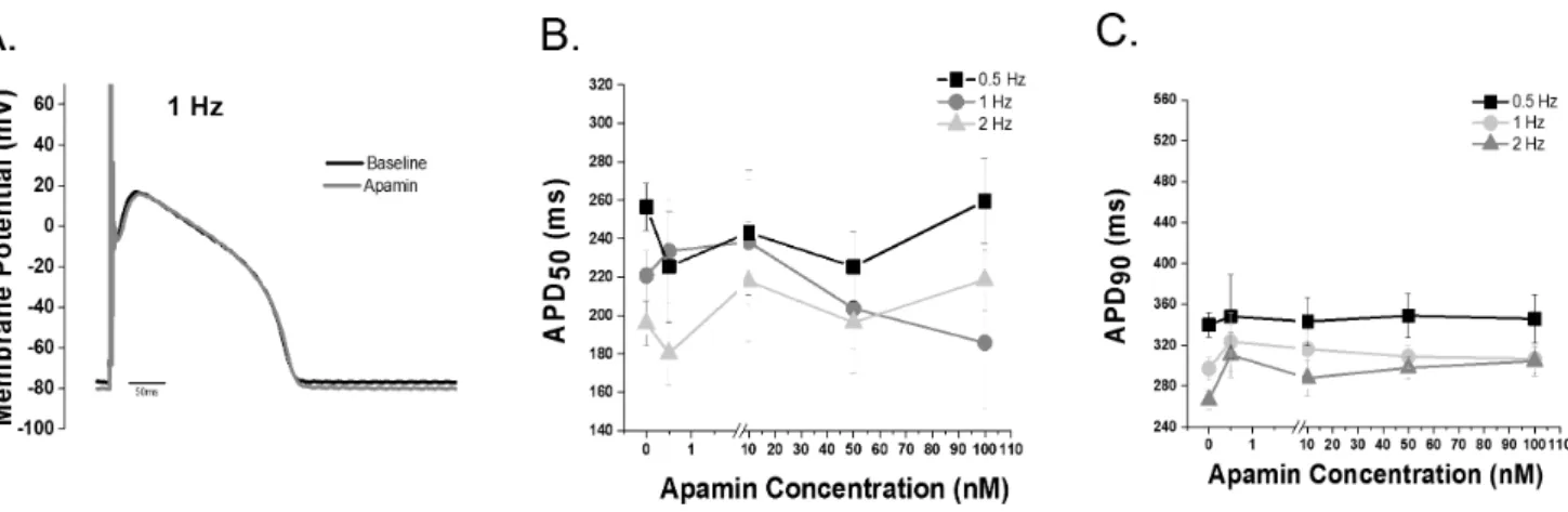
In parallel experiments, failing human cardiac tissue was obtained after written informed consent with the documentation of consent securely stored as approved by the Institutional Review Board approval of The Ohio State University (IRB 2008H0113 Figure 2. IKCainhibition does not alter repolarization in control ventricular cells. A.Representative action potential tracing before and after 100 nM apamin recorded at 1 Hz.B.APD50andC.APD90dose response data (0–100 nM apamin) recorded at stimulation rates of 0.5, 1 and
2 Hz. (p = NS, n = 5–11 cells per group; 8 animals). The Grubb’s test for outlier data was applied and one cell was rejected and is not included in the summary data.
doi:10.1371/journal.pone.0108824.g002
Figure 3. Apamin modulates ventricular repolarization during HF.Representative action potential of a 1 month (A) HF and 4 month HF (B) ventricular cell before and after apamin superfusion.C. Summary data of APD50 in control,1 month and 4 month HF before and after apamin
treatment. No difference between 1 month HF, 4 month HF and control is observed (2–8 animals per group). Apamin treatment of 1 month HF cells causes a prolongation only at 2 Hz(p,0.05), likewise apamin treatment of 4 month HF cells causes a prolongation at 0.5 Hz (p,0.05) and 1 Hz (p, 0.05).D.Apamin prolongs APD90in both 1 and 4 month HF (p,0.05).
and IRB 2012H0197), in accordance with the 1964 Declaration of Helsinki and its later amendments. Additional human cardiac tissue was obtained from the Lifeline of Ohio Organ Procurement program (http://lifelineofohio.org). For these tissues, the Institu-tional Review Board waived the need for consent and these tissues were used according to the Ohio State University guidelines regarding the use of data and/or specimens.
Left ventricular mid-myocardial and left atrial appendage myocytes were isolated and adjacent tissues were collected from explanted end-stage failing hearts (n = 6; obtained from the Ohio State University Wexner Medical Center transplant program). After cannulation of a superficial coronary artery to perfuse the left atrium and/or left ventricle, the methods for myocyte isolation were as described for canine samples above. Left ventricular mid myocardium and left atrial appendege tissues were collected from non-failing heart for Western blotting purposes (n = 8 obtained from Lifeline of Ohio). Non-HF status was confirmed in these tissues by lack of CaMKII pS286 hyperphosphorylation.
Action Potential (AP) Measurements
Amphotericin-B perforated patch clamp techniques with a bath temperature of 3660.5uC were used. The myocytes were placed in a laminin-coated cell chamber (Cell Microcontrols, Norfolk, VA) and superfused (,1 mL/min) with bath solution containing (mM): 135 NaCl, 5 MgCl2, 5 KCl, 10 glucose, 1.8 CaCl2, and 5 HEPES with pH adjusted to 7.40 with NaOH. Borosilicate glass micropipettes with tip resistance of 1.5–3 MV, were filled with pipette solution containing the following (mM): 100 K-aspartate, 40 KCl, 5 MgCl, 5 EGTA, 5 HEPES, pH adjusted to 7.2 with KOH.
APs were recorded in a train of 25 traces at 0.5, 1 and 2 Hz at baseline and after apamin perfusion. The average of the last 10 traces (i.e. from trace 16–25) was used to calculate the action potential duration (APD). APD was calculated at 50 and 90 percent of repolarization (APD50and APD90).
To evaluate repolarization instability, beat to beat variability (BTBV) of APD90was assessed as the standard deviation of the APD90, as previously described. [24,25] Early afterdepolarization (EAD) propensity was assessed as the percentage of cells exhibiting Figure 4. IKca contributes to ventricular repolarization stability in canine HF, and HF increases SK3 expression. A.Beat to beat variability of APD90(BTBV, ms) is unchanged in both 1 month or 4 month HF vs. controls. IKCablock increases the BTBV in the 4 month, but not the 1
month HF group (p,0.05 vs control, 1 month HF and 4 month HF; 2–8 animals per group).B.Representative AP tracings of control, 1 month HF and 4 month HF during superfusion with 100 nm apamin.C.Representative blots of SK2 and SK3.D.SK3 in the 1 and 4 month HF groups is increased (p,0.05 vs control) while SK2 is unchanged (N = 4–5).
doi:10.1371/journal.pone.0108824.g004
EADs. Recordings exhibiting EADs were excluded from APD and BTBV measurements.
Data collection was done at baseline and after superfusion with the IKCa blocker apamin (100 nM), a concentration known to block SK1, SK2 and SK3 encoded-current.[26–28] An Axopatch
200A amplifier with Digidata 1440A (Molecular devices, Sunny-vale, CA) and Clampex 10.2 software was used for data acquisition. At the initiation of each recording the resting potential was examined. For canine cells, atrial cells with a resting membrane potential of$ 255 mV were recorded; for ventricular Figure 5. Apamin modulates ventricular repolarization in end-stage human HF. A.Representative action potential recorded at 1 Hz from an end-stage human HF ventricular myocyte before and after apamin.B.Apamin superfusion prolongs APD50and APD90in end-stage human HF
ventricular myocytes at all rates (p,0.05 vs baseline, n = 7). C. Apamin superfusion increases (p,0.05 vs baseline) BTBV (ms) at 2 Hz. D. Representative action potential showing late phase 3 EADs after apamin superfusion.E.Apamin treatment increases EAD incidence in failing human ventricular myocytes. (p,0.05 vs baseline)F.Representative blots of control and end-stage human HF SK2 and SK3 proteins (SK2 p = 0.556 and SK3 p = 0.141 vs. control). HF: N = 7 (4 male/3 female); age = 52613 years and LV ejection fraction of 14.565.2%; non-failing controls: n = 5 (2 male/3 female); age = 47612 years.
cells those with a resting membrane potential of$ 270 mV were recorded. For the human cells, every cell with complete baseline and apamin-treatment data was included. One apamin-treated ventricular canine cell action potential recording was excluded as an outlier.
Calcium transient Measurements
Calcium transients were recorded using Ca2+
sensitive dye Fluo-4AM (10mM) and an Olympus Fluoview 1000 confocal micro-scope in line scan mode. Myocytes were loaded with dye for 25 minutes at room temperature. Fluo-4 was excited with a 488 nm argon laser and fluorescence collected at wavelength 500–600 nm. Myocytes were paced by extracellular stimulation at 1 Hz with platinum electrodes. External solution contained (mM): 140 NaCl, 5.4 KCl, 2 CaCl2, 0.5 MgCl2, 10 HEPES and 5.6 glucose (pH 7.3).
Immunoblots
Following protein quantification, tissue lysates were analyzed on Mini-PROTEAN tetra cell (BioRad) on a 4–15% precast TGX gel (BioRad) in Tris/Glycine/SDS Buffer (BioRad). Gels were transferred to a nitrocellulose membrane using the
Mini-PROTEAN tetra cell (BioRad) in Tris/Glycine buffer with 20% methanol (v/v, BioRad). Membranes were blocked for 1 hour at room temperature using a 3% BSA solution and incubated with primary antibody overnight at 4uC. Antibodies included: SK2 (Alomone, Santa Cruz), SK3 (Alomone, Santa Cruz), GAPDH (Fitzgerald), and actin (Sigma). Secondary antibodies included donkey anti-mouse-HRP and donkey anti-rabbit-HRP (Jackson Laboratories). Densitometry was performed using Image lab software and all data was normalized to GAPDH or actin levels present in each sample.
Data Analysis
Cellular electrophysiology and Ca2+
imaging data were analyzed using Clampfit 10.3 software (Axon Instruments) and Origin 9.0 software (OriginLab, Northampton, MA, USA). APD data was examined for outliers by application of the Grubb’s test, which rejected one control ventricular myocyte (GraphPad). All APD paired data were compared by paired student t-test. Unpaired data and comparisons between groups were analyzed by one-way ANOVA with post hoc least significant difference testing. Differences in EADs incidence were tested with Pearson’s Chi-Square test. For protein experiments, differences were Figure 6. IKCablock does not affect repolarization in normal or diseased atrial myocytes. A.100 nM apamin does not affect atrial APD50in
any of the studied groups (i.e. control, 4 month HF and 4 month HF+AF, n = 7–9 cells per group)B.100 nM apamin shortened the APD90in controls
at 0.5 and 1 Hz. (p,0.05). HF+AF had a shorter baseline APD90compared to control and HF (p,0.05), however no change in APD90was observed
after apamin treatment in either 4 month HF or 4 month HF+AF groups. (n = 7–9 cells per groups)C and D.Atrial action potential tracings before and after apamin treatment from the 4 month HF group and the 4 month HF+AF group (2–6 animals per group).
doi:10.1371/journal.pone.0108824.g006
assessed with a paired Student’s t test (2-tailed) or ANOVA, as appropriate, for continuous data. The Bonferroni test was used for post-hoc testing. All data are presented as mean6SE and p,0.05 was the criterion for statistical significance for all comparisons.
Chemicals
All chemicals were purchased from Sigma-Aldrich (St. Louis, MO, USA) and Fisher Scientific (Pittsburg, PA, USA), unless otherwise noted. All buffers and solutions were prepared daily.
Results
In vivo cardiac remodeling
Left ventricular fractional shortening (LVFS) was similarly reduced in the 1 month HF, 4 month HF and 4 month HF+AF groups (Figure 1B), consistent with HF. Electrocardiograms (ECGs) in all canines assigned to the HF+AF group demonstrated sustained atrial tachyarrhythmias, evident as the absence of P waves and the irregularly irregular ventricular rate characteristic of AF (Figure 1A). Additionally, atrial contractility, measured as left atrial fractional area change (FAC), was significantly reduced in both the 4 month HF and 4 month HF+AF groups compared to baseline (p,0.05) as shown in Figure 1C. Notably, the presence of sustained AF did not cause a further decrement in atrial contractility compared to HF alone.
IKCainhibition in control ventricular myocytes
Action potentials before and after apamin treatment were recorded from control canine ventricular myocytes. Varying apamin concentrations (0.5–100 nM) were tested in order to generate a concentration response curve. Apamin did not alter APD50or APD90in control ventricular myocytes (Figure 2A).
IKCainhibition and SK expression in failing ventricle Canine. We observed no HF-induced difference in APD50or APD90 in the 1 month HF myocytes compared with control myocytes. However, apamin (100 nM) caused a significant prolongation of the APD90in one month HF (p,0.05; Figure 3 A–D). In contrast to 1 month HF, 4 month HF significantly increased APD90relative to control ventricular myocytes (p,0.05 vs control). Furthermore, when IKCawas blocked in 4 month HF ventricular myocytes (100 nM apamin), there was a significant prolongation of the APD50at lower rates (i.e. p,0.05 vs control at 0.5 and 1 Hz) and a further prolongation in the APD90(p,0.05 vs control and baseline 4 month HF).
In order to assess repolarization instability induced by block of IKCa, we measured the beat to beat variability (BTBV) of APD906 apamin. HF alone did not increase the BTBV in either HF group compared to controls. Block of IKCasignificantly increased BTBV in the 4 month, but not one month, HF group. (Figure 4A,B).
Canine cardiac IKCa encoding proteins SK2 and SK3 were measured in control, 1 month HF and 4 month HF ventricular tissues. No significant change in SK2 protein expression in either HF group (p.0.05 vs control) was found. An,4-fold increase in Figure 7. Atrial SK expression and calcium transients in chronic HF with and without AF. A.Representative Western blots of SK2 and SK3. B.SK2 and SK3 are increased 3- and 2-fold, respectively in the 4 month HF atria. (p,0.05 vs control and 4 month HF+AF) No differences between
control and 4 month HF+AF were found in any of the subunits. (N = 3)C.Representative calcium transient line scans.D.Calcium transient amplitude was decreased in the 4 month HF and 4 month HF+AF groups compared to control (p,0.05 vs control; 3–8 animals per group).
SK3 expression was found in both 1 month and 4 month HF groups. (p,0.05 vs control) (Figure 4C,D).
End-stage human heart failure. In end-stage human HF, inhibition of IKCa(100 nM apamin) caused a significant prolon-gation of both APD50and APD90compared to baseline (p,0.05,
Figure 5 A, B). In addition to AP prolongation, BTBV was significantly increased at 2 Hz in end-stage human HF ventricular cells compared to baseline (Figure 5C). IKCablockade induced late phase 3 early afterdepolarizations (EADs) in,40% of myocytes; while no EADs were observed at baseline (Figure 5D,E). Figure 8. Apamin does not modulate repolarization in end-stage human HF atrial myocytes. A.Representative atrial action potential tracing recorded at 1 Hz.B and C.Apamin did not change APD50or APD90in human atrial myocytes. (n = 3)D.Apamin superfusion did not increase
BTBV (ms).E.Representative SK2 and SK3 Western blots.F.HF increased SK2 and SK3 expression in left atrial tissue (p,0.05 vs non-failing). HF: N = 4 (2 male/2 female); age = 5664 years and LV ejection fraction of 14.561.1%; non-failing controls: n = 4 (2 male/2 female); age = 46614 years. doi:10.1371/journal.pone.0108824.g008
month HF and 4 month HF+AF atrial cells did not cause any change in the APD50or the APD90(Figure 6). In further contrast to what we observed in the ventricle, no change in the BTBV of repolarization was observed in either the control, 4 month HF or the 4 month HF+AF groups after IKCablockade (data not shown). Thus, contrary to what we observed in the ventricle, IKCadoes not modulate repolarization in the atria in our chronic HF model.
The cardiac IKCa encoding proteins SK2 and SK3 were measured in left atrial appendage tissue from the three groups (i.e. Control, 4 month HF and 4 month HF+AF). A 3-fold and 2-fold increase in SK2 and SK3, respectively in the 4 month HF group was observed compared to both control and 4 month HF+AF groups (p,0.05, Figure 7A, B); while the 4 month HF+AF group did not differ from control. HF, with or without sustained AF, caused a similar significant decrease in calcium transient amplitude compared to controls (p,0.05 vs control, Figure 7C, D).
End-stage human heart failure. Human end-stage HF atrial myocytes showed no significant change in either APD50or APD90when treated with apamin (100 nM). Contrary to what we observed in the ventricular cells no difference was observed in BTBV or afterdepolarizations after apamin treatment in human HF atrial cells (Figure 8). SK2 and SK3 were significantly increased in atrial human HF samples compared with atrial samples from non-failing individuals (p,0.05, Figure 8).
Discussion
It is well known that HF is a substrate for AF and these are common co-existing disease states. [1,20] HF patients are at an increased risk for both atrial and ventricular arrhythmias, which contribute to morbidity and mortality. [2] Our main findings were two-fold: first, we did not find any modulation of atrial myocyte repolarization by IKCa in the settings of normal, failing or sustained AF hearts. Secondly, IKCa is activated during HF contributing to stability of ventricular repolarization. Thus, block of IKCain chronic HF ventricular myocytes prolonged repolari-zation and increased repolarirepolari-zation instability; these effects have been shown to predict proarrhythmia. [29] Consistent with our findings, IKCahas been previously suggested to play a protective role in the human ventricle during HF. [14].
One interesting question is how IKCa becomes an important modulator of ventricular repolarization during heart failure. Potential explanations for this finding include 1) increased channel expression; 2) altered channel sensitivity to calcium; 3) increased calcium concentrations; or 4) loss of other repolarizing current(s), thereby unmasking the role of IKCa.
In considering these possibilities, we observed an increase in SK3, but not SK2 in our canine HF model. However, we did not
protein phosphatase A, [7,30] are also known to contribute to the regulation of SK channels, and thus may modulate IKCa during HF.
Since IKCa is a calcium-activated potassium current, HF-induced changes in ventricular calcium handling should directly affect the current. We have previously reported that in our HF model, there is a significant reduction in SR calcium release and calcium transient amplitude, which would reduce rather than augment IKCa. [31] In support of this interpretation, a recent report indicates that SR release is necessary and sufficient for IKCa activation. [32] Considering the HF-induced reduction in calcium cycling, and the lack of apamin effect in control cells where calcium cycling is robust, this suggests that altered calcium cycling is not responsible for the protective role of IKCain heart failure.
Reduced ventricular repolarization reserve may unmask the role of small currents such as IKCa. [33] Decreased repolarization reserve is well-described in the ventricle during HF and attributed to reductions in repolarizing currents such as IK1, IKr and IKs. These changes predispose to repolarization instability and/or arrhythmias.[33–35] Since IKCablockade prolonged the AP only during HF and not in controls, we suggest that the contribution of IKCabecomes evident only in settings of decreased repolarization reserve. Thus we suggest that increased channel expression, altered calcium sensitivity of SK channels, or altered repolariza-tion reserve may contribute to the stabilizing role of IKCa.
IKCa has been suggested as a therapeutic target for AF. [36] IKCais defined pharmacologically as apamin-sensitive current, as apamin blocks SK1, SK2 and SK3-encoded channels [9]. One potential problem with this approach is non-selective effects on other ion currents. However, a recent paper surveying apamin effects on human ion channel protein function has demonstrated a high degree of specificity for SK-encoded IKCa, even at a concentration five-fold higher than in the present study. [10] A potential limitation of previous studies evaluating IKCablockade has been a focus on primarily one cardiac chamber; this is limiting since electrical remodeling during HF is chamber-dependent. Specifically during chronic HF, the atrial action potential is shortened [20,37,38] while the ventricular action potential is prolonged. [39].
One goal of this study was to elucidate the role of IKCain atrial electrophysiology during HF and HF with superimposed AF. Considering HF alone, contrary to previous reports, [15,16] IKCa blockade failed to prolong the atrial action potential in either control or HF atrial myocytes, at physiologic rates. Our findings are different from a recent report where IKCablockade prolonged the atrial action potential in a whole atrial preparation in a reverse rate-dependent fashion; however this only occurred at rates slower than those used in the present study. [16].
The atrial action potential was not prolonged with IKCablock in HF despite increased both SK2 and SK3 expression. Possible explanations include altered protein trafficking, altered channel calcium sensitivity or altered myocyte calcium handling. We previously reported that HF causes a decrease in calcium current in our 4 months HF tachypacing induced canine model, [20,43] and in the present study we report reduced calcium transient amplitude. Surprisingly, even in control myocytes where the calcium transient and current are normal, apamin failed to prolong the action potential.
Since a role for IKCablockers in the treatment of AF has been suggested [11,36] we also evaluated a HF model with superim-posed AF. In a recent report in a canine atrial tachypacing AF model, with preserved LV function, IKCa reduction via a drug which reduced calcium sensitivity of the channel caused a significant prolongation of left atrial action potentials. [15] This contrasts with our AF results in the setting of chronic HF, where IKCablockade failed to prolong the action potential. Notably, we observed that atrial HF myocytes had similar calcium transient amplitudes whether or not AF was superimposed, suggesting that calcium cycling in HF may be insufficient to activate the current. In agreement with a previous study of patients with chronic AF who had decreased expression of SK proteins, we found that AF superimposed on HF caused a decrease in the SK2 and SK3 protein expression relative to HF alone. [42] Thus, the lack of apamin effect in the 4 months HF+AF atrial cells may be explained by a decrease in protein expression and/or a decrease in the calcium available for current activation. Since IKCais a very small current (,14 pS) [44] and repolarization is accelerated in AF (AP is shortened [45]), it may be less likely that a change in IKCa would affect the overall AP duration. The same logic might apply in chronic HF, where atrial repolarization is also accelerated. [20,37].
While we did not find a beneficial role for IKCablock in HF or AF, IKCablockade might have utility in disease states where atrial repolarization is prolonged, or if there is spatial dispersion of atrial repolarization. Additionally, a recent study shows that IKCa blockade in pulmonary veins terminates AF suggesting a potential role for IKCablockers in paroxysmal AF. [15].
Limitations
Several studies have shown a gradient of SK channel expression and IKCacurrent density across the human ventricular wall. [14] However, our experiments used only mid-myocardial myocytes. A similar limitation occurred in the atria, where we only studied cells from the left atrial appendage, and there may be a difference in IKCabetween free wall and appendage. [16] Additionally, we used only single cells which may differ in response compared to coupled cells or intact tissue.
One confounding variable in studying IKCais that the activity varies during the cardiac cycle in a calcium concentration-dependent manner. [32] To assess the role of IKCain an integrated system, we used perforated patch action potential recordings to permit maintenance of intrinsic calcium cycling, rather than conducting voltage clamp studies to assess the current.
We relied on a pharmacologic approach to define IKCa. As with any pharmacologic approach there is a concern about non-specific effects. A recent report evaluated apamin selectivity in multiple human cardiac ion channels including L-type calcium channels, and confirmed the selectivity of apamin for IKCaat 500 nM which is 5-fold higher than the concentration in the present study. [10] However, it has also been reported that apamin inhibits calcium current in neonatal chick and fetal cells. [46] These reported differences of apamin on calcium current may reflect maturation-dependent differences in channel subunit expression. [47] We did not directly evaluate the effects of apamin on calcium current, but our experimental system is closest to that in the recent report by Yu et al. [10], suggesting that block of calcium current was unlikely to mediate the observed effects of apamin in the present study.
Conclusions
These experiments highlight the need to evaluate novel therapeutic targets for arrhythmias in both atria and ventricles. In chronic HF, IKCa plays a protective role in the ventricle and currrent block is proarrhythmic. Notably, in early HF (1 month canine HF), IKCa blockade is not proarrhythmic, perhaps reflecting a relatively preserved repolarization reserve with a diminished dependence on IKCa for repolarization stability compared to chronic HF.
We found that IKCadoes not play a role in repolarization in the atria as current block does not prolong the action potential in either human or canine HF. Similarly, IKCadoes not play a role in repolarization of the atrial AP during sustained AF with concurrent chronic HF, despite increased protein expression.
Collectively, our data do not support a role for IKCablockers for the treatment of atrial arrhythmias. Rather, our findings suggest that therapeutic strategies to reduce IKCamay be unsafe in the setting of atrial arrhythmias with concurrent HF due to potential proarrhythmic effects in the ventricles.
Acknowledgments
Technical support provided by Jeanne Green and Destiny Allen. Human cardiac tissue was obtained through IRB approved protocols and also through collaboration of the Dorothy Davis Heart and Lung Research Institute of the Ohio State University Wexner Medical Center with the Lifeline of Ohio Organ Procurement program.
Author Contributions
Conceived and designed the experiments: IMB SG PJM CC. Performed the experiments: IMB VPL PVP PW AB QL. Analyzed the data: IMB VPL PVP PW AB JY PJM CC. Contributed reagents/materials/analysis tools: KM PFB PMLJ AK PJM. Contributed to the writing of the manuscript: IMB PVP VVF SG PJM CC.
References
1. Kannel WB, Wolf PA, Benjamin EJ, Levy D (1998) Prevalence, incidence, prognosis, and predisposing conditions for atrial fibrillation: population-based estimates. Am J Cardiol 82: 2N–9N.
2. Wang TJ, Larson MG, Levy D, Vasan RS, Leip EP, et al. (2003) Temporal relations of atrial fibrillation and congestive heart failure and their joint influence on mortality: the Framingham Heart Study. Circulation 107: 2920–2925. doi: 10.1161/01.CIR.0000072767.89944.6E.
9. Grunnet M, Jensen BS, Olesen SP, Klaerke DA (2001) Apamin interacts with all subtypes of cloned small-conductance Ca2+-activated K+channels. Pflugers Arch 441: 544–550.
10. Yu CC, Ai T, Weiss JN, Chen PS (2014) Apamin does not inhibit human cardiac Na+current, L-type Ca2+current or other major K+currents. PLoS One 9: e96691. doi: 10.1371/journal.pone.0096691 [doi];PONE-D-14-01991 [pii]. 11. Li N, Timofeyev V, Tuteja D, Xu D, Lu L, et al. (2009) Ablation of a Ca2+
-activated K+channel (SK2 channel) results in action potential prolongation in atrial myocytes and atrial fibrillation. J Physiol 587: 1087–1100. doi: 10.1113/ jphysiol.2008.167718.
12. Gui L, Bao Z, Jia Y, Qin X, Cheng ZJ, et al. (2013) Ventricular tachyarrhythmias in rats with acute myocardial infarction involves activation of small-conductance Ca2+-activated K+channels. Am J Physiol Heart Circ Physiol 304: H118–H130. doi: 10.1152/ajpheart.00820.2011.
13. Chua SK, Chang PC, Maruyama M, Turker I, Shinohara T, et al. (2011) Small-conductance calcium-activated potassium channel and recurrent ventricular fibrillation in failing rabbit ventricles. Circ Res 108: 971–979. doi: 10.1161/ CIRCRESAHA.110.238386.
14. Chang PC, Turker I, Lopshire JC, Masroor S, Nguyen BL, et al. (2013) Heterogeneous upregulation of apamin-sensitive potassium currents in failing human ventricles. J Am Heart Assoc 2: e004713. doi: 10.1161/ JAHA.112.004713.
15. Qi XY, Diness JG, Brundel B, Zhou XB, Naud P, et al. (2013) Role of Small Conductance Calcium-Activated Potassium Channels in Atrial Electrophysiol-ogy and Fibrillation in the Dog. Circulation 430–440. doi: 10.1161/ CIRCULATIONAHA.113.003019.
16. Hsueh CH, Chang PC, Hsieh YC, Reher T, Chen PS, et al. (2013) Proarrhythmic effect of blocking the small conductance calcium activated potassium channel in isolated canine left atrium. Heart Rhythm 10: 891–898. doi: 10.1016/j.hrthm.2013.01.033.
17. Nishijima Y, Feldman DS, Bonagura JD, Ozkanlar Y, Jenkins PJ, et al. (2005) Canine nonischemic left ventricular dysfunction: a model of chronic human cardiomyopathy. J Card Fail 11: 638–644. doi: 10.1016/j.cardfail.2005.05.006. 18. Terentyev D, Gyorke I, Belevych AE, Terentyeva R, Sridhar A, et al. (2008) Redox modification of ryanodine receptors contributes to sarcoplasmic reticulum Ca2+leak in chronic heart failure. Circ Res 103: 1466–1472. doi: 10.1161/CIRCRESAHA.108.184457.
19. Nishijima Y, Feldman DS, Bonagura JD, Ozkanlar Y, Jenkins PJ, et al. (2005) Canine nonischemic left ventricular dysfunction: a model of chronic human cardiomyopathy. J Card Fail 11: 638–644.
20. Sridhar A, Nishijima Y, Terentyev D, Khan M, Terentyeva R, et al. (2009) Chronic heart failure and the substrate for atrial fibrillation. Cardiovasc Res 84: 227–236. doi: 10.1093/cvr/cvp216.
21. Nishijima Y, Sridhar A, Viatchenko-Karpinski S, Shaw C, Bonagura JD, et al. (2007) Chronic cardiac resynchronization therapy and reverse ventricular remodeling in a model of nonischemic cardiomyopathy. Life Sci 81: 1152–1159. doi: 10.1016/j.lfs.2007.08.022.
22. Bonilla IM, Sridhar A, Nishijima Y, Gyorke S, Cardounel AJ, et al. (2013) Differential effects of the peroxynitrite donor, SIN-1, on atrial and ventricular myocyte electrophysiology. J Cardiovasc Pharmacol 61: 401–407. doi: 10.1097/ FJC.0b013e31828748ca.
23. Bonilla IM, Sridhar A, Gyorke S, Cardounel AJ, Carnes CA (2012) Nitric oxide synthases and atrial fibrillation. Front Physiol 3: 105. doi: 10.3389/ fphys.2012.00105.
24. Thomsen MB, Volders PG, Beekman JD, Matz J, Vos MA (2006) Beat-to-Beat variability of repolarization determines proarrhythmic outcome in dogs susceptible to drug-induced torsades de pointes. J Am Coll Cardiol 48: 1268– 1276. doi: 10.1016/j.jacc.2006.05.048.
j.neuron.2004.08.033.
31. Belevych AE, Terentyev D, Terentyeva R, Nishijima Y, Sridhar A, et al. (2011) The relationship between arrhythmogenesis and impaired contractility in heart failure: role of altered ryanodine receptor function. Cardiovasc Res 90: 493–502. doi: 10.1093/cvr/cvr025.
32. Terentyev D, Rochira JA, Terentyeva R, Roder K, Koren G, et al. (2013) Sarcoplasmic reticulum Ca2+release is both necessary and sufficient for SK channel activation in ventricular myocytes. Am J Physiol Heart Circ Physiol In Press. doi: 10.1152/ajpheart.00621.2013.
33. Roden DM (1998) Taking the ‘‘idio’’ out of ‘‘idiosyncratic’’: predicting torsades de pointes. Pacing Clin Electrophysiol 21: 1029–1034.
34. Winslow RL, Rice J, Jafri S, Marban E, O’Rourke B (1999) Mechanisms of altered excitation-contraction coupling in canine tachycardia-induced heart failure, II: model studies. Circ Res 84: 571–586. doi: 10.1161/01.RES.84.5.571. 35. Kaab S, Nuss HB, Chiamvimonvat N, O’Rourke B, Pak PH, et al. (1996) Ionic mechanism of action potential prolongation in ventricular myocytes from dogs with pacing-induced heart failure. Circ Res 78: 262–273. doi: 10.1161/ 01.RES.78.2.262.
36. Nattel S (2009) Calcium-activated potassium current: a novel ion channel candidate in atrial fibrillation. J Physiol 587: 1385–1386. doi: 10.1113/ jphysiol.2009.170621.
37. Workman AJ, Pau D, Redpath CJ, Marshall GE, Russell JA, et al. (2009) Atrial cellular electrophysiological changes in patients with ventricular dysfunction may pred is pose to AF. H eart Rhythm 6: 44 5–451. d oi: 10.1016/ j.hrthm.2008.12.028.
38. Schreieck J, Wang Y, Overbeck M, Schomig A, Schmitt C (2000) Altered transient outward current in human atrial myocytes of patients with reduced left ventricular function. J Cardiovasc Electrophysiol 11: 180–192.
39. Glukhov AV, Fedorov VV, Kalish PW, Ravikumar VK, Lou Q, et al. (2012) Conduction remodeling in human end-stage nonischemic left ventricular cardiomyopathy. Circulation 125: 1835–1847. doi: 10.1161/CIRCULATIO-NAHA.111.047274.
40. Chang SH, Chang SN, Hwang JJ, Chiang FT, Tseng CD, et al. (2012) Significant association of rs13376333 in KCNN3 on chromosome 1q21 with atrial fibrillation in a Taiwanese population. Circ J 76: 184–188. doi: 10.1253/ circj.CJ-11-0525.
41. Ellinor PT, Lunetta KL, Glazer NL, Pfeufer A, Alonso A, et al. (2010) Common variants in KCNN3 are associated with lone atrial fibrillation. Nat Genet 42: 240–244. doi: 10.1038/ng.537.
42. Yu T, Deng C, Wu R, Guo H, Zheng S, et al. (2012) Decreased expression of small-conductance Ca2+-activated K+ channels SK1 and SK2 in human chronic atrial fibrillation. Life Sci 90: 219–227. doi: 10.1016/j.lfs.2011.11.008. 43. Kubalova Z, Terentyev D, Viatchenko-Karpinski S, Nishijima Y, Gyorke I, et
al. (2005) Abnormal intrastore calcium signaling in chronic heart failure. Proc Natl Acad Sci U S A 102: 14104–14109. doi: 10.1073/pnas.0504298102. 44. Park YB (1994) Ion selectivity and gating of small conductance Ca(2+)-activated
K+channels in cultured rat adrenal chromaffin cells. J Physiol 481 (Pt 3): 555– 570.
45. Van Wagoner DR, Pond AL, Lamorgese M, Rossie SS, McCarthy PM, et al. (1999) Atrial L-type Ca2+currents and human atrial fibrillation. Circ Res 85: 428–436. doi: 10.1161/01.RES.85.5.428.
46. Bkaily G, Sculptoreanu A, Jacques D, Economos D, Menard D (1992) Apamin, a highly potent fetal L-type Ca2+ current blocker in single heart cells. Am J Physiol 262: H463–H471.