AR
TIGO ORIGINAL / ORIGINAL AR
TICLE
TRREMS PROCEDURE (TRANSANAL
REPAIR OF RECTOCELE AND RECTAL
MUCOSECTOMY WITH ONE CIRCULAR
STAPLER).
A prospective multicenter trial
José Vinicius
CRUZ
1, Francisco Sergio P.
REGADAS
2, Sthela Maria
MURAD-REGADAS
2,
Lusmar Veras
RODRIGUES
2, Fernando
BENICIO
3, Rogério
LEAL
3, César G.
CARVALHO
4,
Margarete
FERNANDES
5, Lucimar M. C.
ROCHE
5, Antônio Carlos
MIRANDA
6, Lucia
CÂMARA
7,
Joaquim Costa
PEREIRA
8, Antonio Mallén
PARRA
9, and Vilmar Moura
LEAL
10INTRODUCTION
Stapled mucosectomy for treatment of rectal mucosa prolapse and hemorrhoids was initially described in 1997(18) and many publications have
mentioned satisfactory results(11, 14, 16, 27, 28). And
more recently new techniques have been described to treat the anorectal dysfunction such as rectocele and rectal intussusception(2, 3). Rectocele may be
described as a herniation of the anterior anorectal junction and rectal wall through the vaginal lumen. The pathogenesis of rectocele has been extensively
1 School of Medicine. Federal University of Porto Alegre, RS; 2,School of Medicine. Federal University of Ceará, Fortaleza, CE; 3 Barão de Lucena Hospital, Recife, PE; 4 Hospital da Lagoa, Rio de Janeiro, RJ; 5 Hospital do Servidor Público Estadual, São Paulo, SP; 6 Piedade Hospital, Rio de Janeiro, RJ; 7 Hospital Policlínica, Rio de Janeiro, RJ, Brasil; 8 Hospital Distrital Macedo de Cavaleiros, Lisboa, Portugal; 9 Servicio Ambulatorio Cirugia Avanzada. Puerto Ordaz, Estado Bolívar, Venezuela; 10 School of Medicine. Federal University of Piauí, Teresina, PI, Brasil.
Correspondence: Dr. José Vinicius Cruz – Av. Bagé, 1244 - apt.701 – 90460-080 - Porto Alegre, RS, Brazil. E-mail: [email protected]
discussed(7, 8, 9, 10, 12, 13, 15, 17, 24, 25, 26, 29). However, using
anal 3-dimensional ultrasonography (3-DAUS), Regadas et al.(20) demonstrated that the anal canal
is asymmetrical and that the internal anal sphincter is shorter in women. It is formed distally in the anterior upper anal canal weakening the anorectal junction which is devoid of striated muscle or any other anatomic support structure(23). Thus, herniation
starts in the anterior upper anal canal and anorectal junction wall as demonstrated by echodefecography technique(19). Suggesting that these patients have
anorectocele rather than rectocele. Since anorectocele
ABSTRACT – Context – Since anorectocele is usually associated with mucosa prolapse and/or rectal intussusceptions, it was developed a stapled surgical technique using one circular stapler. Objective - To report the results of Transanal Repair of Rectocele and Rectal Mucosectomy with one Circular Stapler (TRREMS procedure) in the treatment of anorectocele with mucosa prolapse in a prospective multicenter trial. Methods - It was conducted by 14 surgeons and included 75 female patients, mean aged 49.6 years, with symptoms of obstructed defecation due to grade 2 (26.7%) and grade 3 (73.3%) anorectocele associated with mucosa prolapse and/or rectal intussusception (52.0%) and an average validated Wexner constipation score of 16. All patients were evaluated by a proctological examination, cinedefecography, anal manometry and colonic transit time. The TRREMS procedure consists of the manual removal of the rectocele wall with circumferential rectal mucosectomy performed with a circular stapler. The mean follow-up time was 21 months. Results - All patients presented obstructed defecation and they persisted with symptoms despite conservative treatment. The mean operative time was 42 minutes. In 13 (17.3%) patients, bleeding from the stapled line required hemostatic suture. Stapling was incomplete in 2 (2.6%). Forty-nine patients (65.3%) required 1 hospitalization day, the remainder (34.7%) 2 days. Postoperatively, 3 (4.0%) patients complained of persistent rectal pain and 7 (9.3%) developed stricture on the stapled suture subsequently treated by stricturectomy under anesthesia (n = 1), endoscopic stricturectomy with hot biopsy forceps (n = 3) and digital dilatation (n = 3). Postoperative cinedefecography showed residual grade I anorectoceles in 8 (10.6%). The mean Wexner constipation score decreased signiicantly from 16 to 4 (0-4: n = 68) (6: n = 6) (7: n = 1) (P<0.0001). Conclusion - Current trial results suggest that TRREMS procedure is a safe and effective technique for the treatment of anorectocele associated with mucosa prolapse. The stapling technique is low-cost as requires the use of a single circular stapler.
is usually associated with mucosa prolapse and/or rectal intussusception, the authors developed a surgical technique called “transanal repair of rectocele and rectal mucosectomy with a single circular stapler (TRREMS)” which makes it possible to remove the anorectal mucosa circumferential and reinforce the anterior anorectal junction wall with the use of a single circular stapler and a new surgical device kit(21, 22). The aim of the present paper was to report the
mid-term functional results of a prospective multicenter trial using TRREMS procedure in the treatment of anorectocele with mucosa prolapse and/or associated rectal intussusception.
METHODS
Between August 2004 and October 2006, 75 adult female patients aged 49.6 years on the average (range 30–70), with symptoms of obstructed defecation due to grade 2 (20%-26.7%) and grade 3 (55%-73.3%) anorectocele associated with mucosa prolapse and/or rectal intussusception (39%-52.0%) and an average validated Wexner constipation score of 16(1),
were prospectively enrolled in a prospective multicenter trial coordinated by Prof J.V.C. Sixty-eight (90.7%) patients were multiparous and 7 (9.3%) were nulliparous. All were operated on with 34 mm (n = 45) and 31 mm (n = 30) EEA staplers (Auto Suture, New Haven, USA) by 14 surgeons from 10 different institutions (8 Brazilian, 1 Portuguese and 1 Venezuelan) using TRREMS procedure. All patients gave their informed consent and were pre and postoperatively submitted to proctological examination followed by cinedefecography, anal manometry and colonic transit time measurement. The mean follow-up time was 21 months (range 4–37). The irst functional results were registered 90 days after the procedure based on clinical symptoms, validated Wexner constipation score, cinedefecography, colonic transit time and anal manometry.
Surgical technique
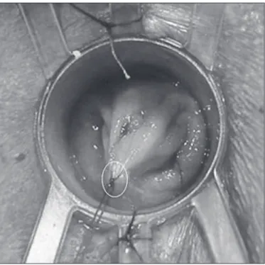
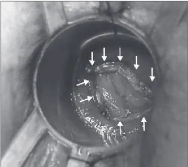
After a full mechanical bowel preparation and under spinal anesthesia, patients were placed in the Lloyd-Davis position. Broad-spectrum antibiotic prophylaxis was given before surgery (1g cephoxitin). A circular anal dilator was inserted into the anal canal and maintained secured to the perianal skin with two stay sutures (anterior and posterior). The rectocele was pushed through the anal canal with a inger inserted into the vagina to identify the apex. The posterior vaginal wall was pulled up with a Babcock forceps, the apex of the rectocele was pulled down (Figure 1) and a running horizontal suture (Greek suture technique) was placed through the base of the rectocele, including mucosa, submucosa and the muscle layer of the whole anorectal junction wall (Figure 2). This suture was placed approximately 2.0 cm above the dentate line, depending on the size of the rectocele. The exceeded prolapsed mucosa and the muscle layer were then excised with an electrical scalpel, keeping the wound open with the edges joined by the previous manual suture. A continuous
FIGURE 1. The apex of the rectocele is identiied and pulled down through a stitch (circle)
FIGURE 2. A running horizontal suture (Greek suture technique) is placed through the base of the rectocele (arrows)
FIGURE 3. The exceeded prolapsed mucosa and the muscular layer were excised, keeping an opened wound with the edges joined by the previous manual suture (arrows). The pursestring suture is tied around the stapler’s center rod
FIGURE 4. The remaining stapled suture line (arrows)
stapler’s center rod (Figure 3), taking care to include the full rectal wall anteriorly, ired and withdrawn, keeping a circular stapled suture (Figure 4).
Statistical analysis
The results were analyzed with Student’s t test. The level of statistical signiicance was set at P<0.05.
RESULTS
All patients presented the marks in rectum on the 3rd day preoperative but showing that 80.0% of the marks had been evacuated on the 5th day. All patients complained of obstructed defecation despite conservative treatment (high iber diet and laxatives) for at least 4 weeks. Anismus was observed in 15 patients (20.0%), all of whom were successfully submitted to preoperative biofeedback therapy. The average time of surgery was 42 minutes. In 13 patients (17.3%), bleeding from the stapled line required hemostatic suture. Stapling was incomplete in 2 (2.6%) patients. Forty-nine (65.3%) required 1 day of hospitalization, the remainder (34.7%) 2 days. Ten (13.3%) patients presented postoperative complications. Of these, 7 (9.3%) developed stricture on the stapled suture subsequently treated by stricturectomy under anesthesia (n = 1), endoscopic stricturectomy with hot biopsy forceps (n = 3) or digital dilatation (n = 3). Three patients (4.0%) complained of persistent rectal pain for 2 weeks (n = 2) and for 3 months (n = 1). Postoperative cinedefecography showed residual anorectoceles (grade I) in 8 patients (10.6 %). No statistically signiicant parameters were observed upon anal manometric evaluation. The mean Wexner constipation score decreased signiicantly from 16 to 4 (0-4: n = 68) (6: n = 6) (7: n = 1) (P<0.0001).
DISCUSSION
Anal manometry showed that anal pressures were not affected by the procedure. Most patients (91.2%) displayed signiicant improvement of constipation symptoms with no changes in anal continence, as demonstrated by the reduction from 16 to 4 in the mean validated Wexner constipation score (P<0.0001) after an average follow-up time of 21 months (range 4–37). Anorectocele has commonly been treated with perineal levatorplasty and transanal techniques, especially Block’s(4) and Sarles’(25) repairs, with successful outcome in
70%–90% of cases. Recently, several new stapling techniques have been tested, such as stapled transanal prolapsectomy(7),
the STARR double-stapling procedure(6, 7), combined perineal
and endorectal repair with circular stapler(2), transanal repair
with linear stapler and stapler resection of the rectocele area(3). The main disadvantages of perineal levatorplasty
is postoperative dyspareunia reported by approximately 25.0% of the patients(5) and failure to treat associated rectal
mucosal prolapse potentially impairing defecation. Sarles’(4)
and Block’s(25) procedures repair the anatomic defect of the
anterior rectal wall but leave the posterior rectal mucosal prolapse untreated. Stapled circular mucosectomy repairs rectal mucosal prolapse but is not extensive enough to repair the
anterior anatomic anorectal wall defect. The double-stapled technique (STARR) is a more costly procedure as it requires the use of two staplers, one for repairing the anterior rectal mucosal prolapse and rectocele and one for removing the posterior mucosal prolapse. However, the European STARR Registry has reported a signiicant reduction (from 15.8 to 5.8) in obstructed defecation scores at 12 months of follow-up(12). Similar results may be achieved at a smaller cost with
the TRREMS procedure, using a single stapler. The anterior anorectal junction wall defect is excised by manual resection, followed by stapled full rectal mucosectomy and anopexy. In addition, the vaginal-anorectal septum becomes straight and reinforced by the ibrous tissue produced during healing. To avoid injury, the posterior vaginal wall should always be pulled up with a Babcock forceps during manual horizontal suture placement and stapler closure.
In conclusion, TRREMS procedure is a safe, effective and relatively inexpensive technique for the treatment of anorectocele without dyspareunia. In addition, it offers the advantage of restoring anatomical integrity using a single circular stapler. Further investigation is required to evaluate the long-term follow up results.
REFERENCES
1. Agachan F, Chen T, Pfeifer J, Reissman P, Wexner SD. A constipation scoring system to simplify evaluation and management of constipated patients. Dis Colon Rectum. 1996;39:681-5.
2. Altomare DF, Rinaldi M, Veglia A, Petrolino M, De Fazio M, Sallustio P. Combined perineal and endorectal repair of rectocele by circular stapler: a novel surgical technique. Dis Colon Rectum. 2002;45:1549-52.
3. Ayav A, Bresler L, Brunaud L, Boissel P. Long-term results of transanal repair of rectocele using linear stapler. Dis Colon Rectum. 2004;47:889-94.
CruzJV, Regadas FSP, Murad-Regadas SM, Rodrigues LV, Benicio F, Leal R, Carvalho CG, Fernandes M, Roche LMC, Miranda AC, Câmara L, Pereira JC, Parra AM, Leal VM. Procedimento TRREMS (reparo transanal de retocele e mucosectomia retal com um grampeador circular). Estudo multicêntrico prospectivo. Arq Gastroenterol. 2011;48(1):3-7.
RESUMO – Contexto - Como a anoretocele está sempre associada a prolapso mucoso e/ou intussuscepção retal, foi desenvolvida uma técnica cirúrgica grampeada, utilizando somente um grampeador mecânico. Objetivo - Demonstrar os resultados do estudo multicêntrico que realizou o tratamento cirúrgico de pacientes com anorretocele associado a prolapso mucoso, utilizando o reparo transanal da retocele e mucosectomia com grampeador circular mecânico. Método - Foram incluídos 75 pacientes, média de idade 49,6 anos, com sintomas de evacuação obstruída, apresentando escore médio de constipação de Wexner de 16 e diagnóstico de anorretocele grau II (26.7%), grau III (73,3%) associado a prolapso mucoso e intussuscepção (52%). Todos foram avaliados com exame proctológico, defecograia, manometria anorretal e tempo de trânsito colônico. O procedimento cirúrgico foi realizado por 14 cirurgiões e consiste na remoção manual da parede do reto no local da retocele e mucosectomia circunferencial com um grampeador circular mecânico. O seguimento médio foi de 21 meses. Resultados - Os pacientes apresentavam sintomas de evacuação obstruída, mesmo após tratamento clinico. O tempo operatório médio foi de 42 minutos. Houve sangramento transanal na linha de sutura em 13 (17,3%) pacientes, sutura grampeada incompleta em 2 (2,6%) e dor retal persistente em 3 (4,0%). O tempo médio de internação hospitalar foi de 1 dia em 49 (65,3%) e 2 dias em 34,7%. Ocorreu redução na linha de sutura em 7 (9,3%), sendo necessário estricturotomia cirúrgica sob anestesia (n = 1), utilizando “hot biopsy” (n = 3) e dilatação anal (3). Defecograia no pós-operatório demonstrou anorretocele residual grau I em 8 (10.6%). O escore de constipação de Wexner reduziu 16 para 4 (0-4: n = 68) (6: n = 6) (7: n = 1) (P<0.0001). Conclusão - O resultado do estudo multicêntrico demonstrou que a técnica cirúrgica apresentada é segura e efetiva para tratamento da anorretocele associada a prolapso mucoso. Apresenta baixo custo pois utiliza um grampeador circular mecânico.
DESCRITORES – Retocele, cirurgia. Mucosa intestinal, cirurgia. Prolapso retal. Incontinência fecal. Grampeamento cirúrgico.
4. Block IR. Transrectal repair of rectocele using obliterative suture. Dis Colon Rectum. 1986;29:707-11.
5. Boccasanta P, Venturi M, Calabrò G, Trompetto M, Ganio E, Tessera G, Bottini C, Pulvirenti D’Urso A, Ayabaca S, Pescatori M. Which surgical approach for rectocele? A multicentric report from Italian coloproctologists. Tech Coloproctol. 2001;5:149-56.
7. Dodi G, Pietroletti R, Milito G, Binda G, Pescatori M. Bleeding, incontinence, pain and constipation after STARR transanal double stapling rectotomy for obstructed defecation. Tech Coloproctol. 2003;7:148-53.
8. Fritsch H. The connective tissue sheath of uterus and vagina in the human female fetus. Ann Anat. 1992;174:261-6.
9. Fritsch H. Topography and subdivision of the pelvic connective tissue in human fetuses and in the adult. Surg Radiol Anat. 1994;16:259-65.
10. Fritsch H, Hotzinger H. Tomographical anatomy of the pelvis, visceral pelvic connective tissue and its compartments. Clin Anat. 1995;8:17-24.
11. Habr-Gama A, Sousa AH Jr, Roveló JM, Souza JV, Benício F, Regadas FS, Wainstein C, da Cunha TM, Marques CF, Bonardi R, Ramos JR, Pandini LC, Kiss D. Stapled hemorrhoidectomy: initial experience of a Latin American group. J Gastrointestinal Surg. 2003;7:809–13.
12. Jayne DG, Schwandner O, Stuto A. Stapled transanal rectal resection for obstructed defecation syndrome: one-year results of the European STARR Registry. Dis Colon Rectum. 2009;52:1205-14.
13. Kovacs EJ, DiPietroa LA. Fibrogenic cytokines and connective tissue production. FASEB J. 1994;8:854-61.
14. Longo A. Treatment of hemorrhoid disease by reduction of mucosa and hemorrhoidal prolapse with a circular suturing device: a new procedure. In: Proceedings of the 6th World Congress of Endoscopic Surgery; 1998 June 3–6; Rome. Bologna: Monduzzi Editore; 1998. p.777-84.
15. Milley PS, Nicholls DH. A correlative investigation of the human rectovaginal septum. Anat Rec. 1969;163:443-51.
16. Nahas SC, Borba MR, Brochado MC, Marques CF, Nahas CS, Miott-Neto B. Stapled hemorrhoidectomy for the treatment of hemorrhoids. Arq Gastroenterol. 2003;40:35-9.
17. Nicholls DH, Milley PS. Surgical signiicance of the rectovaginal septum. Am J Obstet Gynecol. 1970;15:215-20.
18. Pescatori M, Favetta V, Dedola S, Orsini S. Stapled transanal excision of rectal mucosa prolapse. Tech Coloproctol. 1997;1:96–8.
19. Regadas FS, Regadas SM; Rodrigues LV, Misici R, Tramujas I, Barreto JB, Lins MA, Silva FR, Filho FS. New devices for stapled rectal mucosectomy: a multicenter experience. Tech Coloproctol. 2005;9:243-6.
20. Regadas FS, Regadas SM, Rodrigues LV, Misici R, Silva FR, Regadas-Filho FS. Transanal repair of rectocele and full rectal mucosectomy with one circular stapler: a novel surgical technique. Tech Coloproctol. 2005;9:63-6.
21. Regadas FS, Murad-Regadas SM, Lima DM, Silva FR, Barreto RG, Souza MH, Regadas Filho FS. Anal canal anatomy showed by three-dimensional anorectal ultrasonography. Surg Endosc. 2007;21:2207-11.
22. Regadas FS, Murad-Regadas SM, Rodrigues LV, Wexner SD, Souza MH, Silva FR, Lima DM, Regadas-Filho FS. Anorectal three-dimensional endosonography and anal manometry in assessing anterior rectocele in women. A new pathogenesis concept and the basic surgical principle. Colorectal Dis. 2007;9:80-5.
23. Regadas SM, Regadas FS, Rodrigues LV, Silva FR, Lima DM, Regadas-Filho FS. Importância do ultra-som tridimensional na avaliação anorretal. Arq Gastroenterol. 2005;42:226-32.
24. Richardson AC. The rectovaginal septum revisited: its relationship to rectocele and its importance in rectocele repair. Clin Obstet Gynecol. 1993;36:976-83. 25. Sarles JC, Arnaud A, Selezneff I, Olivier S. Endo-rectal repair of rectocele. Int
J Colorectal Dis. 1989;4:167-71.
26. Smith AR. Role of connective tissue and muscle in pelvic loor dysfunction. Curr Opin Obstet Gynecol. 1994;6:317-9.
27. Sobrado CW, Cotti GC, Coelho FF, Rocha JR. Initial experience with stapled hemorrhoidopexy for treatment of hemorrhoids. Arq Gastroenterol. 2006;43: 238-42.
28. Wilson MS, Pope V, Doran HE, Fearn SJ, Brough WA. Objective comparison of stapled anopexy and open hemorrhoidectomy: a randomized, controlled trial. Dis Colon Rectum. 2002;45:1437–44.
29. Zbar AP, Lienemann A, Fritsch H, Beer-Gabel M, Pescatori M. Rectocele: pathogenesis and surgical management. Int J Colorectal Dis. 2003;18:369-84.