LETTER TO THE EDITOR
Gastric wall abscess – an uncommon condition
treated by an alternative form
Wander C. Marcos, Bruno G. Petrini, Rafael L. Xavier, Rosaˆngela M. Starling, Jose´ C. Couto, Guilherme J. Ribeiro
Vera Cruz Hospital, Endoncopy and Surgery - Barbacena, Belo Horizonte/MG, Brasil.
Email: [email protected] Tel.: 55 31 32901000
INTRODUCTION
Phlegmonous gastritis or suppurative gastritis is a rare condition characterized by a purulent inflammatory process involving the gastric wall that can occur in a diffuse, localized, or mixed form. The most common type, the diffuse form, is characterized by initial involvement of the gastric submucosa with posterior extension to all layers of the gastric wall, resulting in extensive gangrene of the stomach. Although the exact pathogenesis of phlegmonous gastritis is unknown, it is likely caused by a bacterial infection of the stomach wall seeded either directly by invasion through a breach in the gastric mucosa or secondary to hematogenous spread from a distant source of infection.6,9,10Predisposing conditions associated with suppurative gastritis include alcoholism, diabetes, ingestion of a foreign body, gastric surgery, endoscopic polypectomy, ingestion of corrosive agents, gastritis, chronic peptic ulcers, gastric carcinoma, and human immunodeficiency virus infection.5,9,12,17–19Until recently, the recommended therapy for an intramural gastric abscess was surgical drainage in combination with antibio-tics. However, technical advances now allow both radiologic and endoscopic intervention. In this case report, we describe an intramural gastric abscess that was successfully treated with antibiotics and endoscopic drainage.
CASE REPORT
A 48-year-old man presented to the hospital with a 5-day history of fever and dull epigastric pain radiating to the back and left upper quadrant of the abdomen. His medical history included a large hiatal hernia and erosive gastritis, both documented by a recent upper endoscopy. He did not have a history of alcohol abuse or pancreatitis and was currently taking omeprazole for gastroesophageal reflux. On admission, he was febrile and a tender, 8-cm mass was palpable in his upper epigastrium. His laboratory tests were all within normal limits, with the exception of a white blood cell count of 126103/mm3(normal: 3,5 a 10,56103/mm3).
An abdominal ultrasound was performed, and it showed a thickened parietal gastric wall (1.3 cm) at the gastric antrum in the great bend, and pylorus. Intravenous antibiotics (ampicillin and sulbactam) were initiated empirically. His
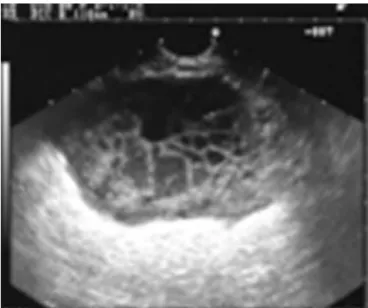
fever remitted within three days, but his pain persisted. A computed tomography scan was performed that revealed an abscess, 10 cm in length, within the gastric wall and adjacent to the large curvature, near the gastric antrum (Figures 1 and 2). Endoscopic ultrasound showed a well-circumscribed intramural mass of mixed echogenicity in the gastric wall, and the presence of internal fluid and debris confirmed the presence of an abscess (Figure 3). Endoscopic drainage was performed with a needle knife, and abundant purulent discharge was removed. A pigtail catheter was put in place for drainage (Figure 4). Bacterioscopy of the drained material revealed gram-positive cocci present in pairs and chains. Candida albicans was the organism cultured. The patient was discharged from the hospital, and an endoscopic ultrasound performed one month after the drainage demonstrated no residual fluid collection. At that time, the pigtail catheter was removed, and the patient continues to remain asymptomatic.
DISCUSSION
A localized abscess of the gastric wall is one variety of phlegmonous or suppurative gastritis,9 a purulent inflam-matory process involving the gastric wall that can occur in diffuse, localized, and mixed forms. The localized form accounts for 5 to 15% of all cases.1–4 The more common diffuse form is characterized by involvement of the gastric submucosa, with posterior extension into all layers of the
Copyrightß2010CLINICS– This is an Open Access article distributed under the terms of the Creative Commons Attribution Non-Commercial License (http:// creativecommons.org/licenses/by-nc/3.0/) which permits unrestricted non-commercial use, distribution, and reproduction in any medium, provided the
original work is properly cited. Figure 1- Computed tomography showing the gastric wall abscess. CLINICS 2010;65(8):819-821 DOI:10.1590/S1807-59322010000800016
gastric wall, and results in extensive gangrene of the stomach.1,9 Microscopically, the stomach exhibits marked submucosal thickening, with flattening of the overlying mucosa and loss of the normal folds, an appearance that mimics Gastro intestinal stromal tumors.9
The exact pathogenesis of phlegmonous gastritis is unknown. It is probably caused by bacterial infection of the gastric wall by direct invasion through a breach in the gastric mucosa or secondary to hematogenous spread from a distant infectious source.6,9,10Streptococciare the most common bacteria isolated in cultures from gastric abscesses (75% of cases).1Other pathogens that have been isolated includeStaphylococci,E. coli,
Haemofilus influenzae, Proteus species, Clostridium welchii,
Pseudomonas aeruginosa, andBacillus subtilis.6,11–16In this case,
Candida albicanswas the organism cultured.
Predisposing conditions associated with suppurative gastritis include alcoholism, diabetes, ingestion of a foreign body, gastric surgery, endoscopic polypectomy, ingestion of corrosive agents, gastritis, chronic peptic ulcers, gastric carcinoma, and human immunodeficiency virus
infec-tion.5,9,12,17–19 The condition affects men more often than women, in a ratio of 31, and the average age of patients ranges from 30 to 60 years old.9,12,14,20 In this case, the patient was a 48-year-old man who suffered from gastritis, but without other predisposing conditions.
To our knowledge, only 19 cases of gastric abscesses have been reported.5 The majority of patients present with abdominal pain,1but other symptoms may include nausea, vomiting, and, rarely, fever.1 Two specific but seldom present clinical signs are Deininger’s sign (decreased pain when changing from a supine to a sitting position)6 and vomiting of frank pus.7
Until recently, the recommended therapy for an intra-mural gastric abscess was surgical drainage in combination with antibiotics. However, technical advances now allow both radiologic and endoscopic intervention. Endoscopic drainage with or without antibiotics has been shown to be effective.1,5,19From the 19 cases of gastric abscess that have been reported, 12 underwent surgery, 4 were drained endoscopically, 2 were drained percutaneously, and 1 patient took antibiotics alone without drainage. The surgical approach in the majority of cases is probably due to the fact that a gastric abscess is seldom diagnosed preoperatively and is often misdiagnosed. In prior studies, all patients were successfully treated, with the exception of the death of the one patient treated merely with antibiotics.1,21
In our case, the abscess was treated with antibiotics and endoscopic drainage of the purulent fluid through the gastric lumen. A pigtail catheter was left in place for one month to allow for additional drainage of pus.
This case highlights many of the salient points regarding the presentation, evaluation and management of intramural gastric abscesses. The initial presentation and CT findings were suggestive of a gastric abscess, and an endoscopic ultrasound with drainage of the purulent secretion by conventional endoscopy confirmed the diagnosis and permitted treatment of the condition.
REFERENCES
1. Choong NW, Levy MJ, Rajan E, Kolars JC. Intramural gastric abscess. Gastrointestinal Endoscopy. 2003;58:627–9.
2. Gerster JC. Phlegmonous gastritis. Ann Surg 1927;85:668–82, doi: 10. 1097/00000658-192705000-00006.
3. Nevin NC, Eakins D, Clarke SD, Carson DJ. Acute phlegmonous gastritis. Br J Surg. 1969; 56:268–70, doi: 10.1002/bjs.1800560407. Figure 2- Computed tomography showing the gastric wall abscess.
Figure 3 - Endoscopic ultrasonography showing debris in the fluid collection.
Figure 4- Computed tomography showing the pigtail catheter left in the gastric wall abscess for drainage.
Gastric wall abscess
Marcos WC et al. CLINICS 2010;65(8):819-821
4. Miller AI, Smith B, Rogers AI. Phlegmonous gastritis. Gastroenterology. 1975;68:231–8 PMID:1116671.
5. Will U, Masri R, Bossekert H, Knopke A, Schonlebe J, Justus J. Gastric wall abscess, a rare endosonographic differential diagnosis of intramural tumors: successful endoscopic treatment. Endoscopy. 1998;30:432–5, doi: 10.1055/s-2007-1001297.
6. Murphy JF, Graham DY, Frankel NB, Spjut HJ. Intramural gastric abscess.AM J Surg.1976;131:618–21, doi: 10.1016/0002-9610(76)90028-3. 7. Lifton LJ, Schlossberg D. Phlegmonous gastritis after endoscopic
polypectomy. Ann Inter Med. 1982;97:373–5.
8. Seidel RH, Burdick JS. Gastric leyomiosarcoma presenting as a gastric wall abscess. The American Journal of Gastroenterology. 1998;93:2241, doi: 10.1111/j.1572-0241.1998.00625.x.
9. Kiil C, Rosenberg J. Gastric intramural abscess successfully drained during gastroscopy. Gastrointestinal Endoscopy. 2001;53;231–3, doi: 10. 1067/mge.2001.111559.
10. Muller SC. Totale magenwandphlegmone. Chirurg. 1980;51:102–5. 11. Miler AI, Smith B, Rogers AI. Phlegmonous gastritis. Gastroenterology.
1975;68:231–8.
12. Starr A, Wilson JM. Phlegmonous gastritis. Ann Surg 1957;145:88–93, doi: 10.1097/00000658-195701000-00009.
13. Smith GE. Subacute phlegmonous gastritis simulating intramural neoplasm: case report and review. Gastrointestinal Endosc. 1972;19:23– 6, doi: 10.1016/S0016-5107(72)73938-3.
14. Guzzeta PC, Southwick HW. Acute phlegmonous gastritis. Surgery. 1947;22:453–7.
15. Eley A, Richards HGH. A case of localized gastric abscess. Br J Surg 1959;47:97–8, doi: 10.1002/bjs.18004720119.
16. Rosen Y, Won OH. phlegmonous enterocolitis. Dig dis. 1978;23:248–56, doi: 10.1007/BF01072325.
17. Wilkinson LH, Simms AG, Floyd VT. Acute phlegmonous gastritis. Am Surg. 1960;26:160–5.
18. Eliason EL, Murray Wright VW. Phlegmonous gastritis. Surg Clin North Am. 1938;18:1553–64.
19. Avile´s JF, Ferna´ndez-Seara J, Ba´rcena R, Domı´inguez F, Ferna´ndez C, Ledo L. Localized phlegmonous gastritis: endoscopy view. Endoscopy. 1988;20:38–9, doi: 10.1055/s-2007-1018124.
20. Weiner C, Kumpe DA, Diaconis JN. Idiopathic gastic abscess – a bizarre intramural lesion. Am J Gastroenterol. 1975;64;452–9.
21. Ashin Asrani, Robert Novelline, Hani Abujedeh, James Lawrason, Rathachai Kaewlai. Intramural gastric abscess – preoperative diagnosis with CT. Emerg Radiol. 2007;14:253–6, doi: 10.1007/s10140-007-0589-x.
CLINICS 2010;65(8):819-821 Gastric wall abscess
Marcos WC et al.