CLINICAL SCIENCE
Abnormal sensory integration affects balance control
in hemiparetic patients within the first year after
stroke
Clarissa B. Oliveira,I,III´talo R. T. Medeiros,IIIMario G. Greters,III Norberto A. F. Frota,IVLeandro Tavares Lucato,VIMilberto Scaff,IIAdriana B. ConfortoII,V
IAACD - Adult Physiotherapy, Sa˜o Paulo/SP, Brazil.IIHospital das Clı´nicas da Faculdade de Medicina da Universidade de Sa˜o Paulo, Neurology Department,
Sa˜o Paulo/SP, Brazil.IIIHospital das Clı´nicas da Faculdade de Medicina da Universidade de Sa˜o Paulo, Otorhinolaryngology Department, Sa˜o Paulo/SP, Brazil.IVFortaleza University - School of Medicine, Fortaleza/CE, Brazil.VInstituto Israelita de Ensino e Pesquisa Albert Einstein, Sa˜o Paulo/SP, Brazil. VIHospital das Clı´nicas da Faculdade de Medicina da Universidade de Sa˜o Paulo, Radiology Department, Sa˜o Paulo/SP, Brazil.
OBJECTIVE:Impairments in balance can be a consequence of changes in the motor, sensory, and integrative aspects of motor control. Abnormal sensory reweighting, i.e., the ability to select the most appropriate sensory information to achieve postural stability, may contribute to balance impairment. The Sensory Organization Test is a component of Computerized Dynamic Posturography that evaluates the impact of visual, vestibular, and somatosensory inputs, as well as sensory reweighting, under conditions of sensory conflict. The aim of this study is to compare balance control in hemiparetic patients during the first year post-stroke and in age-matched neurologically normal subjects using the Berg Balance Scale and Computerized Dynamic Posturography.
METHODS: We compared the Berg Balance Scale and Sensory Organization Test scores in 21 patients with hemiparesis after first-ever ischemic stroke and in 21 age-matched, neurologically normal subjects. An equilibrium score was defined for each Sensory Organization Test condition.
RESULTS: Berg Balance Scale scores were significantly lower in the patients than in the neurologically normal subjects. Equilibrium scores were significantly lower in the patients than in the neurologically normal subjects for those Sensory Organization Test conditions that did not provide appropriate somatosensory information and under conditions of sensory conflict. A history of falls was more frequent in patients with lower equilibrium scores.
CONCLUSION:During the first year after a stroke, defective sensory reweighting significantly impacts balance control in hemiparetic patients. These results are important for the planning of effective rehabilitation interventions.
KEYWORDS: Posture; Rehabilitation, Cerebrovascular accident; Equilibrium; Posturography.
Oliveira CB, Medeiros IRT, Greters MG, Frota NAF, Lucato LT, Scaff M, et al. Abnormal sensory integration affects balance control in hemiparetic patients within the first year after stroke. Clinics. 2011;66(12):2043-2048.
Received for publication onAugust 10, 2011;First review completed onAugust 23, 2011;Accepted for publication onAugust 23, 2011
E-mail: [email protected]
Tel.: 55 11 5576-0924
INTRODUCTION
Falls occur in up to 73% of patients within the first six months after a stroke1 due to balance abnormalities associated with impairments in the motor, sensory, cogni-tive, or integrative aspects of movement control. Falls can lead to complications, such as hip fractures, that have high individual and social costs.2-10 Even in high-functioning post-stroke populations, falls are common and are asso-ciated with limitations on activity and participation.11
Reductions in muscle strength and range of movement, abnormal muscle tone and loss of motor coordination and sensory organization can contribute to balance disturbances to varying degrees.12 The difficulties in determining the specific causes of balance impairments in hemiparetic patients are related to the diversity of mechanisms involved and the variety of strategies used to adapt to these changes.13
Clinical scales or laboratory measurements can be used to evaluate balance control. Laboratory measurements of pos-tural reactions are more sensitive for investigating balance during standing and functional activities13-15 than clinical scales such as the Berg Balance Scale (BBS).16One of the most widely used laboratory tools for evaluating balance is posturography,12 which is capable of quantifying pos-tural asymmetry, instability17 and sensory contributions to balance control. Computerized Dynamic Posturography (CDP) is a specific type of posturography that challenges
Copyrightß2011CLINICS– This is an Open Access article distributed under
the terms of the Creative Commons Attribution Non-Commercial License (http:// creativecommons.org/licenses/by-nc/3.0/) which permits unrestricted non-commercial use, distribution, and reproduction in any medium, provided the original work is properly cited.
postural stability by providing different levels of sensory deprivation and conflict as part of the Sensory Organization Test. In this test, a subject stands on a force platform with sensors that provide kinetic data regarding postural reactions both during normal conditions and after the manipulation of visual, vestibular and somatosensory inputs.18This manip-ulation allows for the quantification of the patient’s relative reliance on different sensory inputs that are required to maintain balance, as well as their integration.
Decreased multisensory integration, with excessive reli-ance on visual information and consequent poor balreli-ance control, has been demonstrated during the chronic stages (.1 year) after a stroke.12It is known that rehabilitation is more effective when delivered within the first months after a stroke, but the ways in which defective selection and integration of sensory input affect balance at this stage are not well understood. If the factors that influence postural impairment within the first year post-stroke can be determined, then rehabilitation programs can be tailored to target specific components of balance control and, hence, decrease the risk of falls.
To better understand the mechanisms underlying balance disturbances at an earlier stage, within the first year after a stroke, we used an innovative approach that employed not only a clinical tool (the Berg Balance Scale)16 but also a sophisticated laboratory test, the CDP. In addition, we investigated the relationship between CDP results and a history of falls in post-stroke patients.
MATERIALS AND METHODS
Subjects
Stroke patients
Patients diagnosed with stroke at Hospital das Clı´nicas/ Sa˜o Paulo University were consecutively screened between July 2006 and April 2008. The following inclusion criteria were used: hemiparesis after a first-ever ischemic stroke in a cerebral hemisphere one to twelve months previously, confirmed by computed tomography or magnetic resonance imaging; ability to stand unassisted for 30 seconds; no severe systemic disorders; ability to understand and cooperate with testing; normal visual fields; visual acuity of at least 20/60; and availability for evaluations. The exclusion criteria were as follows: other neurological diseases; brain stem or cerebellar stroke; vertigo; vestibular dysfunction in neurootological examination (electrooculo-graphy with positional tests and caloric tests with water at 44˚C and 30˚C); lower limb orthopedic disorders; hemi-neglect (evaluated using the cancellation test);19and pusher syndrome, defined according to the Clinical Assessment Scale for Contraversive Pushing (the subject should present a contralesional tilted posture, a tendency to push toward the paretic side with the nonaffected limbs, and resistance to external correction of the tilted posture, in either a seated or standing position).20
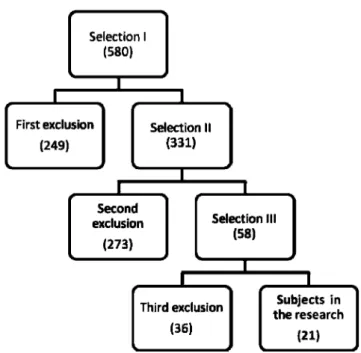
The selection of participants was performed in three stages: evaluation of medical charts (I), telephone inter-views (II), and personal interinter-views/evaluations at the hospital (III). Figure 1 illustrates the exclusions at each stage of research. The main reasons for non-inclusion were as follows: recurrent stroke (26.1% - 146 subjects), no hemiparesis (18.2% - 102 subjects), inability to be contacted by phone for screening (8.9% - 50 subjects), and brainstem
or cerebellar stroke (7.5% - 45 subjects). In addition, 19 patients (3.4%) were not able to stand on the platform for at least 30 seconds. In total, 21 patients (3.6%) were included in the study. All patients provided informed consent, and the protocol was approved by the institutional ethics committee.
Table 1 shows the patients’ characteristics. Only patients with mild to moderate hemiparesis and neurological impairment were included because severe hemiparesis precludes complete posturographic evaluation. Motor and sensory impairments were characterized using the motor and sensory examination components of the Fugl-Meyer Assessment for the lower limbs.21Neurological impairment was evaluated using the National Institute of Health Stroke Scale (NIHSS),22 and disability was evaluated using the Barthel Index23 and the Functional Ambulatory Category score.24Ankle proprioception (evaluated as part of the Fugl-Meyer Assessment) was abnormal in three patients. Of the patients, seven (33.3%) had a history of at least one fall after stroke, and 19 (90.5%) were right-handed (laterality before the stroke was asked in the interview). Lesions encom-passed the following cerebral areas/structures: frontal lobe Figure 1 - Selection of participants. The number of patients included at each stage of the research is shown.
Table 1 -Characteristics of the patients.
Age (years) 55.9¡13.9
Time from stroke (months) 4.8¡2.5
Men/women 13/8
Hemiparesis (R/L) 14/7
Handedness (R/L) 19/2
Barthel Index 95 (80–100)
NIHSS 2 (0–8)
Fugl-Meyer Assessment for the lower limbs 31 (17-33)
Functional Ambulatory Category 4 (2-5)
(61.9% - 13 subjects), insula (52.4% - 11 subjects), parietal lobe (47.6% - 10 subjects), temporal lobe (19% - 4 subjects), internal capsule (19% - 4 subjects), basal ganglia (14.3% - 3 subjects), and thalamus (4.8% - 1 subject).
Neurologically normal volunteers
For the control group, 21 age-matched neurologically normal subjects were recruited. The inclusion criteria were the absence of neurological disorders and visual acuity of at least 20/60. The exclusion criteria were a history of vertigo and orthopedic disorders in the lower limbs. In addition, subjects with abnormalities in the vestibular evaluation of the Sensory Organization Test in posturography were excluded because it is known that individuals with peripheral vestibular disorders cannot perform normally on Sensory Organization Test (SOT) conditions 5 and 6.25
Balance evaluation
BBS and posturography were performed in the patients (the mean time after stroke ¡ standard deviation was
4.8¡2.5 months) and neurologically normal volunteers. The BBS16is a simple test of functional balance composed of 14 items based on activities of daily living. Each item is rated using an ordinal scale, ranging from 0 to 4, and the maximum score is 56.
Posturography was performed with the SOT and the Equitest protocol (NeuroCom International) of the CDP. During testing, the subjects stood barefoot in the upright position, protected by a harness, with their arms alongside their body and their feet on a predesignated site. A dual-force platform, with four transducer outputs, measured vertical forces and shear forces exerted in the anteroposter-ior direction, separately for each leg.
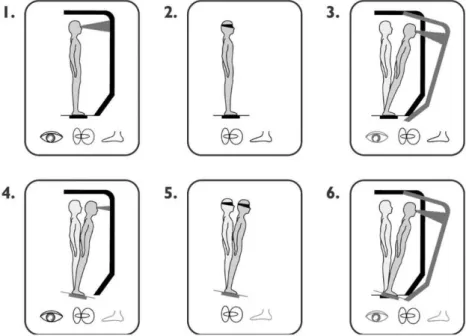
The SOT assesses the three sensory components of balance under different visual and support-surface condi-tions. Figure 2 shows the six different conditions that were studied: 1. Eyes open, fixed platform surface and back-ground (in this situation, the subject relies on visual, vestibular and somatosensory inputs); 2. Eyes closed, fixed
platform surface and background (in this situation, the subject uses mainly vestibular and somatosensory inputs to maintain balance because there is no visual information about his or her position in relation to the environment); 3. Eyes open, fixed platform surface and sway-referenced visual background (in this situation, the subject should rely mainly on vestibular and somatosensory information and not on visual inputs, which are not providing accurate information about his or her position in relation to the environment); 4. Eyes open and sway-referenced surface (in this situation, the subject uses mainly visual and vestibular inputs to maintain balance because the somatosensory information is distorted); 5. Eyes closed and sway-refer-enced surface (in this situation, the subject uses mainly vestibular inputs to maintain balance because there is no visual information about his or her position in relation to the environment, and the somatosensory information is dis-torted); and 6. Eyes open, sway-referenced surface and visual background (in this situation, the subject should use mainly vestibular inputs to maintain balance, because the somatosensory and visual information are distorted). Conditions 4, 5, and 6 provide challenging sensory inputs, and Conditions 3 and 6 introduce visual conflicts.
For each subject, three trials of 20 seconds were performed with each condition. Sensory functions were assessed as the ratio between the average of the results of the following conditions and the average of Condition 1:
N
Somatosensory function (Condition 2);N
Visual function (Condition 4); andN
Vestibular function (Condition 5).Visual preference was evaluated by comparing the sum of the equilibrium scores for Conditions 3 and 6 with the sum of the equilibrium scores for Conditions 2 and 5 (Conditions 3+6/Conditions 2+5). Visual preference reflects the ability
to suppress visual information perceived as incorrect. An equilibrium score (ES) was calculated for each of the six SOT conditions. An ES is a measure of postural stability,
based on sway during a 20-second SOT trial. A score of 100 indicates no sway, whereas 0 indicates sway beyond the limits of stability (8.5˚anteriorly and 4˚posteriorly; 12.5˚is the theoretical limit of sway in the sagittal plane for normal stance). The score was calculated using the following formula: ES = (12.5˚- [hmax – hmin]) 6100/12.5˚, where
12.5˚is the normal limit of anteroposterior sway, andhis the
angle between a line extending vertically from the center of foot support and a line extending from the center of foot support through the patient’s center of gravity.26
A composite equilibrium score (ES) was generated for each of the 21 patients. This ES represents the overall evaluation of all six Conditions (maximum score = 100) and was calculated by independently averaging all trial scores from Conditions 1 and 2, adding these two average scores to the individual trial scores from conditions 3 through 6 and then dividing the sum by 14.26
Statistical analysis
For data normally distributed according to the Kolmogorov-Smirnov test, the means and standard devia-tions are given, and the stroke patients and neurologically normal volunteers were compared using unpaired t-tests. For categorical or non-normally distributed data, the median, minimum, and maximum values are reported. Comparisons between groups were performed using Mann-Whitney tests, which were also used for the post-hoc
comparison of ES’s between patients with normal and impaired ankle proprioception, as evaluated using the Fugl-Meyer Assessment. Univariate logistic regression was performed to examine the relationship between ES (inde-pendent variable) and a history of falls (de(inde-pendent variable). p- values #0.05 were considered significant. Minitab 15.0 and SPSS 10.0 were used for the statistical analysis.
RESULTS
Subjects
There were no differences in age (p= 0.76), sex (p= 0.76) or manual preference (p= 0.66) between the neurologically normal volunteers and patients with stroke.
Berg Balance Scores
BBS scores were 53 (42-56) in the patients with stroke and 56 (55-56) in the neurologically normal volunteers. The difference between scores in the two groups was small but statistically significant (p= 0.01).
Computerized Dynamic Posturography
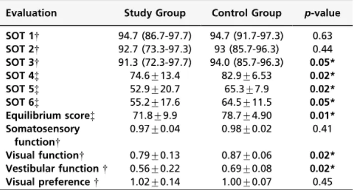
The CDP results are shown in Table 2. There were no significant differences between the groups while standing on a fixed platform with eyes open (Condition 1) or eyes closed (Condition 2).
However, ES’s were significantly lower in the patients than in the neurologically normal volunteers while standing on a fixed platform with sway-referenced vision (Condition 3), on a sway-referenced platform with their eyes either open (Condition 4) or closed (Condition 5) and with sway-referenced vision and a sway-sway-referenced platform (Con-dition 6). Post-hoc analysis revealed that patients with impaired ankle proprioception had lower composite ES’s than those with normal ankle proprioception (p =0.02).
In summary, the patients performed worse in conditions that provided challenging somatosensory information (Conditions 4, 5, and 6) or that introduced visual conflicts (Conditions 3 and 6). Somatosensory information was able to compensate for absent visual information (Condition 2), but it was not completely effective in a more demanding situation of visual and somatosensory conflict (Condition 3). Composite ES’s were significantly lower in the patients than in the neurologically normal subjects (Table 2). Composite ES’s were inversely associated with falls (odds ratio = 0.63, confidence interval = 0.41-0.99,p =0.043), i.e., a history of falls was more frequent in patients with lower scores.
DISCUSSION
The main finding of this study was that patients with mild to moderate hemiparesis performed better on the SOT in the presence of accurate somatosensory input, suggesting greater reliance on this sensorial modality to maintain their balance. Also, postural reactions were not sufficient for maintaining balance when the patients were challenged by the manipulation of somatosensory and visual information. Under these conditions, the patients performed worse than the neurologically normal subjects.
BBS scores were significantly different between the groups, but the magnitude of the difference was small (three points, on average). This finding may be explained by a ceiling effect in the BBS, which does not offer challenges to postural control, unlike the SOT. In the BBS, there is only one condition involving the manipulation of sensory input, i.e., standing with eyes closed. Our results are consistent with the BBS ceiling effect reported in patients with stroke and mild neurological deficits by Garland and colleagues.27 In this study, the patients’ postural adjustments were only comparable to those of the neurologically normal volunteers under less challenging conditions (Conditions 1 and 2). The patients’ reliance mostly on vestibular and visual functions to maintain balance was inefficient, despite the absence of visual or vestibular impairments in clinical or otoneurolo-gical evaluations. This result indicates the presence of abnormal vestibular and visual integration or difficulties in ‘‘choosing’’ the more reliable sensorial modality in each condition (sensory reweighting).
Table 2 -Posturography scores and sensory analysis in stroke patients and neurologically normal volunteers.
Evaluation Study Group Control Group p-value
SOT 1{ 94.7 (86.7-97.7) 94.7 (91.7-97.3) 0.63
SOT 2{ 92.7 (73.3-97.3) 93 (85.7-96.3) 0.44
SOT 3{ 91.3 (72.3-97.7) 94.0 (85.7-96.3) 0.05*
SOT 4{ 74.6¡13.4 82.9¡6.53 0.02*
SOT 5{ 52.9¡20.7 65.3¡7.9 0.02*
SOT 6{ 55.2¡17.6 64.5¡11.5 0.05*
Equilibrium score{ 71.8¡9.9 78.7¡4.90 0.01*
Somatosensory function{
0.97¡0.04 0.98¡0.02 0.41
Visual function{ 0.79¡0.13 0.87¡0.06 0.02*
Vestibular function{ 0.56¡0.22 0.69¡0.08 0.02*
Visual preference{ 1.02¡0.14 1.00¡0.07 0.45
NOTE:
{Mann-Whitney test; {
Student’s unpaired t test; *
Although the patients relied more on somatosensory input than the control subjects to maintain their balance, and although most patients had normal ankle proprio-ception, somatosensory input was not completely effec-tive, because the patients performed worse than the control subjects under conditions of visuovestibular conflict, characterized by a moving background, even in the presence of appropriate somatosensory input (Condition 3).
The importance of sensory input in balance control in the stroke group was reinforced by the higher ES’s measured in patients with normal ankle proprioception. All except one of the published studies12 have found positive correlations between ankle proprioception and balance control in stroke patients.15,28 Tyson and collea-gues29 found that changes in proprioception, along with motor impairments, were the best predictors of balance abnormalities.
Sensory reweighting is defined as the ability to select the most appropriate sensory information to achieve postural stability. When multisensory integration is intact, the weight of one type of sensory input is enhanced to compensate for a decrease in or absence of information from another sensory channel. Thus, a neurologically normal subject does not lose balance during visual deprivation because the information provided from vestibular and somatosensory input is integrated to guide postural adjustments. Our results indicate that this ability is impaired in patients after one month, but to a less extent than one year after stroke, similarly to that described at an earlier phase (within one month).30
Our patients presented worse visuovestibular integration compared with patients at a more chronic stage after stroke.12 More than one year after stroke, patients have been reported to perform worse than control subjects when somatosen-sory input is more critical to guiding postural adjustments (Conditions 5 and 6) but not under less demanding circumstances (Conditions 3 and 4). Differences between our results and those obtained in patients in the chronic stage with comparable neurological impairments are likely to reflect different degrees of compensation or adaptive plastic changes due to injury at different phases after stroke. It is known that recovery from a stroke evolves dynamically.31 Over time, different neurophysiological mechanisms are likely to be used for sensory reweighting; most studies have shown greater reliance on visual input during more chronic stages.12,32
This study has some limitations. First, we included patients at early (one month - six months) and later stages (six months - one year) after stroke. Despite this hetero-geneity in the time from lesion onset, we were able to find significant differences between the patient and control groups. Second, the sample size did not allow for an investigation into the specific roles of age or lesion location on sensory reweighting. Larger, multicenter studies will be necessary to further examine the roles of these factors on postural control. Another interesting subject for future studies is whether misperception of verticality, which has been reported to correlate with poor balance after stroke,33 interacts with abnormal sensory integration in patients at different stages and with various degrees of motor impairment and function after stroke. Third, we found a significant association between SOT score and a history of falls but did not determine the relationship between
postural impairments and the risk of future falls. However, our results provide the basis for prospective studies that measure SOT scores over time, from the acute to chronic phases, to investigate this clinically relevant issue.
The conclusions of the present study cannot be extended to patients with more severe motor impairments who obviously cannot undergo posturography. Our patients had mild neurological impairments and disabilities, as evidenced by low NIHSS scores and high BBS, Fugl-Meyer, Functional Ambulation Category and Barthel Index scores. Nevertheless, their balance was not normal, as evidenced by the posturography results. This finding is in line with previous reports in patients with comparable functional levels at either earlier or later stages after stroke.12,34 In addition, one-third of the patients had a history of falls at the time they were included in this study, and a history of falls was significantly correlated with composite ES’s. Altogether, these findings highlight the need for balance evaluation and, if necessary, rehabilitation in all patients with strokes, even those with mild neurolo-gical deficits.
Sensory integration can improve after specific training. Manipulation of the standing surface by instructing patients to walk on soft surfaces and manipulation of visual input by eye closure, for instance, can improve somatosensory integration and have positive effects on postural stability in neurologically intact elderly people35,36 and in hemi-paretic subjects at least six months post-stroke.37,38Whether more specific programs based on posturography results can improve balance outcomes and decrease the risk of falls in patients at an earlier stage after stroke remains to be determined. In the first months after stroke, treatment strategies are likely to benefit from efforts to improve multisensory integration and to address the reweighting of sensory information. As a result, preferential reliance on somatosensory information or on visual input in situations of visual and somatosensory conflicts would become less critical for balance control.
Hemiparetic stroke patients are heterogeneous in terms of their lesions, degrees of impairment, disabilities, and recovery potentials. Biomechanical components, sensory inputs, integration and reweighting, as well as motor strategies, cognitive processing and perception of vertical-ity, contribute to balance control to different extents in different patients. Posturography sheds light on the choice and integration of afferent inputs for balance control in patients with mild motor deficits. We showed, for the first time, that sensory reweighting differs between neurologi-cally normal controls and patients during the first year after stroke and that posturography results correlate with a history of falls. These results have important implications for the future design and assessment of balance-rehabilita-tion intervenbalance-rehabilita-tions in patients with stroke.
AUTHOR CONTRIBUTIONS
REFERENCES
1. Forster A, Young J. Incidence and consequences of falls due to stroke: a systematic inquiry. BMJ. 1995;311:83–4.
2. Wing MA, Goodrich S, Virji-Babul N, Jenner J, Clapp S. Balance evaluation in hemiparetic stroke patients using lateral forces applied to the hip. Arch Phys Med Rehabil. 1993;74:292-9.
3. Chen IC, Cheng PT, Hu AL, Liaw MY, Chen LR, Hong WH, et al. Balance evaluation in hemiplegic stroke patients. Chan Gung Med J. 2000;23:339-46.
4. De Haart M,Geurts AC,Huidekoper SC,Fasotti L,Van Limbeek J.
Recovery of standing balance in postacute stroke patients: a rehabilita-tion cohort study. Arch Phys Med Rehabil. 2004;85:886-95, doi: 10.1016/j. apmr.2003.05.012.
5. Lamb SE,Ferrucci L,Volapto S,Fried LP,Guralnik JM,Gustafson Y.Risk factors for falling in home-dwelling older women with stroke: the Women’s Health and Aging Study. Stroke. 2003;34:494-500, doi: 10.1161/ 01.STR.0000053444.00582.B7.
6. Harris JE, Eng JJ, Marigold DS, Tokuno CD, Louis CL. Relationship of balance and mobility to fall incidence in people with chronic stroke. Phys Ther. 2005;85:150-8.
7. Belgen B, Beninato M, Sullivan PE, Narielwalla K. The association of balance capacity and fall self-efficacy with history of falling in community dwelling people with chronic stroke. Arch Phys Med Rehabil. 2006;87:554-61, doi: 10.1016/j.apmr.2005.12.027.
8. Chen IC, Cheng PT, Chen CL, Chen SC, Chung CY, Yeh TH. Effects of balance training on hemiplegic stroke patients. Chan Gung Med J. 2002;25:583-90.
9. Ikai T, Kamikubo T, Takehara I, Nishi M, Miyano M. Dynamic postural control in patients with hemiparesis. Am J Phys Med Rehabil. 2003;82:463-9.
10. Hyndman D, Ashburn A, Stack E. Fall events among people with stroke living in the community: circumstances of falls and characteristics of fallers. Arch Phys Med Rehabil 2002;83:165-70, doi: 10.1053/apmr.2002. 28030.
11. Schmid AA, Rittman M. Consequences of Poststroke Falls: Activity Limitation, Increased Dependence, and the Development of Fear of Falling. Am J Occ Ther. 2009;63:310-6.
12. Bonan IV, Colle FM, Guichard JP, Viacut E, Eisenfisz M, Huy TB, et al. Reliance on visual information after stroke. Part I: Balance on Dynamic Posturography. Arch Phys Med Rehabil. 2004;85:268-73.
13. Horak FB, Henry SM, Shumway-Cook A. Postural perturbations: new insights for treatment of balance disorders. Phys Ther. 1997;77:517-33. 14. Helbostad JL,Askim T,Moe-Nilssen R.Short-term repeatability of body
sway during quiet standing in people with hemiparesis and in frail older adults. Arch Phys Med Rehabil. 2004;85:993-9, doi: 10.1016/j.apmr.2003. 07.020.
15. Niam S, Cheung W, Sullivan PE, Kent S, Gu X. Balance and physical impairments after stroke. Arch Phys Med Rehabil. 1999;80:1227-33, doi: 10.1016/S0003-9993(99)90020-5.
16. Berg K, Wood-Dauphinee SL, Williams J, Gayton D. Measuring balance in the elderly: preliminary development of an instrument. Physioter. 1989;41:304-8.
17. Genthon N, Gissot AS, Froger J, Rougier P, Pe´rennou D. Posturography in patients with stroke: estimating the percentage of body weight on each foot from a single force platform. Stroke. 2008;39:489. Epub 2008 Jan 3, doi: 10.1161/STROKEAHA.107.493478
18. Benvenuti F, Mecacci R, Gineprari I, Bandinelli S, Benvenuti E, Ferrucci L, et al. Kinematic characteristics of standing disequilibrium: reliability and validity of a posturographic protocol. Arch Phys Med Rehabil. 1999;80:278-87, doi: 10.1016/S0003-9993(99)90138-7.
19. Mesulan MM. Principles of behavioral neurology. Philadelphia: F.A. Davis Company; 1985.
20. Karnath HO, Ferber S, Dichgans J. The origin of contraversive pushing: evidence for a second graviceptive system in humans. Neurology. 2000;55:1298-304.
21. Fugl-Meyer AR, Jaasko L, Leyman I, Olsson S, Steglind S. The post stroke hemiplegic patient. I. A method for evaluation of physical performance. Scand J Rehabil Med. 1975;7:13-31.
22. Goldstein LB, Samsa GP. Reliability of the National Institutes of Health Stroke Scale. Extension to non-neurologists in the context of a clinical trial. Stroke. 1997;28:307-10, doi: 10.1161/01.STR.28.2.307.
23. Mahoney Fl, Barthel DW. Functional evaluation: the Barthel Index. Md State Med J. 1965;14:61-5
24. Holden MK, Gill KM, Magliozzi MR, Nathan J, Piehl-Baker L. Clinical gait assessment in the neurologically impaired. Reliability and mean-ingfulness. Phys Ther. 1984;64:35-40.
25. Black FO. What can posturography tell us about vestibular function? Ann N Y Acad Sci. 2001;942:446-64, doi: 10.1111/j.1749-6632.2001. tb03765.x.
26. NeuroCom Internacional Inc. Equitest system operator’s manual. Clackamas (OR): NeuroCom International; 1998.
27. Garland SJ, Ivanova TD, Mochizuki G. Recovery of standing balance and health-related quality of life after mild or moderately severe stroke. Arch Phys Med Rehabil. 2007;88:218-27, doi: 10.1016/j.apmr.2006.11.023. 28. Keenan M, Perry J, Jordan C. Factors affecting balance and ambulation
following stroke. Clin Orthop. 1984;182:165-71.
29. Tyson SF, Hanley M, Chillala J, Selley A, Tallis RC. Balance disability after stroke. Phys Ther. 2006;86:30-8.
30. Al-Zamil. Dynamic posturography findings in stroke patients. Saudi Med J. 1997;18:64-9.
31. Ward NS, Cohen LG. Mechanisms Underlying Recovery of Motor Function After Stroke. Arch Neurol. 2004;61:1844-8, doi: 10.1001/ archneur.61.12.1844.
32. Rode G, Tiliket C, Boisson D. Predominance of postural imbalance in left hemiparetic patients. Scand J Rehab Med. 1997;29:11-6.
33. Bonan IV, Hubeaux K, Gellez-Leman MC, Guichard JP, Vicaut E, Yelnik AP. Influence of subjective visual vertical misperception on balance recovery after stroke. J Neurol Neurosurg Psychiatry. 2007;78:49-55, doi: 10.1136/jnnp.2006.087791.
34. Shumway-Cook A, Woollacott MH. Motor control: theory and practical applications. 2d ed. Philadelphia: Lippincott Willians & Willians, 2001. 35. Hu MH, Woollacott MH. Multisensory training of standing balance
in older adults: I. Postural stability and one-leg stance balance. J Gerontol, 49:M52–M61.
36. Hu MH, Woollacott MH. Multisensory training of standing balance in older adults: II. Kinematic and electromyographic postural responses. J Gerontol 49:M62–M71.
ERRATA
CLINICS 2011;66(12):2043-48
Page 2043
Replace Clarissa B. Oliveira,I,II I´talo R. T. Medeiros,III Mario G. Greters,III Norberto A. F. Frota,IV Leandro Tavares Lucato,IIMilbertoScaff,IAdriana B. ConfortoI
For Clarissa B. Oliveira,I,II I´talo R. T. Medeiros,III Mario G. Greters,III Norberto A. F. Frota,IV Leandro Tavares
Lucato,VIMilbertoScaff,IIAdriana B. ConfortoII,V
Replace IAACD – Adult Physiotherapy, Sa˜o Paulo/SP, Brazil. IIHospital das Clı´nicas da Faculdade de Medicina da
Universidade de Sa˜o Paulo, Neurology, Sa˜o Paulo/SP, Brazil. IIIHospital das Clı´nicas da Faculdade de Medicina da
Universidade de Sa˜o Paulo, Otorhinolaryngology Department, Sa˜o Paulo/SP, Brazil. IVFortaleza University – Schoolof
Medicine, Fortaleza/CE, Brazil.
For IAACD – Adult Physiotherapy, Sa˜o Paulo/SP, Brazil. IIHospital das Clı´nicas da Faculdade de Medicina da
Universidade de Sa˜o Paulo, Neurology Department, Sa˜o Paulo/SP, Brazil.IIIHospital das Clı´nicas da Faculdade de Medicina
da Universidade de Sa˜o Paulo, Otorhinolaryngology Department, Sa˜o Paulo/SP, Brazil.IVFortaleza University – Schoolof
Medicine, Fortaleza/CE, Brazil.VInstituto Israelita de Ensino e Pesquisa Albert Einstein, Sa˜o Paulo/SP, Brazil.VIHospital das
Clı´nicas da Faculdade de Medicina da Universidade de Sa˜o Paulo, Radiology Department, Sa˜o Paulo/SP, Brazil.
CLINICS 2011;66(09):1563-68
Page 1567
Include
ACKNOWLEDGMENTS
The authors would like to thank FAPESP.
CLINICS 2011;66(04):691-700
Page 691
ReplaceEsteves SC, Miyaoka R, Agarwal A. 2011;66(4):PAGES An update on the clinical assessment of the infertile male.
Clinics. 2011;66(4):691-700.