DOI: 10.1590/0004-282X20150128 ARTICLE
Sensory deficits in ipsilesional
upper-extremity in chronic stroke patients
Déicit sensorial na extremidade superior ipsilateral em pacientes com AVE crônico
Núbia Maria Freire Vieira Lima1, Karina Cândido Menegatti2, Érica Yu2, Natália Yumi Sacomoto2, Thais Botossi Scalha3, Illia Nadinne Dantas Florentino Lima1, Saionara Maria Aires da Camara1, Marcelo Cardoso de Souza1, Roberta de Oliveira Cacho1, Enio Walker de Azevedo Cacho1, Donizeti Cesar Honorato4
Dysfunction after a stroke in the ipsilesional body has
been studied by neuroscientists in recent years1,2,3,4. However,
in clinical practice, little or no attention has been given to
ip-silesional neurological deicits. his is supported by the fact
that the hemibody ipsilateral to the stroke has been named
“healthy,” “intact” or “unafected,” while the hemibody contra
-lateral to the lesion has been named “afected” or “involved.”
Clinical signs such as paresis, reduced dexterity, slow dig-ital movements, impaired interarticular coordination and dysdiadochokinesia in stroke patients are widely listed in the
literature3,4,5,6, whereas the description of sensory changes is
still limited. he involvement of sensory function in the ip -silesional hand may result in reduced manual task accuracy
or impairment of motor function 7.
1Universidade Federal do Rio Grande do Norte, Faculdade de Ciências da Saúde, Departamento de Fisioterapia, Santa Cruz RN, Brazil;
2Universidade Estadual de Campinas, Campinas SP, Brazil;
3Universidade de Sorocaba, Departamento de Fisioterapia, Sorocaba SP, Brazil;
4Universidade Estadual de Campinas, Departamento de Neurologia, Campinas SP, Brazil.
Correspondence: Núbia Maria Freire Vieira Lima; Faculdade de Ciências da Saúde do Trairí (FACISA/UFRN); Avenida Rio Branco, s/n Centro; 59200-000 Santa Cruz RN, Brasil; E-mail: nubiavl@yahoo.com.br
Conlict of interest: There is no conlict of interest to declare.
Support: The study had the support of CAPES (Comissão de Aperfeiçoamento de Pessoal de Nível Superior) Received 06 January 215; Received in inal form 08 May 2015; Accepted 29 May 2015.
ABSTRACT
Objective: To investigate somatosensory deicits in the ipsilesional wrist and hand in chronic stroke patients and correlate these deicits with contralesional sensorimotor dysfunctions, functional testing, laterality and handedness. Method: Fifty subjects (twenty-two healthy volunteers and twenty-eight stroke patients) underwent evaluation with Semmes-Weinstein monoilaments, the sensory and motor Fugl-Meyer Assessment, the Nottingham Sensory Assessment in both wrists and hands and functional tests. Results: Twenty-ive patients had sensory changes in the wrist and hand contralateral to the stroke, and eighteen patients (64%) had sensory deicits in the ipsilesional wrist and hand. The most signiicant ipsilesional sensory loss was observed in the left-handed patients. We found that the patients with brain damage in the right hemisphere had better scores for ipsilesional tactile sensation. Conclusions: A reduction in ipsilesional conscious proprioception, tactile or thermal sensation was found in stroke subjects. Right hemisphere damage and right-handed subjects had better scores in ipsilesional tactile sensation.
Keywords: stroke, tactile sensation, proprioception, ipsilesional upper extremity.
RESUMO
Objetivo: Investigar déicits somatossensoriais no punho e mão ipsilesional em pacientes com acidente vascular encefálico (AVE) crônico e correlacionar esses déicits com disfunções sensório-motoras contralesional, testes funcionais, lateralidade e preferência manual.
Método: Cinquenta indivíduos (vinte e dois voluntários saudáveis e vinte e oito pacientes com AVE) foram submetidos à avaliação com monoilamentos de Semmes-Weinstein, Avaliação Fugl-Meyer (sensorial e motora), Avaliação Sensorial Nottingham em punhos e mãos, e testes funcionais. Resultados: Vinte e cinco pacientes apresentaram alterações sensoriais no punho e mão contralateral ao AVE, e dezoito pacientes (64%) apresentaram déicits sensoriais no punho e mão ipsilesional. A perda sensorial ipsilesional mais signiicativa foi observada nos pacientes canhotos. Pacientes com lesão cerebral no hemisfério direito tiveram melhores pontuações para sensação tátil ipsilesional. Conclusões: A redução da propriocepção consciente ipsilesional, da sensibilidade tátil e térmica foi encontrada em indivíduos com AVE. Lesão no hemisfério direito e indivíduos destros apresentaram melhores pontuações na sensação tátil ipsilesional.
Early studies, dating from 1960, showed ipsilesional
sen-sory deicits in individuals with unilateral brain injury se -quelae8,9,10,11,12. Baskett et al.13 analyzed 20 subjects with acute
stroke and found slow responses in tests that measured sen-sorimotor function of the ipsilesional hemibody. Kim and
Choi-Know14 found bilateral deicits in discriminative touch
(17 of 39 patients) and astereognosis (7 of 38 patients) in
sub-jects in the acute phase of stroke.
Previous studies have analyzed the presence of sensory
deicits in the ipsilesional upper extremity (UE) in subjects
with chronic sequelae of stroke1,2,7 through a modiied Von
Frey device, joint position sense and scores of the Moving
Touch-Pressure test position and found proprioceptive and
tactile disturbances. Carey and Matyas15 found that
approx-imately 19% of chronic post-stroke patients showed altera-tions in conscious proprioception in the ipsilesional hand.
Given the functional importance of sensory loss after a stroke, the aim of this study was to investigate the
somato-sensory deicits in the wrist and hand ipsilateral to the chron
-ic stroke and to correlate these de-icits with contralesional
sensorimotor dysfunction, scores on functional tests (with and without visual deprivation), laterality and handedness.
METHOD
his prospective clinical study was approved by the Ethics Committee of the State University of Campinas (num
-ber 697/2011). he study was conducted in the outpatient Physical and Occupational herapy department of the hos -pital clinic of the institution. Participants and their families
were informed about the study objectives and the procedures
to be performed.
Participants
he control group consisted of twenty-two healthy volun -teers. Exclusion criteria included the presence of cardiovas-cular or peripheral vascardiovas-cular disease, diabetes, neurological or musculoskeletal disease, recent trauma, reduced sensitivity in the hands, pregnancy and adverse reactions to cold, in-cluding Raynaud’s phenomenon.
Twenty-eight male and female stroke patients were
se-lected from a list of 41 outpatients. he individuals who were
included could understand prompts and exhibited hemipa-resis, which was secondary to a single stroke, for more than 1 year (chronic stage). Patients were included regardless of
stroke cause (ischemic or hemorrhagic) and afected hemi -sphere. We used the following exclusion criteria: orthopedic and neurological comorbidities, Wernicke’s aphasia,
cogni-tive problems (Mini-Mental State Examination < 24 points),
patients with wounds at the site of application test, individu-als with hypertension uncontrolled by treatment, cardiovas-cular or peripheral vascardiovas-cular disease, diabetes, recent trauma, pregnancy and adverse reactions to cold, including Raynaud’s
phenomenon. All individuals who were invited to participate in the study signed an informed consent form.
Measures
An evaluation form addressing personal physiotherapy, clinical diagnosis, personal and family history and a history
of present and past illnesses was completed. Measurement
instruments were applied in a single day.
Sensorimotor evaluation
he sensory evaluation was performed using a
Semmes-Weinstein kit (Smiles®). he kit contains a set of six
nylon monoilaments (esthesiometry) of the same length, which exert force on the speciic area tested. Each monoila -ment is represented by a color and diameter: Green (0.05 g), blue (0.2 g), violet (2 g), red (4 g), orange (10 g) and magenta
red (300 g) 16. he sensitivity test was performed on the C6, C7
and C8 dermatomes (end of the irst, third and ifth ingers) of each hand. he scores ranged from seven (green monoila
-ment) to 1 (magenta red monoila-ment). hese scores were
indicative of a normal range of sensitivity for each hand. A score of “no answer” was indicative of a loss of sensitivity to deep pressure where pain cannot be felt. Sensory evaluation
using monoilaments was applied to both hands. he sum
of the esthesiometry scores ranged from 21 to 3 points for each hand per patient, with higher scores representing bet-ter function.
he Fugl-Meyer Assessment (FMA)17 measures
sensorim-otor recovery in patients. Each item was scored 0-2, with a
higher score indicating better patient function. he following
sections were used: exteroceptive and proprioceptive
sensi-tivity of the wrist and hand of both upper extremities (UE) (maximum score for each UE, 8) and UE motor function, with a maximum score of 66. Mild impairment was represented by a score ≥ 50, a moderate to severe score was 50-20, and a se
-vere score was < 2018.
he Nottingham Sensory Assessment (NSA)19 is an
in-strument that identiies sensory deicits after stroke and
includes four subscales: tactile, conscious proprioception, stereognosis and two-point discrimination. A subset of the tactile sensation subscale was used (e.g., light touch,
pres-sure, prick, temperature and location touch). he items were
scored 0, 1 or 2, which represented the absence of sensation, altered sensation and normal sensation, respectively. Tactile sensation was tested in wrist and hand of ipsilesional and
contralesional hemibodies. he materials used included a pin, blindfold, lannel and cup of ice. he sum of the wrist and hand NSA score was ranged 0 to 20, with a higher num
-ber relecting better function.
Sensory-motor function was tested using a modiication of functional tests described by Smania et al.20. he function
pouring water into a glass and (7) putting on a glove with the paretic hand. One point was awarded for each task if it was
completed within 15 seconds. he score (maximum, 7) was
obtained by summing the points obtained in each trial. FT tests were initially performed with the aid of vision (with
vi-sion - WV), but patients were subsequently blindfolded and
assigned scores for each condition.
Procedures
he subjects underwent evaluation and treatment from January 2012 to March 2012 between 14:00 and 18:00 hours.
he control and stroke subject groups were assessed by FMA sensory scale, esthesiometry and the NSA items light
touch, pressure, prick, temperature and location touch in
both wrists and hands. he FMA motor scale (contralesional UE) and functional tests were applied only to stroke patients.
Statistical analysis
Data were analyzed using SAS (Statistical Analysis System) for Windows, v. 9.2, SAS Institute Inc, 2002-2008,
Cary, NC, USA and GraphPad Prism for Windows v. 5.0, Inc, CA, USA. here was no normal sample distribution
(Kolmogorov-Smirnov’s test). A descriptive analysis was performed. Frequency tables for categorical variables are presented, while measurements of location and dispersion are represented by numeric variables. We used a Spearman’s
correlation to compare diferent variables. To verify an
association or to compare frequencies between groups of
subjects, the chi-square test or Fisher’s exact test was used. he Mann-Whitney test was used to compare numerical measurements between the two groups. he signiicance
level for all statistical tests was set at 5%.
RESULTS
No signiicant diferences were observed between the
control and stroke groups in terms of distribution of sex,
age and hand dominance. he results indicate that measure
-ments in the stroke group (ipsilesional side) were signiicantly diferent (p < 0.05) from those in the control group (dominant side), except for the NSA ipsilesional UE scores. here was no
association between the scores on the measurement instru-ments, age and time post-stroke.
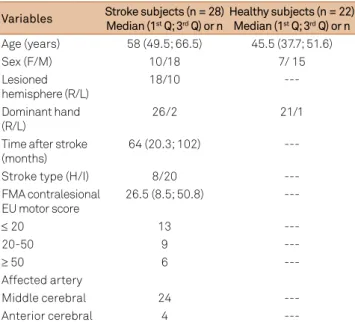
Table 1 shows the demographics data and measurement scores of the stroke group, and Table 2 summarizes the
mea-surement scores. Table 3 shows the scores of monoilaments, and Table 4 summarizes the FMA sensory score and the sum of NSA scores for each stroke patient.
All participants in the control group showed a maxi-mum score (7) in the dermatomes C6, C7 and C8 of both hands, a maximum score on the sensory scale and on the
FMA and NSA items in both wrists and hands. One or more
instruments detected sensory changes in the wrist and
hand contralateral to the stroke in twenty-ive (89%) stroke patients, and eighteen subjects (64%) had sensory deicits in
the ipsilesional wrist and hand.
According to the sum of the scores for esthesiometry, 60% showed ipsilesional and 85% showed contralesional
sensory loss. Approximately 10%, 14% and 14% of subjects had a score ≤ 5 on the ipsilesional dermatomes C6, C7 and
C8, respectively.
Table 1. Demographic characteristics of healthy and stroke subjects.
Variables Stroke subjects (n = 28) Median (1st Q; 3rd Q) or n
Healthy subjects (n = 22) Median (1st Q; 3rd Q) or n
Age (years) 58 (49.5; 66.5) 45.5 (37.7; 51.6)
Sex (F/M) 10/18 7/ 15
Lesioned hemisphere (R/L)
18/10
---Dominant hand (R/L)
26/2 21/1
Time after stroke (months)
64 (20.3; 102)
---Stroke type (H/I) 8/20
---FMA contralesional EU motor score
26.5 (8.5; 50.8)
---≤ 20 13
---20-50 9
---≥ 50 6
---Affected artery
Middle cerebral 24
---Anterior cerebral 4
---F: female; M: male; R: right; L: left; I: ischemic; H: hemorrhagic; Y: yes; N: no; FMA: Fugl-Meyer Assessment; UE: upper extremity; Q: quartile.
Table 2. Measurement scores of healthy and stroke subjects.
Stroke subjects (n = 28) Healthy subjects (n = 22)
Variables Contralesional UE
Median (1st Q; 3rd Q)
Ipsilesional UE Median (1st Q; 3rd Q)
Non dominant UE Median (1st Q; 3rd Q)
Dominant UE Median (1st Q; 3rd Q)
Sum of esthesiometry score 11.5 (6.25; 18) 20 (18; 21) § 21 (21; 21)* 21 (21; 21)**
Sum of NSA score 20 (20; 20) 18 (7.25; 20) § 20 (20; 20) 20 (20; 20)
FMA sensory score 6 (4; 7.75) 8 (8; 8) § 8 (8; 8)* 8 (8; 8)
Functional tests with visual guidance 2 (0; 7)
---Functional tests without visual guidance 1 (0; 4)
According to the FMA scores, 75% of the stroke patients had light touch and hand conscious proprioceptive deicits
on the contralesional wrist and 17% showed these character-istics on the ipsilesional side. With regard to the sum of the
scores on the NSA, 7% had sensory changes only in the ipsile
-sional wristand 64% had sensory changes in the wrist and
hand contralateral to the lesion.
Approximately 78% of the subjects had mild or mod
-erate motor impairment, according to the FMA score. The comparison of the scores for the sensory FMA, NSA
and esthesiometry assessments between the ipsilesional and contralesional limbs showed significant differences
(p < 0.001 for the three measuring instruments), while no
correlation between the ipsilesional and contralesional sensory deficits was found. The ipsilesional sensory def-icits showed no significant correlation with the contral-esional motor deficits. There was no correlation between
the scores for esthesiometry, the NSA and the ipsilesion
-al sensory FMA with the performance on function-al tests
(with and without visual guidance). We found a significant difference between functional tests with and without
vi-sual orientation scores (p = 0.002).
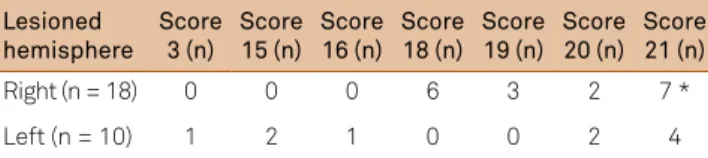
Table 5 shows the diference in esthesiometry scores (ipsilesional UE) in right and left lesions; no signiicant diferences between lesion laterality and other measuring instruments were found. he most signiicant ipsilesion -al sensory loss was observed in the 2 left-handed patients
(p = 0.05), according to NSA (one subject had right hemi -spheric damage and the other had left neurologic damage).
DISCUSSION
In the present study, the reduction of conscious pro-prioception, tactile sensation (light touch, pressure or lo-cation) or thermal sensation was found in the wrist and
hand ipsilateral to neurological injury in 64% of subjects
after a stroke.
In this study, ipsilateral sensory deicits showed no sig
-niicant correlation with contralesional motor deicits, con
-irming the indings of Brasil-Neto and Lima1. he authors
Table 3. Esthesiometry scores of stroke patients (n = 28).
Case C6 IL C6 CL C7 IL C7 CL C8 IL C8 CL Total Score IL
Total Score CL
Case 1 7 3 7 4 6 4 20 11
Case 2 6 4 7 4 7 4 20 12
Case 3 6 4 6 4 6 4 18 12
Case 4 6 1 6 2 6 2 18 5
Case 5 7 7 6 7 7 6 20 20
Case 6 6 5 6 2 6 4 18 11
Case 7 7 6 7 6 7 6 21 18
Case 8 6 6 6 6 7 6 19 18
Case 9 7 2 5 3 4 5 16 10
Case 10 5 3 5 2 5 2 15 7
Case 11 7 1 7 1 6 1 20 3
Case 12 7 1 7 1 7 1 21 3
Case 13 6 4 6 3 6 1 18 8
Case 14 7 7 7 7 7 7 21 21
Case 15 6 6 6 5 6 6 18 17
Case 16 7 7 7 7 7 7 21 21
Case 17 7 1 6 1 6 1 19 3
Case 18 7 5 7 5 7 5 21 15
Case 19 6 1 6 1 6 1 18 3
Case 20 7 3 7 3 7 3 21 9
Case 21 7 7 7 7 7 7 21 21
Case 22 7 5 7 6 7 7 21 18
Case 23 7 6 7 6 7 6 21 18
Case 24 5 5 5 5 5 5 15 15
Case 25 7 7 7 7 7 7 21 21
Case 26 7 1 7 4 7 1 21 6
Case 27 7 1 6 1 6 1 19 3
Case 28 1 2 1 3 1 3 3 8
IL: ipsilesional upper extremity; CL: contralesional upper extremity. The scores ranged from 7 (green monoilament) to 1 (magenta red monoilament).
Table 4. FMA sensory scores and sum of NSA scores in stroke patients (n = 28).
Case
FMA sensory score Ipsilesional UE
FMA sensory score Contralesional
UE
ASN Score Ipsilesional
UE
ASN score Contralesional
UE
Case 1 8 8 20 20
Case 2 4 4 20 6
Case 3 7 6 20 19
Case 4 8 4 20 7
Case 5 8 7 20 20
Case 6 8 7 20 8
Case 7 8 8 20 19
Case 8 8 8 20 20
Case 9 6 6 20 8
Case 10 8 7 20 11
Case 11 8 2 20 4
Case 12 8 2 20 3
Case 13 8 2 20 10
Case 14 8 6 20 20
Case 15 8 8 20 20
Case 16 8 7 20 20
Case 17 8 2 20 5
Case 18 8 4 20 20
Case 19 8 0 20 0
Case 20 8 4 20 6
Case 21 8 8 20 20
Case 22 8 0 20 18
Case 23 8 6 20 20
Case 24 7 6 16 11
Case 25 8 8 20 20
Case 26 6 7 20 16
Case 27 8 4 18 18
Case 28 8 8 20 19
analyzed a sample of 25 chronic post-stroke patients and observed a reduction in tactile sensation of the
ipsilesion-al hand through the Moving Touch-Pressure test in com
-parison with normal subjects. However, Son et al.2 found
a positive correlation between the sense of position of the
metacarpophalangeal joint ipsilesional with contralesional
tracking task.
No correlation between ipsilesional and contralesional sensory deicits was found in contrast to the Essing et al.7
study. Essing et al.7 evaluated 30 subjects post-stroke and
found 30% of them with bilateral tactile hypoesthesia. he
authors noted that there was a correlation between the de-gree of sensory loss in the ipsilesional and contralesional limb, so that individuals with tactile anesthesia in the hand contralateral to the stroke were more likely to exhibit
ipsilat-eral sensory deicits.
The finding of reduced conscious proprioception in the ipsilesional upper limb replicates the results found by
Sartor-Glittenberg and Powers21 andSon et al.2. Son et al.2
investigated joint position sense in 50 stroke subjects. The
tests were conducted in the hand ipsilateral to the dam-aged hemisphere. Higher error scores were found in the
joint reposition test in the stroke group. The authors sug -gest that it is possible that the transcallosal transfer dis-turbance after unilateral brain damage may lead to ipsi-lateral sensory deficits.
Dannenbaum et al.22 indicated that the tactile
senso-ry evaluation using the Semmes-Weinstein test is not re-lated to normal function. However, we found a maximum
score (normal function) in all healthy subjects and sensory changes in the upper limb in post-stroke subjects. In this
study, over 10% of the post-stroke sample exhibited a score less than or equal to 5 for esthesiometry in the ipsilesional hand, which indicates a reduced protective sensation,
dif-iculty with the discrimination of shape and temperature,
vulnerability to skin lesions and, in some cases, loss of hot/cold discrimination.
In the present study, it was found that the subjects
with brain damage in the right hemisphere exhibited
bet-ter scores in ipsilesional tactile sensation (veriied by es
-thesiometry), conirming the indings of Vaughn and
Costa8. Boll11 found worse sensory function in patients
with brain damage in the right hemisphere. According
to Desrosiers et al.23, subjects with lesions in the left
hemisphere have a double disadvantage: in addition to
higher contralesional UE deicits, they are obliged to use their non-dominant UE (ipsilesional).
here are justiications for the existence of ipsilesional
sensory disturbances. Sherwood24 noted that there are few
aferent neuronal ibers following the ipsilateral cerebral
cortex. Another plausible explanation is the existence of a bilateral representation of the body in the secondary so-matosensory area, in contrast to the representation of the
contralateral primary somatosensory area25.
A third justification for the existence of ipsilesional
sensory loss is the activation of both cerebral hemispheres during unimanual motor tasks, considering the contribu-tion of interhemispheric interaccontribu-tions with excitatory or inhibitory effects of a cerebral hemisphere on the
oppo-site hemisphere26. In turn, the posterior parietal cortex is
connected with the frontal motor areas and the primary somatosensory area ensuring a close relationship between
sensory and motor function27. Moreover, it is speculated
that there can be problems with the corpus callosum after a stroke, leading to issues with neural information transfer
across hemispheres1.
he indings of this study reinforce the need, as revealed
by other researchers, to not consider the ipsilesional
up-per extremity as “unafected” or “normal” 1,7,28. Kitsos et al.28
conducted a review of the scientiic literature on the sensory-motor deicits in ipsilesional UE after stroke and recommended the use of the terms “most afected” for the contralesional upper limb and “less afected” for the ipsile -sional upper limb.
In conclusion, reduction of conscious proprioception, tactile sensation or thermal sensation was found in the wrist
and hand ipsilateral to neurological injury in 64% of subjects after stroke. he esthesiometry through the monoilament
was a more sensitive tool for detection of sensory disturbanc-es in the ipsildisturbanc-esional wrist and hand.
We found that the subjects with brain damage in the right
hemisphere exhibited better scores on the ipsilesional tactile
sensation assessment (veriied by esthesiometry). he most signiicant ipsilesional sensory loss was observed in the 2
left-handed patients.
Comparisons of scores of the sensory FMA scale, the NSA and the esthesiometry between ipsilesional and con
-tralesional extremities showed signiicant diferences, and
no correlation between ipsilesional and contralesional
sensory deicits was found. Ipsilesional sensory deicits showed no signiicant correlation with contralesional motor deicits.
here was no correlation between the scores of ipsile
-sional esthesiometry, NSA and sensory FMA scale with the performance on functional tests. We found signiicant dif -ferences between functional tests with and without visual orientation scores.
Table 5. Lesioned hemisphere and esthesiometry score of ipsilesional upper extremity.
Lesioned hemisphere
Score 3 (n)
Score 15 (n)
Score 16 (n)
Score 18 (n)
Score 19 (n)
Score 20 (n)
Score 21 (n)
Right (n = 18) 0 0 0 6 3 2 7 *
Left (n = 10) 1 2 1 0 0 2 4
References
1. Brasil-Neto JP, Lima AC. Sensory deicits in the unaffected hand of hemiparetic stroke patients. Cogn Behav Neurol. 2008;21(4):202-5. doi:10.1097/WNN.0b013e3181864a24
2. Son SM, Kwon YH, Lee NK, Nam SH, Kim K. Deicits of movement accuracy and proprioceptive sense in the ipsi-lesional upper limb of patients with hemiparetic stroke. J Phys Ther Sci. 2013;25(5):567-9. doi:10.1589/jpts.25.567
3. Kwon YH, Kim CS, Jang SH. Ipsi-lesional motor deicits in hemiparetic patients with stroke. NeuroRehabilitation. 2007;22(4):279-86.
4. Schaefer SY, Haaland KY, Sainburg RL. Ipsilesional motor deicits following stroke relect hemispheric specializations for movement control. Brain. 2007;130(8):2146-58. doi:10.1093/brain/awm145
5. Hermsdörfer J, Goldenberg G. Ipsilesional deicits during fast diadochokinetic hand movements following unilateral brain damage. Neuropsychologia, 2002;40(12):2100-15. doi:10.1016/S0028-3932(02)00048-9
6. Nowak DA, Grefkes C, Dafotakis M, Küst J, Karbe H, Fink GR. Dexterity is impaired at both hands following unilateral subcortical middle cerebral artery stroke. Eur J Neurosci. 2007;25(10):3173-84. doi:10.1111/j.1460-9568.2007.05551.x
7. Essing JP, Gersten JW, Yarnell P. Light touch thresholds in normal persons and cerebral vascular disease patient: bilateral deicit after unilateral lesion. Stroke. 1980;11(5):528-33. doi:10.1161/01.STR.11.5.528
8. Vaughan HG, Costa LD: Performance of patients with lateralized cerebral lesions II: Sensory and motor tests. J Nerv Ment Dis. 1962;134(3):237-43. doi:10.1097/00005053-196203000-00004
9. Corkin S, Milner B, Taylor L. Bilateral sensory loss after unilateral cerebral lesion in man. Trans Am Neurol Assoc. 1973;98:118-22.
10. Carmon A. Disturbances of tactile sensitivity in patients with unilateral cerebral lesions. Cortex. 1971;7(1):83-97. doi:10.1016/S0010-9452(71)80024-2
11. Boll TJ. Right and left cerebral hemisphere damage and tactile perception: performance of the ipsilateral and contralateral sides of the body. Neuropsychologia. 1974;12(2):235-8. doi:10.1016/0028-3932(74)90008-6
12. Gainotti G, Tiacci C. [Homolateral and contralateral disturbances in the tactile discrimination of the hemispheric lesions]. Rev Neurol. 1975;45(3):339-52. Italian.
13. Baskett JJ, Marshall HJ, Broad JB, Owen PH, Green G. The good side after stroke: ipsilateral sensory-motor function needs careful assessment. Age Ageing. 1996;25(3):239-44. doi:10.1093/ageing/25.3.239
14. Kim JS, Choi-Kwon S. Discriminative sensory dysfunction after unilateral stroke. Stroke. 1996;27(4):677-82. doi:10.1161/01.STR.27.4.677
15. Carey LM, Matyas TA. Frequency of discriminative sensory loss in the hand after stroke in a rehabilitation setting. J Rehabil Med. 2011;43(3):257-63. doi:10.2340/16501977-0662
16. Moreira D, Alvarez R. Utilização dos monoilamentos de Semmes-Weinstein na avaliação de sensibilidade dos membros superiores de pacientes hansenianos atendidos no Distrito Federal. Hansenol Int. 1999;24(2):121-8.
17. Maki T, Quagliato EMAB, Cacho EWA, Paz LPS, Nascimento NH, Inoue MMEA, Viana MA. Estudo de coniabilidade da aplicação da escala de Fugl-Meyer no Brasil. Rev Bras Fisiot. 2006;10(2):177-83. doi:10.1590/S1413-35552006000200007
18. Michaelsen SM, Dannenbaum R, Levin MF. Task-speciic training with trunk restraint on arm recovery in stroke: randomized control trial. Stroke. 2006;37(1):186-92. doi:10.1161/01.STR.0000196940.20446.c9
19. Lima DHF, Queiroz AP, Salvo G, Yoneyama SM, Oberg TD, Lima NMFV. Versão brasileira da Avaliação Sensorial de Nottingham: validade, concordância e coniabilidade. Rev Bras Fisioter. 2010;14(2):166-74. doi:10.1590/S1413-35552010005000006
20. Smania N., Montagnana B., Faccioli S., Fiaschi A., Aglioti SM. Rehabilitation of somatic sensation and related deicit of motor control in patients with pure sensory stroke. Arch Phys Med Rehabil. 2003;84(11):1692-702. doi:10.1053/S0003-9993(03)00277-6
21. Sartor-Glittenberg C, Powers R. Quantitative measurement of kinesthesia following cerebral vascular accident. Physiother Canada. 1993;45:179-86.
22. Dannenbaum RM, Michaelsen SM, Desrosiers J, Levin MF. Development and validation of two new sensory tests of the hand for patients with stroke. Clin Rehabil. 2002;16(6):630-9. doi:10.1191/0269215502cr532oa
23. Desrosiers J, Bourbonnais D, Bravo G, Roy PM, Guay M. Performance of the “unaffected” upper extremity of elderly stroke patients. Stroke. 1996;27(9):1564-70. doi:10.1161/01.STR.27.9.1564
24. Sherwood L. Human physiology: from cells to systems. Belmont Wadsworth; Company, 1997.
25. Whitsel BL, Petrucelli LM, Werner G. Symmetry and connectivity in the map of the body surface in somatosensory area II of primates. J Neurophysiol. 1969;32(2):170-83.
26. Murase N, Duque J, Mazzocchio R, Cohen LG. Inluence of interhemispheric interactions on motor function in chronic stroke. Ann Neurol. 2004;55(3):400-9. doi:10.1002/ana.10848
27. Gardner EP, Martin JH, Jesell TM. As sensações corporais. In: Kandel ER, Schwartz JH, Jessel TM. Princípios da neurociência. Barueri: Manole; 2003; 430-450p.