Artigo Original
Jaqueline Medeiros de Mello1
Valter Augusto Della-Rosa2
Renata Mota Mamede Carvallo3
Keywords
Hearing tests Hearing Genetics Mutation
Descritores
Testes auditivos Audição Genética Mutação
Correspondence address: Jaqueline Medeiros de Mello
Rua Vaz Caminha, 633, Zona 2, Maringá (PR), Brasil, CEP: 87010-420. E-mail: [email protected] Received: 01/09/2013
Study carried out at the Laboratory of Speech Therapy and Human Hearing, Department of Physical Therapy, Speech and Occupational Therapy, Medical School of Universidade de São Paulo – USP – São Paulo (SP), Brazil. (1) Faculdade Ingá – UNINGÁ – Maringá (PR), Brazil.
(2) Programa de Pós-graduação (Doutorado) em Ciências Biológicas e Genética, Universidade Estadual de Maringá – UEM – Maringá (PR), Brazil.
(3) Faculdade de Medicina, Universidade de São Paulo – USP – São Paulo (SP), Brazil. Conlict of interests: nothing to declare.
ultra-high frequencies in parents of individuals with
autosomal recessive hearing loss
Emissões otoacústicas evocadas por produto de distorção
em frequências ultra-altas em pais de indivíduos com
deiciência auditiva autossômica recessiva
ABSTRACT
Purpose: To evaluate the cochlear function of parents of individuals with autosomal recessive gene Gap Junction Protein Beta-2 hearing loss by ultra-high frequencies distortion-product otoacoustic emissions (DPOAEs), compared with responses of a control group matched for age and gender. Methods: We studied 56 subjects aged from 20 to 58 years, divided into two groups. The study group comprised 28 parents of hearing-impaired patients due to autosomal recessive inheritance, 14 females aged 20.0–55.0 years (mean 32.8 years) and 14 males aged 20.0–58.0 years (mean 35.2 years). Control group was composed of normal hearing individuals, 14 males and 14 females age-matched to the study group. The subjects underwent tests for audiometry, tympanometry, and DPOAE in the frequency range of 9.000–16.000 Hz. Results: We found 64.3% of normal results of DPOAE in the study group compared to 91.1% in the control. There were signiicant differences between groups in the ears and DPOAE responses, and the mean level of response was in 10 dBNPS in study group and 14 dBNPS in the control. The Pearson’s correlation between age and DPOAE in ultra-high frequencies showed no statistical signiicance. Conclusion: DPOAE at ultra-high frequencies were able to identify individuals from both groups, suggesting that heterozygous individuals for the Gap Junction Protein Beta-2 gene mutation may have damage to the cochlear function before clinical manifestation in audiometry.
RESUMO
INTRODUCTION
Hearing impairment may be caused by environmental and genetic factors. Among genetic-based cases, 70% may manifest only hearing loss (nonsyndromic hearing loss) and 30% may be associated with other anomalies (syndromic hear-ing loss)(1,2). Autosomal recessive inheritance is predominant among nonsyndromic cases, with rates of 75–80%, followed by autosomal dominant (15%), X-linked (3%), mitochondrial, or maternal inheritance(2,3). Among mutations with autosomal recessive transmission leading to hearing loss, those related to the Gap Junction Beta-2 Protein gene (GJB2) are prominent, which is responsible for the synthesis of Connexin 26, espe-cially 35delG, characterized by a guanine deletion among six at the position 30–35 from the nucleotide 1, encoder region in exon 2 of the gene GJB2, 35delG(3).
Connexins are proteins that act in communication between cells named gap junctions. In the cochlea, they play an impor-tant role in the physiology related to ionic homeostasis, lead-ing to a change in structure and function of gap junctions that promote the maintenance of high potassium concentrations, which may cause cell intoxication(3,4).
The phenotype–genotype relationship has been investigat-ed in patients with autosomal recessive inheritance of hearing loss, usually sensorineural, nonprogressive, severe, and pro-found, with onset in the pre-linguistic phase of language(5-7). There are a few studies(8-10) about the phenotype–genotype relationship in heterozygotic individuals, that is, listeners with one normal and one mutated allele who usually become parents of deaf patients presenting autosomal recessive hear-ing loss and passhear-ing their genes to populations. Therefore, studies to identify even minimal hearing disorders in het-erozygotic parents are justiied because there is an attempt to associate such changes with genotypes. The detection of minimal changes in hearing function by conventional audiologic evaluation, including tone hearing threshold and immittance testing, has not been effective(6,7). By contrast, otoacoustic emissions testing is an effective method to early identify discrete changes in the hearing function of parents of autosomal recessive hearing-impaired subjects(8-10).
After the emergence of ultra-high frequencies distortion-product otoacoustic emissions (DPOAEs) in the frequen-cy range of 9,000–16,000 Hz, hearing changes preceding hearing loss in conventional frequencies could be identiied in early stages, before the cochlear damage reaches low frequencies(11). The investigation of cochlear function at high frequencies in parents of autosomal recessive hearing-impaired patients is a means of early diagnosis of hearing disorders, besides being potentially useful in identifying patients at risk of presenting mutated alleles in genes related to hearing loss.
This study is therefore aimed at evaluating the cochlear function in heterozygotic parents of deaf patients presenting GJB2 mutation by means of DPOAE at the frequencies of 9,000–16,000 Hz and compare results with a control group matched in gender and age.
METHODS
This study was carried out at the Laboratory of Speech Language Therapy and Human Hearing of the Department of Physical, Speech and Occupational Therapy from Universidade de São Paulo, and approved by the Research Committee (CAPPesq), protocol 170/10.
The sample was composed of 56 individuals aged be-tween 20 and 58 years, and divided into two groups. The study group (SG) had 28 parents of autosomal recessive hearing-impaired patients, 14 females aged 20–55 years old (mean 32.8 years) and 14 males aged 20–58 years old (mean 35.2 years). Control group (CG) had subjects without hearing disorders or complaints: 14 males and 14 females with ages matching SG.
The inclusion criteria for both groups were: SG help mother and fathers carrying recessive mutated alleles of the gene GJB2, including normal and mutated alleles in 35delG identiied by molecular investigation. Couples without hearing complaints and presenting at least one autosomal recessive hearing-impaired child were also included.
CG had 28 subjects without hearing complaints, all of them being matched in age and gender with SG to avoid bias in result analysis. The inclusion criteria for CG was absence of factors related to congenital or acquired hearing loss, history of exposure to high-intensity noise, alcohol and drug consumption and ototoxicity, as well as past history of changes in the middle ear.
Because of these inclusion criteria, molecular analysis was not performed in CG to avoid extra costs. Moreover, blood collection is considered an invasive procedure to use in a population without hearing complaints. Patients with integ-rity of the middle ear evidenced by type A tympanogram(12), contralateral acoustic relexes at 500–4,000 Hz, and normal hearing thresholds (≤25 dBNA) were included in the sample. All patients were submitted to anamnesis, edge of auditory meatus assessment, tone threshold audiometry, immitanci-ometry, and ultra-high frequency DPOAE after signing the informed consent form.
DPOAE was performed with the patients in an acoustic enclosure wearing an olive adapted to the probe of the equip-ment for otoacoustic emissions DP2000 (Distortion Product Otoacustic Emission Measurement System Starkey) to the outer auditory meatus.
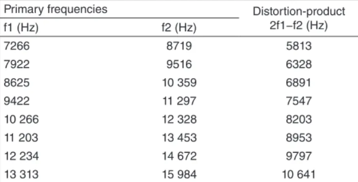
To register DPOAE, we followed the patterns established by the manufacturer, as proposed by Gorga et al.(13), such as intensity of different inputs (L1>L2), that is, L1=65 dBNPS and L2=55 dBNPS, and presentation of two primary tones f1 and f2 (f1<f2), with relation f2/f1=1,2 in two octaves. Responses for DPOAE were generated in the region 2f1−f2 of the bands of ultra-high frequencies from 9,000 to 16,000 Hz, as shown in Chart 1.
deviation of the noise. The background noise was always equal or inferior to -5 dBNPS for each frequency, and the DPOAE amplitude was always ≥0 dB.
The duration of the sound emitted was 2 seconds so it could be received in the outer auditory meatus, only one automatic fre-quency scanning being performed. However, when the response
achieved. This typical pattern with asset decline curve start-ing from 10,000 Hz can be seen in Figure 1.
RESULTS
The i ndings of tone threshold audiometry showed that mean auditory thresholds at 250–8,000 Hz were 10 dB in SG and 8 dB in CG, without statistically signii cant differences between ears and groups.
Results show the qualitative analyses of DPOAE responses generated for the region 2f1−f2 at ultra-high frequencies. The distributions of normal and altered results for DPOAE, as well as the Z test for proportions, are shown in Table 1. The quantitative analysis of DPOAE at ultra-high frequencies by ear is presented in Figure 2, and box-plots by group are shown in Figure 3.
Inferential analysis of DPOAE was made by 5% and 95% percentile to dei ne a 90% reference interval for DPOAE at each frequency in order to verify the mean variation of a coni dence interval(14). The correlation between ultra-high frequency in SG and reference thresholds for DPOAE estimated in CG are presented in Figure 4.
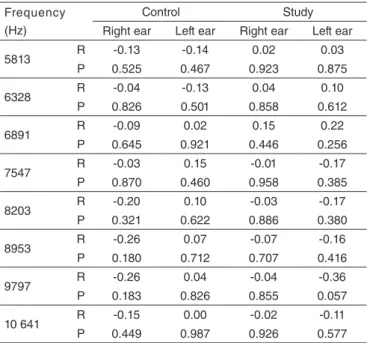
To measure the correlation between age and DPOAE at each frequency, Pearson’s correlation values were calculated for each group and ear. Values are shown in Table 2.
DISCUSSION
The audiological proi le of 28 parents carrying heterozy-gosis of the mutated allele GJB2, which causes hearing loss,
Chart 1. Primary frequencies (f1 and f2) of stimulus and response of distortion-product in 2f1−f2 for ultra-high frequency distortion-product otoacoustic emissions
Primary frequencies Distortion-product
2f1−f2 (Hz)
f1 (Hz) f2 (Hz)
7266 8719 5813
7922 9516 6328
8625 10 359 6891
9422 11 297 7547
10 266 12 328 8203
11 203 13 453 8953
12 234 14 672 9797
13 313 15 984 10 641
to a frequency was absent or decreased, it would be individually retested in the same register at least three times for coni rmation. Tests presenting absence of response would be interrupted when noises exceeded -5 dBNPS and level of response were lower than 0 dB. This procedure was chosen to prevent real responses from being jeopardized by physiological movements or noises.
Before DPOAE, an automatic calibration of the probe was performed in situ, after selecting an olive in an adequate size to insert in the outer auditory meatus. Afterwards, the test would be initiated. Two curves with signii cant superpo-sition in visual analysis were reported in order to verify test representation in the frequency band of 9,000–16,000 Hz, as shown in Figure 1. Probe calibration for frequencies 9,000– 16,000 Hz requires time and caution, sometimes needed to reinsert it to the outer auditory meatus many times, until a pattern of calibration demanded by the examination is
Table 1. Distribution of normal and changed results in ultra-high fre-quency distortion-product otoacoustic emissions between groups
Otoacoustic emissions Study group Control group Z test
n % n % p-value
Changed Ultra-high
Frequency 20 35.7 5 8.9 0.255
Normal Ultra-high
Frequency 36 64.3 51 91.1 0.002*
*Statistically significant result
Figure 1. Probe calibration in situ before registration of distortion-product otoacoustic emissions at 9,000–16,000 Hz
110 Left Ear
-2 dB SPL
/Volt
Transducer Sensitivity 100
90 80 70 60 50
-1 -5 1 2 5 10 20
Frequency kHz
Ch 1
Ch 2
110 Right Ear
-2 dB SPL
/Volt
Transducer Sensitivity 100
90 80 70 60 50
-1 -5 1 2 5 10 20
Frequency kHz
Ch 1
Ch 2 DPOAE In-The-Ear Calibration
was investigated in this study. Although these subjects had no hearing complaints, we raised the hypothesis that they could present discrete changes in hearing function, showed by decrease in response of DPOAE at ultra-high frequencies compared to asymptomatic parents without risk factors for hearing loss, once the cochlear outer hair cells go through subtle changes.
The analysis showed a statistically signiicant difference between responses for otoacoustic emissions, as the SG presented a lower level of response compared to the CG. We can, thus, attest that ultra-high frequency DPOAEs were able to distinguish subjects from SG and CG, suggesting that the changes in the outer hair cells resulting from the gap junctions caused by the mutation in GJB2 may be noted at otoacoustic emissions in heterozygotic parents carrying this mutation. The investigation of hearing patterns in parents of hear-ing-impaired patients presenting GJB2 mutation is useful to identify discrete hearing disorders. This characterization may indicate future members of the family who may carry the recessive genes causing hearing loss, especially in cases where molecular tests are not yet available(8,15).
In DPOAE, SG presented fewer normal occurrences com-pared to CG (Table 1). Although conventional audiological tests were similar, there were differences in DPOAE between heterozygotic subjects presenting GJB2 mutation and the others, which show that the cochlear outer hair cells are more susceptible to the negative effects of this mutation.
The fact that parents of autosomal recessive hearing-im-paired patients carry the recessive allele causing the mutation of GJB2 may indicate changes in ionic homeostasis and in the endocochlear processes, which in turn leads to changes in the structure and functioning of gap junctions, with conse-quent maintenance of high potassium (K+) concentrations and intoxication of the spiral organ(3,4).
Table 2. Pearson’s correlation values as to age and distortion-product otoacoustic emissions at ultra-high frequencies by group and ear
Frequency (Hz)
Control Study
Right ear Left ear Right ear Left ear
5813 R -0.13 -0.14 0.02 0.03
P 0.525 0.467 0.923 0.875
6328 R -0.04 -0.13 0.04 0.10
P 0.826 0.501 0.858 0.612
6891 R -0.09 0.02 0.15 0.22
P 0.645 0.921 0.446 0.256
7547 R -0.03 0.15 -0.01 -0.17
P 0.870 0.460 0.958 0.385
8203 R -0.20 0.10 -0.03 -0.17
P 0.321 0.622 0.886 0.380
8953 R -0.26 0.07 -0.07 -0.16
P 0.180 0.712 0.707 0.416
9797 R -0.26 0.04 -0.04 -0.36
P 0.183 0.826 0.855 0.057
10 641 R -0.15 0.00 -0.02 -0.11
P 0.449 0.987 0.926 0.577
Caption: R = Pearson’s correlation; P = statistically significant result
Figure 4. Dispersions of otoacoustic emissions related to frequencies in the study group and reference thresholds for distortion-product otoacoustic emissions estimated for control group at ultra-high frequencies
Percentil 5%
30
25
20
15
10
5
0
Frequency (Hz)
DPO
AE (dBNPS)
5,813 6,328 6,891 7,547 8,203 8,953 9,797 10,641
Percentil 95% Study
Caption: DPOAE = distortion-product otoacoustic emissions
Figure 2. Box-plots for distortion-product otoacoustic emissions (dBNPS) at each frequency by group and ear
Rigth Ear Left Ear Group
30
25
20
15
10
5
0
*
D
PO
A
E
(dBNPS)
Control Study
*
*
* ** * *
Frequência (Hz)
5,
81
3
6,
328
6,
891 7,547
8,
203
8,
953
9,
797
10
,641 5,813 6,328
6,
891 7,547
8,
203
8,
953
9,
797
1,
0541
Caption: DPOAE = distortion-product otoacoustic emissions
Figure 3. Box-plots and median profiles for product-distortion otoacoustic emissions (dBNPS) by groups at ultra-high frequencies
30
25
20
15
10
5
0
Frequency (Hz) *
DPO
AE (dBNPS)
5,813 6,328 6,891 7,547 8,203 8,953 9,797 10,641
*
** ** *
* *
*
Group Control Study
As to the laterality of DPOAE, when we compared results of ears between subjects from a same group, we found no statistically signiicant difference. Although results were not signiicant, we saw an advantage of the right ear (RE) over the left ear as to mean values (Figures 2 and 3).
Most studies report better responses for otoacoustic emis-sions in RE(16,17). Some explanations to these indings are related to differences in ears associated with the slight advantage of right aural awareness and the dominance of the left hemisphere in the perception of speech and language function, besides the effect of higher suppression of otoacoustic emissions in the RE, which proves that there is an asymmetric activity between ears and favors the acoustic signal detection and morphological asymmetry between the right and left craniofacial regions(16,17). However, some studies have not shown signiicant differences in responses to otoacoustic emissions as to ear side(18).
Because there are no statistically signiicant differences between ear sides in a same group, we performed a comparative analysis of results between SG and CG, grouping the values ob-tained to identify the signiicant values in all frequencies in the comparison between groups, with advantage to CG (Figures 2 and 3). Although both groups had no hearing complaints and presented similar audiological proiles, heterozygotic subjects for GJB2 mutation were reported to present more damage to the active process of outer hair cells.
A lower level of response to ultra-high frequencies DPOAE was expected due to the cochlear tone organization, for the ultra-high frequencies are set at the basis of the cochlea and are therefore more prone to damage, hence the possibility to detect them early before the onset of typical effects in the conventional frequency range(19).
Another factor that may have contributed with the lower level of responses was the dificulty to calibrate the probe before the registration of the otoacoustic emissions (Figure 1) because of the short wavelength of the highest frequencies and of the com-plex interactions between them and the outer auditory meatus dimensions(20). In the future, the problem of probe calibration will be solved with the registration of DPOAE using two micro-phones, one being the reference microphone inserted near the tympanic membrane to actually calibrate the inputs and another one used to capture the responses by the outer hair cells(21).
Studies about DPOAE using ultra-high emissions also showed problems with calibration in this frequency range and pointed out that the great variability in responses may be inlu-enced by it(11,22-26). Dreisbach et al.(23) reported that this may be minimized by the exact calibration, using a microphone in the outer auditory meatus correctly, resetting the frequency, and repositioning the probe to conirm if the response is exclusively due to the cochlear activity.
To avoid problems in calibration due to stationary waves, especially in ultra-high frequencies, Dreisbach and Siegel(22) created a speciic probe for their study containing three tubes. The irst one would send the input; the second one was the microphone, and the third one was an optic iber to visualize the position of the probe in relation to the tympanic membrane (roughly 10–15 mm) to assure the same level of acoustic sound pressure during the assessment at all frequencies.
The leading problems found in measuring ultra-high oto-acoustic emissions were calibration limitations(25,26). Because of that, the authors created a technique named incident pressure calibration to prevent the effect of the stationary waves at high frequencies by using an 8-mm cylindrical tube inserted to the phone, which would function as an anechoic chamber.
Trying to establish if the inputs at ultra-high frequencies could distinguish individuals from the SG and CG, we used a 90% conidence interval test. Figure 4 shows that many indi-viduals from SG are below the lower reference threshold es-timated for CG, while other are above the highest threshold. The hypothesis raised was that parents of autosomal recessive hearing-impaired patients would present more impairment of the cochlear basis and, therefore, ultra-high frequencies DPOAE would have a due pattern in these individuals. However, as this tool has been only made available very recently, there are very few studies assessing the frequency range of 9,000–16,000 Hz as to DPOAE and to hearing perception at these frequencies.
Some dificulties lead to the lack of studies on the subject. The irst one is that most speech sounds are in the frequency range of up to 8,000 Hz, and ultra-high frequencies related to the decodiication of background sounds and music are not usually clinically concerning, once the speech perception is more relevant for human communication(25). The recent avail-ability of equipment in the market is another reason, as well as the lack of standardization of DPOAE responses in the range of 9,000–16,000 Hz.
When there is equipment available, there is also dificulty in the correct and safe calibration of probes. There are a few studies on ultra-high frequencies DPOAE precisely because the calibration in this frequency range is very dificult, which makes researchers quit studies before completing them(21).
However, the recent advances in technology and in the knowledge about audiology and acoustic physics relations are expected to improve and solve the issue of calibration pat-terns for ultra-high frequencies because of their applicability, for example, when monitoring patients suspected for or with hearing pathologies such as ototoxicity, otitis media sequela, presbycusis, noise exposure, and renal failure(27,28).
Vallejo et al.(29) added that one of the clinical applications of ultra-high frequency research is hearing impairment in families with members presenting genetic hearing loss, as in the case of asymptomatic heterozygotic parents with GJB2 mutation.
In this context, the investigation of ultra-high frequencies in parents of autosomal recessive hearing-impaired children is a tool to identify early discrete hearing changes.
In agreement with other studies, we found that DPOAE tend to decrease with aging, probably due to changes in cochlear biomechanics or to the loss and deterioration of Outer Hair Cells (OHC) that occur throughout life, thus increasing hearing thresholds at the level of frequency(30,31).
55–57 years, probably because of the hearing impairment re-sulting from aging (presbycusis), where there are changes in the cochlea such as loss of sensory cells, atrophy of the stria vascularis, and loss of cells of spiral ganglion(30).
Both groups would then have the same chances to present hearing disorders due to the age factor. However, the greatest incidence among subjects from SG may be explained by the recessive allele in heterozygosis, which causes hearing loss in autosomal inheritance.
Hearing impairment at ultra-high frequencies starts in childhood and is accompanied by a decrease in the response to transient and DPOAEs, as well as the absence of spontane-ous otoacspontane-oustic emissions. So, the evaluation of ultra-high frequencies may help to detect hearing loss early, for it may not be identiied by conventional audiological assessment(31). Loss of hearing perception in the range of 15,000–18,000 Hz starts around 20 years old. Hearing loss is usually a result of changes related to metabolism age, cochlear nerve degeneration, and mechanical changes in the spiral organ, with adverse effects to the peripheral and central hearing function(32).
Scatterplots correlating age and DPOAE at ultra-high frequencies in both groups showed no trend in score, and Pearson’s correlation values for each group and ear did not show signiicant negative correlation in any group (Table 2).
The little knowledge about hearing perception at ultra-high frequencies associated with the no correlation to age and DPOAE at ultra-high frequencies may be explained by the small sample size, so further studies with bigger samples are needed to assure accuracy to audiological data.
Studies on autosomal recessive hearing loss are usually conducted with small samples because it is relatively dificult to gather a large number of individuals presenting these char-acteristics in Brazil, where it is not easy to distinguish genetic from environmental hearing loss because molecular tests are mostly inaccessible. Even if it were easier to identify autoso-mal recessive hearing loss etiology, this number would still be reduced, because this type of hearing disorder affects 15% of the deaf population(3,4). Engel-Yeger et al.(9) pointed out that very few studies tried to determine the clinical picture of autosomal recessive hearing loss, because only a few families/patients are actually examined.
When it comes to the dificulty in gathering subjects for the sample, we mention refusal of patients in performing audiologi-cal tests, especially because they are commonly “harassed” to participate in hearing loss studies, besides claiming the exams would not solve their problem, and that they were already sub-mitted to such tests periodically in order to update documents such as driver license and public transportation insurance.
It is worth mentioning the dificulty in inding couples presenting the requirements for the study if we consider the number of divorces that take place currently. The family was often composed of a mother and a stepfather, and vice-versa. When the researchers oriented potential participants as to mo-lecular analysis, which is a synonym with DNA analysis for the layman, many mothers refused to participate in the study facing the possibility of paternity suspicion by their partners.
To verify the possibility of decrease in response of cochlear function of heterozygotic parents for GJB2 mutation, we ad-opted an eletroacoustic methodology capable of conirming the initial hypothesis, even though further studies are needed, once technological advances allow more accuracy in the registration of ultra-high frequency DPOAE.
CONCLUSION
Ultra-high frequency DPOAEs were able to distinguish indi-viduals from the SG and the CG, suggesting that heterozygotic in-dividuals for the GJB2 mutation may present damage to the active process of outer stereocilia before the clinical onset of the disorder based on the results of conventional audiological evaluation.
*JMM was responsible for data collection and tabulation, and writing; VADR monitored data collection and helped in the analysis; RMMC monitored data collection and inal discussion.
REFERENCES
1. Cohen MM, Gorlin RJ. Epidemiology, etiology and genetic patterns. In: Gorlin RJ, Toriello HV, Cohen MM (eds.). Hereditary hearing loss and its syndromes. Oxford: Oxford University Press; 1995. p.9-21. 2. Alves FRA, Ribeiro FAQ. Roteiro diagnóstico e de conduta frente à
perda auditiva sensorioneural genética. Rev Bras Otorrinolaringol. 2007;73(3):412-7.
3. Lefebvre PP, Van De Water TR. Connexins, hearing and deafness: clinical aspects of mutations in the connexin 26 gene. Brain Res Brain Res Rev. 2000;32(1):159-62.
4. Shibata Y, Kumai M, Nishii K, Nakamura K. Diversity and molecular anatomy of gap junctions. Med Electron Microsc. 2001;34(3):153-9. 5. Bitner-Glindzicz M. Hereditary deafness and phenotyping in humans. Br
Med Bull. 2002;63:73-94.
6. Han J, Li F, Zhao M, Zhang Z, Ni D. Study on distortion otoacoustic emissions and expanded high frequency audiometry in noise exposure workers. Lin Chuang Er Bi Yan Hou Ke Za Zhi. 2003;17(1):16-9. 7. Carvallo RMM. Audição em altas freqüências: repercussões no
reconhecimento de fala no ruído e nas emissões otoacústicas [tese]. São Paulo: Faculdade de Medicina da Universidade de São Paulo; 2002. 8. Engel-Yeger B, Zaaroura S, Zlotogora J, Shalev S, Hujeirat Y,
Carrasquillo M, et al. The effects of a connexin 26 mutation-35delG on otoacoustic emissions and brainstem evoked potentials: homozygotes and carriers. Hear Res. 2002;163:93-100.
9. Engel-Yeger B, Zaaroura S, Zlotogora J, Shalev S, Hujeirat Y, Carrasquillo M, et al. Otoacoustic emissions and brainstem evoked potentials in compound carriers of connexin 26 mutations. Hear Res. 2003;175(1-2):140-51.
10. Franzé A, Caravelli A, Di Leva F, Marciano E, Auletta G, D’Aulos F, et al. Audiometric evaluation of carriers of the connexin 26 mutation 35delG. Eur Arch Otorhinolaryngol. 2005;262(11):921-4.
11. Dunckley KT, Dreisbach LE. Gender effects on high frequency distortion product otoacoustic emissions in humans. Ear Hear. 2004;25(6):554-64. 12. Jerger J. Clinical experience with impedance audiometry. Arch
Otolaryngol. 1970;92(4):311-24.
13. Gorga MP, Neely ST, Ohlrich B, Hoover B, Redner J, Peters J. From laboratory to clinic: a large scale study of distortion product otoacoustic emissions in ears with normal hearing and ears with hearing loss. Ear Hear. 1997;18(6):440-55.
15. Silva LS, Mingroni Netto RC, Sanches SGG, Carvallo RMM. Medidas de audição de pais de indivíduos com deiciência auditiva de herança autossômica recessiva. Pró-Fono R Atual Cient. 2010;22(4):403-8. 16. Bassetto MCA, Chiari BM, Azevedo MF. Emissões otoacústicas evocadas
transientes (EOAET): amplitude da resposta em recém-nascidos a termo e pré-termo. Rev Bras Otorrinolaringol. 2003;69(1):84-92.
17. Durante AS, Carvallo RMM, Costa FS, Soares JC. Características das emissões otoacústicas por transientes em programa de triagem auditiva neonatal. Pró-Fono. 2005;17(2):133-40.
18. Carvallo CU, Carvallo RMM. Latência das EOAPD em milissegundos e número de ondas. Rev Bras Otorrinolaringol. 2005;71(6):784-90. 19. Silva IMC, Feitosa MAG. Audiometria de alta freqüência em adultos
jovens e mais velhos quando a audiometria convencional é normal. Rev Bras Otorrinolaringol. 2006;72(5):665-72.
20. Barret AR. The contribution of ultra-high frequency hearing on distortion product otoacoustic emission amplitudes [dissertation]. Harrisonburg: James Madison University; 2007.
21. Middlesworth LV. Advanced otoacoustic emissions testing in the clinic. In: Anais do 27º Encontro Internacional de Audiologia (EIA); 2012. Bauru. 22. Dreisbach LE, Siegel JH. Level dependence of distortion-product
otoacoustic emissions measured at high frequencies in humans. J Acoust Soc Am. 2005;117(5):2980-8.
23. Dreisbach LE, Long KM, Lees SE. Repeatability of high-frequency distortion-product otoacoustic emissions in normal-hearing adults. Ear Hear. 2006;27(5):466-79.
24. Kei J, Brazel B, Crebbin K, Richards A, Willeston N. High frequency distortion product otoacoustic emissions in children with and without middle ear dysfunction. Int J Pediatr Otorhinolaryngol. 2007;71(1):125-33.
25. Goodman SS, Fitzpatrick DF, Ellison JC, Jesteadt W, Keefe DH. High-frequency click-evoked otoacoustic emissions and behavioral thresholds in humans. J Acoust Soc Am. 2009;125(2):1014-32. 26. Keefe DH, Goodman SS, Ellison JC, Fitzpatrick DF, Gorga MP.
Detecting high-frequency hearing loss with click-evoked otoacoustic emissions. J Acoust Soc Am. 2011;129(1):245-61.
27. Carvallo RMM. Audição em altas frequências: repercussões no reconhecimento de fala no ruído e nas emissões otoacústicas [tese]. São Paulo: Faculdade de Medicina de Universidade de São Paulo; 2002. 28. Klagenberg KF, Oliva FC, Gonçalves CGO, Lacerda ABM, Garofani
VG, Zeigelboim BS. Audiometria de altas frequências no diagnóstico complementar em audiologia: uma revisão da literatura nacional. Rev Soc Bras Fonoaudiol. 2011;16(1):109-14.
29. Vallejo JC, Silva MN, Oliveira JAA, Carneiro JJ, Rocha LSO, Figueiredo JFC, et al. Detecção precoce de ototoxicidade usando emissões otoacústicas produto de distorção. Rev Bras Otorrinolaringol. 2001;67(6):845-51.
30. Uchida Y, Sugiura S, Ando F, Shimokata H, Yoshioka M, Nakashima T. Analyses of factors contributing to hearing aids use and both subjective and objective estimates of hearing. Nihon Jibiinkoka Gakkai Kaiho. 2008;111(5):405-11.
31. Groh D, Pelanova J, Jilek M, Popelar J, Kabelka Z, Syka J. Changes in otoacoustic emissions and high-frequency hearing thresholds in children and adolescents. Hear Res. 2006;212(1-2):90-8.