Hemodynamic and ventilatory response to different
levels of hypoxia and hypercapnia in carotid
body-denervated rats
Joa˜o Paulo J. Sabino, Mauro de Oliveira, Humberto Giusti, Mogens Lesner Glass, Helio C. Salgado, Rubens Fazan Jr.
Universidade de Sa˜o Paulo, School of Medicine of Ribeira˜o Preto, Department of Physiology, Sa˜o Paulo/SP, Brazil.
OBJECTIVE:Chemoreceptors play an important role in the autonomic modulation of circulatory and ventilatory responses to changes in arterial O2and/or CO2. However, studies evaluating hemodynamic responses to hypoxia and hypercapnia in rats have shown inconsistent results. Our aim was to evaluate hemodynamic and respiratory responses to different levels of hypoxia and hypercapnia in conscious intact or carotid body-denervated rats. METHODS:Male Wistar rats were submitted to bilateral ligature of carotid body arteries (or sham-operation) and received catheters into the left femoral artery and vein. After two days, each animal was placed into a plethysmographic chamber and, after baseline measurements of respiratory parameters and arterial pressure, each animal was subjected to three levels of hypoxia (15, 10 and 6% O2) and hypercapnia (10% CO2). RESULTS:The results indicated that 15% O2decreased the mean arterial pressure and increased the heart rate (HR) in both intact (n = 8) and carotid body-denervated (n = 7) rats. In contrast, 10% O2did not change the mean arterial pressure but still increased the HR in intact rats, and it decreased the mean arterial pressure and increased the heart rate in carotid body-denervated rats. Furthermore, 6% O2increased the mean arterial pressure and decreased the HR in intact rats, but it decreased the mean arterial pressure and did not change the HR in carotid body-denervated rats. The 3 levels of hypoxia increased pulmonary ventilation in both groups, with attenuated responses in carotid body-denervated rats. Hypercapnia with 10% CO2increased the mean arterial pressure and decreased HR similarly in both groups. Hypercapnia also increased pulmonary ventilation in both groups to the same extent.
CONCLUSION:This study demonstrates that the hemodynamic and ventilatory responses varied according to the level of hypoxia. Nevertheless, the hemodynamic and ventilatory responses to hypercapnia did not depend on the activation of the peripheral carotid chemoreceptors.
KEYWORDS: Arterial Pressure; Heart Rate; Pulmonary Ventilation; Chemoreceptor Cells.
Sabino JPJ, Oliveira M, Giusti H, Glass ML, Salgado HC, Fazan Jr R. Hemodynamic and ventilatory response to different levels of hypoxia and hypercapnia in carotid body-denervated rats. Clinics. 2013;68(3):395-399.
Received for publication onSeptember 28, 2012;First review completed onNovember 21, 2012;Accepted for publication onNovember 25, 2012 E-mail: [email protected]
Tel.: 55 16 3602-3331
& INTRODUCTION
Peripheral arterial chemoreceptors consist of aortic bodies situated in the aortic arch and carotid bifurcations (1), while central chemoreceptors are located on the ventral surface of the medulla (2). The chemoreceptors play an important role in the regulation of ventilation and circulation in response to changes in arterial O2and/or CO2(2-4).
It has been well established that activation of the chemoreceptors under hypoxia or hypercapnia elicits an
increase in pulmonary ventilation (PV) (5-8). Nevertheless, studies in rats have shown conflicting hemodynamic responses to hypoxia. While some studies have demon-strated that hypoxia elicits a decrease in the mean arterial pressure (MAP) combined with an increase in heart rate (HR) (9-15), others have shown an increase in MAP combined with a decrease in HR (16,17).
In contrast, while there is general agreement that hypercapnia promotes a hypertensive response (18-21), there is no agreement concerning the HR response to hypercapnia. Studies have reported that bradycardia (22,23) or tachycardia (18) was elicited by hypercapnia.
Accordingly, the evident disagreement in the literature concerning the hemodynamic and ventilatory responses to hypoxia and hypercapnia requires further investigation and characterization of the role of the peripheral carotid chemoreceptors in these responses. Therefore, this study aimed to evaluate the hemodynamic and ventilatory
Copyrightß2013CLINICS– This is an Open Access article distributed under the terms of the Creative Commons Attribution Non-Commercial License (http:// creativecommons.org/licenses/by-nc/3.0/) which permits unrestricted non-commercial use, distribution, and reproduction in any medium, provided the original work is properly cited.
No potential conflict of interest was reported.
responses to different levels of hypoxia and hypercapnia in conscious intact or carotid body-denervated (CBD) rats.
& MATERIALS AND METHODS
Experiments were performed on male Wistar rats (270-300 g) housed individually with free access to food and water and maintained on a 12:12-h light-dark cycle. All experimental procedures were performed in accordance with the Guide for the Care and Use of Laboratory Animals [Dept. of Health, Education and Welfare, Publication No. (NIH) 85-23, Revised 1985. Office of Science and Health Reports, DRR/NIH, Bethesda, MD]. The experimental protocols used in this research were approved by the Committee of Ethics in Animal Research of the School of Medicine of Ribeira˜o Preto, University of Sa˜o Paulo (protocol 053/2010).
Two days before the experiments, the rats were anesthe-tized with tribromoethanol (250 mg.kg-1, i.p., Sigma, St. Louis, MO, USA), and bilateral ligature of the carotid body arteries was performed as described elsewhere (24). Briefly, the carotid artery bifurcations were identified and isolated bilaterally. Then, the carotid body arteries were identified and tightly ligated with a suture line to block the arterial supply to the carotid bodies. The procedure was similar for rats that received the sham operation (intact), except that they did not undergo the ligature. After artery ligation, polyethylene catheters were inserted into the left femoral artery and vein for the direct measurement of arterial pressure (AP) and administration of potassium cyanide (KCN, 160mg.kg-1, Merck, Darmstadt, Germany),
respec-tively. Both catheters were tunneled subcutaneously and exteriorized through the back of the neck, and the surgical incision sites were closed by sutures.
For the experiments, the rats were placed into a 3.9 L PlexiglasH chamber (with sealed exit ports for catheters), which allowed them to move freely. The arterial catheter was connected to a pressure transducer (MLT0380/D, ADInstruments, Sydney, Australia), and the amplified AP signal was fed to an IBM/PC connected to a Power Lab system (ML866, ADInstruments, Sydney, Australia) and continuously sampled (2 kHz). Different gas mixtures were allowed to easily pass through the chamber to change the gas concentration. The gas concentration inside the chamber was continuously measured with an O2
analy-zer (GasAlertMax XT, Canada) and a CO2 analyzer
(microCapStar CO2Monitor, PA, USA). A flowmeter
gas-mixing pump (Cameron, Canada) allowed for the injection of different gas concentrations into the chamber. To measure respiratory parameters, the airflow through the chamber was interrupted for a short time (,1 min), and the inlet and
outlet ports were sealed. Pressure oscillations caused by respiratory movements were detected by a differential pressure transducer (ML141, ADInstruments, Sydney, Australia) and were digitally recorded simultaneously with AP. Volume calibration was performed during each measurement by injecting a known air volume (1 mL) into the chamber. The tidal volume (TV) was calculated offline using the formula described by Malan (25). The respiratory frequency (RF) was calculated from the extrapolation of the plethysmographic signal. Pulmonary ventilation (PV) was calculated as the product of TV and RF. MAP and HR were calculated from the arterial pulse pressure.
Experimental Protocol
Each animal was individually placed into the plethysmo-graphic chamber at 25
˚
C and allowed to move freely while the chamber was flushed with humidified air (21% O2and79% N2, 1.2 L.min-1) for 40-50 min of acclimatization. After
the baseline measurements of respiratory parameters and AP, each animal was subjected to 3 levels of hypoxia (15, 10 and 6% of inhaled O2) and combined hypercapnia and
hyperoxia (10% CO2+31% O2). The order of each episode of
hypoxia or hypercapnia was chosen randomly, and the episodes were spaced 30 min apart.
Hypoxia was achieved by switching the gas flushed through the chamber to 100% N2. Nitrogen flow was
adjusted (2, 3.5 or 5 L.min-1) to achieve the desired level of hypoxia (15, 10 or 6%) within approximately 45 s. Hypercapnia plus hyperoxia (10% CO2+30% O2) was
achieved by flushing the chamber with 50% CO2and 50%
O2 (2 L.min-1) for 45 s. Each period of chemoreceptor
stimulation was held for 1 minute, and the respiratory parameters, MAP and HR were measured. Subsequently, the chamber was opened to atmospheric air, and the animals were given a 30-min recovery period. At the end of the experiment, intravenous KCN was administered to verify the efficacy of carotid body denervation, as described elsewhere (17).
Statistical Analysis
The results are expressed as means¡standard errors of
the mean (SEM). Changes in the MAP and PV in response to hypoxia, hypercapnia and KCN were evaluated by the paired t-test. The comparison between CBD and the intact rats was evaluated by Student’s t-test. p,0.05 was con-sidered to be statistically significant.
& RESULTS
Hemodynamic responses to KCN in intact and CBD rats
The baseline values for MAP and HR were similar between the intact (n = 8) and CBD rats (n = 7) and are shown in Table 1. KCN produced a hypertensive response combined with bradycardia in intact rats but did not change the MAP or HR in CBD rats (Figure 1).
Hemodynamic responses to different levels of hypoxia and hypercapnia
Hypoxia with 15% O2 elicited a small but significant
hypotensive and tachycardic response that was similar between intact and CBD rats (Figure 2). Hypoxia with 10% O2 did not change the MAP but elicited a tachycardic
response in intact rats. In CBD rats, 15% O2 elicited a
marked decrease in AP and increased HR (Figure 2). The strongest hypoxia (6% O2) elicited a hypertensive and
bradycardic response in intact rats and a decrease in MAP with no change in HR in CBD rats (Figure 2).
Hypercapnia (10% CO2+31% O2) promoted an increase in
the MAP and a decrease in HR, which was similar in intact and CBD rats (Figure 3).
Response of pulmonary ventilation to hypoxia and hypercapnia
increased PV in both groups (Figure 2). However, the PV response elicited by 15% inhaled O2 was smaller than the
responses elicited by 10% or 6% O2 in both groups.
Moreover, peripheral chemoreceptor denervation attenu-ated the ventilatory responses in CBD rats (Figures 2).
Hypercapnia (10% CO2+31% O2) promoted a similar
increase in PV in both groups (Figure 3).
& DISCUSSION
The major finding of this study is that the hemodynamic response varies markedly according to the level of hypoxia (i.e., AP and HR responses can change markedly depending on the level of hypoxia applied). Moreover, this study demonstrated that the hemodynamic and ventilatory responses to the combination of hypercapnia and hyperoxia did not depend on the peripheral carotid chemoreceptors.
Hypoxia with 15% O2 produced slight but significant
hypotension and tachycardia in intact and CBD rats. This
finding suggests that the hemodynamic response to this level of hypoxia in rats does not depend on carotid chemoreceptor activation because a similar response was
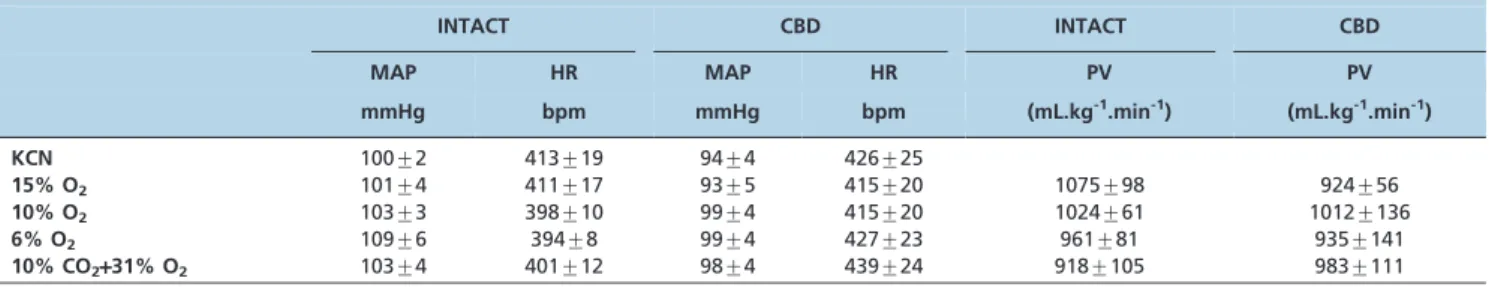
Table 1 -Baseline levels of mean arterial pressure (MAP), heart rate (HR) and pulmonary ventilation (PV) in intact and carotid body-denervated (CBD) rats before chemoreceptor stimulation with KCN, hypoxia and hypercapnia. The data are expressed as means¡SEM.
INTACT CBD INTACT CBD
MAP HR MAP HR PV PV
mmHg bpm mmHg bpm (mL.kg-1.min-1) (mL.kg-1.min-1)
KCN 100¡2 413¡19 94¡4 426¡25
15% O2 101¡4 411¡17 93¡5 415¡20 1075¡98 924¡56
10% O2 103¡3 398¡10 99¡4 415¡20 1024¡61 1012¡136
6% O2 109¡6 394¡8 99¡4 427¡23 961¡81 935¡141
10% CO2+31% O2 103¡4 401¡12 98¡4 439¡24 918¡105 983¡111
Figure 1 -Bar graph showing the changes in the mean arterial pressure (MAP) and heart rate (HR) in response to KCN in intact and carotid body-denervated (CBD) rats. *p,0.05 compared to intact rats.
Figure 2 -Bar graph showing the changes in the mean arterial pressure (MAP, top), heart rate (HR, middle) and pulmonary ventilation (PV, bottom) in response to 3 levels of hypoxia (15%, 10% and 6% of inhaled O2) in intact and carotid
observed in CBD rats. In line with this study, Marshall and Metcalfe (10-12) also reported hypotension and tachycardia in response to 15% inhaled O2. The hypotensive response
with 15% O2might be explained by the release of adenosine
from the skeletal muscle fibers (26) or blood vessel walls (27,28). Adenosine acts on endothelial A1-receptors to
stimulate the synthesis and release of NO, which ultimately induces vasodilation (29). The tachycardia observed in intact and CBD rats exposed to 15% O2 might be due to
the baroreflex, which is triggered by a drop in AP (30-32). Hypoxia with 10% O2produced a similar hemodynamic
response compared to 15% O2 (i.e., hypotension and
tachycardia, except that the hypotensive response did not achieve statistical significance in the intact rats). Nevertheless, the decrease in AP in response to 10% O2in
the CBD rats was markedly greater compared to 10% O2in
the intact rats. The lack of a significant hypotensive response in the intact rats might be explained by sympa-thetic activation elicited by the peripheral chemoreceptors
to counteract a decrease in total peripheral resistance triggered by the effect of hypoxia on skeletal muscle fibers (26) or vasodilation of the blood vessel walls (27,28). The tachycardia in the intact rats could be due to the hypoxia-induced activation of the peripheral chemoreflex, which increases sympathetic activity and enhances the release of plasma catecholamines (33). Studies on conscious intact rats have shown that hypoxia with 8 or 10% O2 for 30 min
decreased the MAP and increased HR (9,13,14). In addition, Sugimura et al. (34) also observed that 10% inhaled O2for
10 min elicited hypotension combined with tachycardia in conscious rats. However, the difference between the results from previous studies (9,13,14,34) and this study might be explained by the difference in the timeframe of exposure to hypoxia among the studies (1 minvs. 10 or 30 min). The
hypotensive response caused by 10% O2 in the CBD rats
might be explained by attenuation of the sympathetic vasoconstrictor outflow due to the removal of the carotid chemoreceptor drive. It has been shown that hypoxia increases adrenal sympathetic nerve activity and catechola-mine secretion (33), while sympathetic activation is not triggered in the absence of peripheral chemoreceptors in CBD rats. The tachycardia observed in the CBD rats exposed to 10% O2may be due to the baroreflex, which is triggered
by a fall in AP (30-32). Although the CBD rats do not have an active peripheral chemoreflex, their baroreflex is well preserved, which explains this reflex tachycardia.
Hypoxia with 6% O2in the conscious intact rats produced
marked hypertension and bradycardia, while in the CBD rats, 6% O2produced hypotension without any change in
HR. The findings of this study in conscious intact rats are in line with previous studies (16,17), indicating that maximal peripheral chemoreceptor activation is attained with 6% to 7% of inhaled O2. Previous studies in anesthetized rats have
demonstrated that hypoxia promotes hypotension and tachycardia (10-12). Nevertheless, the undesirable effect of anesthesia and the longer exposure of 3 min of hypoxia might explain the discrepancy in these results (10-12) compared to this study.
Hypoxia with 6% O2 in conscious CBD rats produced
significant hypotension without any change in HR. These findings were similar to the hemodynamic responses observed with 10% O2, except that the increase in baseline
HR did not reach statistical significance. Thus, due to the lack of activation of the peripheral chemoreceptors in CBD rats, the hypotensive response might be explained by the direct effect of hypoxia in the blood vessel wall (27,28) and endothelial cells to promote vasodilation (28,35).
Hypercapnia (10% CO2+31% O2) increased MAP and
decreased HR in intact and CBD rats. These findings indicate that the hemodynamic response to hypercapnia does not involve the peripheral carotid chemoreceptors but involves the central chemoreceptors. These results are in line with the previous findings of Walker and Brizzee (23) in conscious rats.
Ventilatory responses
Hypoxia with 15, 10 and 6% O2increased PV in the intact
rats but attenuated the ventilatory response in CBD rats. These results indicate that in intact rats, PV is partially dependent on the peripheral carotid chemoreceptors because in the absence of this mechanism, the central chemoreceptors may have triggered the blunted response. It is worth mentioning that the findings concerning the PV responses observed in this study are in line with previous
Figure 3 -Bar graph showing the changes in the mean arterial pressure (MAP, top), heart rate (HR, middle) and pulmonary ventilation (PV, bottom) in response to hypercapnia (10% CO2+31% O2) in intact and carotid body-denervated (CBD) rats.
studies in intact and CBD rats (6,7,23). In addition, recent studies have suggested a significant role for the peripheral chemoreceptors as an important mechanism for the ventila-tory response during exposure to hypercapnia (36,37).
Moreover, the response of PV to hypercapnia (10% CO2+31% O2) was similar in intact and CBD rats. Thus, the
PV response to hypercapnia depends mainly on the central chemoreceptors. It is possible that the severity of hypercapnia makes the peripheral chemoreceptor less important in modulating the ventilatory response. A similar response was observed in previous studies on conscious intact rats, which indicated that hypercapnia with 6 to 7% CO2increased
PV but did not produce a significant difference in the ventilatory response compared to CBD rats (7-8).
In summary, this study shows that the hemodynamic and ventilatory responses varied according to the level of hypoxia. Nevertheless, the hemodynamic and ventilatory responses to hypercapnia (10% CO2+31% O2) did not
depend on peripheral carotid chemoreceptors.
& ACKNOWLEDGMENTS
Fundac¸a˜o de Amparo a` Pesquisa do Estado de Sa˜o Paulo (FAPESP), Conselho Nacional de Desenvolvimento Cientifico e Tecnolo´gico (CNPq) and Coordenadoria de Aperfeic¸oamento de Pessoal de Nı´vel Superior (CAPES).
& AUTHOR CONTRIBUTIONS
Sabino JP collected the data for this manuscript. Oliveira M and Giusti H provided technical support concerning the surgical procedures for vessel cannulation (Oliveira M) and carotid body denervation (Giusti H) and provided support during data acquisition. Glass ML provided valuable insight into data interpretation and manuscript revision. Fazan Jr R provided a substantial contribution to protocol design and data analysis. Salgado HC was the mentor of Sabino JP and was responsible for the conception of the rationale for the development of the study.
& REFERENCES
1. Chugh SS, Chua TP, Coats AJS. Peripheral chemoreflex in chronic heart failure: Friend and foe. Am Heart J. 1996;132(4):900-4, http://dx.doi.org/ 10.1016/S0002-8703(96)90333-6.
2. Schultz HD, Sun SY. Chemoreflex function in heart failure. Heart Fail Rev. 2000;5(1):45-56, http://dx.doi.org/10.1023/A:1009846123893. 3. Somers VK, Mark AL, Abboud FM. Interaction of baroreceptor and
chemoreceptor reflex control of sympathetic nerve activity in normal humans. J Clin Invest. 1991;87(6):1953-7, http://dx.doi.org/10.1172/JCI115221. 4. Pitsikoulis C, Bartels MN, Gates G, Rebmann RA, Layton AM, De
Meersman RE. Sympathetic drive is modulated by central chemoreceptor activation. Respir Physiol Neurobiol. 2008;164(3):373-9, http://dx.doi. org/10.1016/j.resp.2008.08.010.
5. Grisk O, Exner J, Schmidt M, Honig A. Effects of acute hypoxia and hyperoxia on ventilation in spontaneously hypertensive and normoten-sive rat. J Auton Nerv Syst. 1996;57(3):177-80, http://dx.doi.org/10. 1016/0165-1838(95)00079-8.
6. Roux JC, Peyronnet J, Pascual O, Dalmaz Y, Pequignot JM. Ventilatory and central neurochemical reorganisation of O2 chemoreflex after carotid sinus nerve transection in rat. J Physiol. 2000;522(Pt 3):493-501, http:// dx.doi.org/10.1111/j.1469-7793.2000.t01-4-00493.x.
7. Serra A, Brozoski D, Hedin N, Franciosi R, Forster HV. Mortality after carotid body denervation in rats. J Appl Physiol. 2001;91(3):1298-306. 8. Mouradian GC, Forster HV, Hodges MR. Acute and chronic effects of
carotid body denervation on ventilation and chemoreflexes in three rat strains. J Physiol. 2012;590(Pt 14):3335-47, http://dx.doi.org/10.1113/ jphysiol.2012.234658.
9. Walker BR. Role of vasopressin in the cardiovascular response to hypoxia in the conscious rat. Am J Physiol. 1986;251(6 Pt 2):H1316-23. 10. Marshall JM, Metcalfe JD. Cardiovascular changes associated with
augmented breaths in normoxia and hypoxia in the rat. J Physiol. 1988;400:15-27.
11. Marshall JM, Metcalfe JD. Analysis of the cardiovascular changes induced in the rat by graded levels of systemic hypoxia. J Physiol. 1988;407:385-403.
12. Marshall JM, Metcalfe JD. Influences on the cardiovascular response to graded levels of systemic hypoxia of the accompanying hypocapnia in the rat. J Physiol. 1989;410:381-94.
13. Hirakawa H, Nakamura T, Hayashida Y. Effect of carbon dioxide on autonomic cardiovascular responses to systemic hypoxia in conscious rats. Am J Physiol. 1997;273(2 Pt 2):R747-54.
14. Murasato Y, Hirakawa H, Harada Y, Nakamura T, Hayashida Y. Effects of systemic hypoxia on R-R interval and blood pressure variabilities in conscious rats. Am J Physiol. 1998;275(3 Pt 2):H797-804.
15. Marcus NJ, Olson EB Jr., Bird CE, Philippi NR, Morgan BJ. Time-dependent adaptation in the hemodynamic response to hypoxia. Respir Respir Physiol Neurobiol. 2009;165(1):90-6, http://dx.doi.org/10.1016/j. resp.2008.10.013.
16. Bao G, Randhawa PM, Fletcher EC. Acute blood pressure elevation during repetitive hypocapnic and eucapnic hypoxia in rats. J Appl Physiol. 1997;82(4):1071-8.
17. Barros RC, Bonagamba LG, Okamoto-Canesin R, de Oliveira M, Branco LG, Machado BH. Cardiovascular responses to chemoreflex activation with potassium cyanide or hypoxic hypoxia in awake rats. Auton Neurosci. 2002;97(2):110-5, http://dx.doi.org/10.1016/S1566-0702(02)00050-4. 18. Richardson DW, Wasserman AJ, Patterson JL, Jr. General and regional
circulatory responses to change in blood pH and carbon dioxide tension. J Clin Invest. 1961;40:31-43, http://dx.doi.org/10.1172/JCI104234. 19. Kollai M, Koizumi K. Reciprocal and non-reciprocal action of the vagal
and sympathetic nerves innervating the heart. J Auton Nerv Syst. 1979;1(1):33-52, http://dx.doi.org/10.1016/0165-1838(79)90004-3. 20. Rose Jr CE, Althaus JA, Kaiser DL, Miller ED, Carey RM. Acute
hypoxemia and hypercapnia: increase in plasma catecholamines in conscious dogs. Am J Physiol. 1983;245(6):H924-9.
21. Somers VK, Mark AL, Zavala DC, Abboud FM. Contrasting effects of hypoxia and hypercapnia on ventilation and sympathetic activity in humans. J Appl Physiol. 1989;67(5):2101-6.
22. Fukuda Y, Sato A, Suzuki A, Trzebski A. Autonomic nerve and cardiovascular responses to changing blood oxygen and carbon dioxide levels in the rat. J Auton Nerv Syst. 1989;28(1):61-74, http://dx.doi.org/ 10.1016/0165-1838(89)90008-8.
23. Walker BR, Brizzee BL. Cardiovascular responses to hypoxia and hypercapnia in barodenervated rats. J Appl Physiol. 1990;68(2):678-86. 24. Franchini KG, Krieger EM. Cardiovascular responses of conscious rats to
carotid body chemoreceptor stimulation by intravenous KCN. J Auton Nerv Syst. 1993;42(1):63-9, http://dx.doi.org/10.1016/0165-1838(93)90342-R. 25. Malan A. Ventilation measured by body plethysmography in
hibernat-ing mammals and in poikilotherms. Respir Physiol. 1973;17(1):32-44, http://dx.doi.org/10.1016/0034-5687(73)90108-4.
26. Marshall JM, Thomas T, Turner L. A link between adenosine, ATP-sensitive k+channels, potassium and muscle vasodilatation in the rat in systemic hypoxia. J Physiol. 1993;472:1-9.
27. Mian R, Marshall JM. The role of adenosine in dilator responses induced in arterioles and venules of rat skeletal muscle by systemic hypoxia. J Physiol. 1991;443:499-511.
28. Mian R, Marshall JM. The role of adenosine in mediating vasodilatation in mesenteric circulation of the rat in acute and chronic hypoxia. J Physiol. 1995;489( Pt 1):225-34.
29. Ray CJ, Marshall JM. Measurement of nitric oxide release evoked by systemic hypoxia and adenosine from rat skeletal muscle in vivo. J Physiol. 2005;568(Pt 3):967-78, http://dx.doi.org/10.1113/jphysiol.2005.094854. 30. Kirchheim H R. Systemic arterial baroreceptor reflexes. Physiol Rev.
1976;56(1):100-77
31. Chapleau MW, Hajduczok G, Abboud FM. Mechanisms of resetting of arterial baroreceptors: an overview. Am J Med Sci. 1988;295(4):327-34. 32. Krieger EM, Salgado HC, Michelini LC. Resetting of the baroreceptors.
In: International Review of Physiology, University Park Press, Baltimore, ed. Guyton AC, Hall JE. 1982;26:119-46.
33. Biesold D, Kurosawa M, Sato A, Trzebski A. Hypoxia and hypercapnia increase the sympathoadrenal medullary functions in anesthetized, artificially ventilated rats. Jpn J Physiol. 1989;39(4):511-22.
34. Sugimura M, Hirose Y, Hanamoto H, Okada K, Boku A, Morimoto Y, et al. Influence of acute progressive hypoxia on cardiovascular variability in conscious spontaneously hypertensive rats. Auton Neurosci. 2008;141(1-2):94-103, http://dx.doi.org/10.1016/j.autneu.2008.05.008. 35. Marshall JM. Adenosine and muscle vasodilatation in acute systemic
hypoxia. Acta Physiol Scand. 2000;168(4):561-73, http://dx.doi.org/10. 1046/j.1365-201x.2000.00709.x.
36. Forster HV, Martino P, Hodges M, Krause K, Bonis J, Davis S, et al. The carotid chemoreceptors are a major determinant of ventilatory CO2
sensitivity, and PaCO2 during eupneic breathing. Adv Exp Med Biol. 2008;905:322-26, http://dx.doi.org/10.1007/978-0-387-73693-8_56. 37. Forster HV, Smith CA. Central CO2chemoreception in cardiorespiratory
control contributions of central and peripheral chemoreceptors to the ventilator response to CO2/H+. J Appl Physiol. 2010;108(4):989-94,