Case Report
Key words
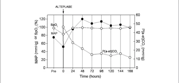
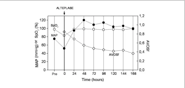
Pulmonary embolism; capnography; thrombolytic therapy. This is the first report of a patient submitted to chemical thrombolysis due to massive pulmonary embolism (PE) during the postoperative period of neurosurgery, in whom due to the lack of adequate clinical conditions, no imaging assessment was performed. Clinical, gasometric and capnographic data allowed the decision to perform the thrombolysis with safety. The P(a-et)CO2 gradient decreased from 46.4 mmHg to 11.8 mmHg (normal < 5 mmHg) and the end-tidal alveolar dead space fraction decreased from 0.85 to 0.37 (normal < 0.15) from the pre-thrombolysis period to the 7th day
post-thrombolysis. We conclude that the volumetric capnography (VC) was useful in the patient’s diagnosis and clinical follow-up.
Thrombolysis in Massive Pulmonary Embolism Based on the
Volumetric Capnography
Marcos Mello Moreira, Renato Giuseppe Giovanni Terzi, Ilma Aparecida Paschoal, Luiz Cláudio Martins, Evandro Pinto
da Luz Oliveira, Antonio Luis Eiras Falcão
Faculdade de Ciências Médicas da Universidade Estadual de Campinas - Unicamp - Campinas, SP - Brazil
Mailing address: Marcos Mello Moreira •
Rua Celso Egídio de Souza Santos, 181 - Jardim Chapadão - 13070-570 - Campinas, SP - Brazil
E-mail: [email protected]
Manuscript received March 30, 2009; revised manuscript received August 06, 2009; accepted December 17, 2009.
Case Report
A 22-year-old female patient, admitted at the Intensive Care Unit (ICU) of a tertiary hospital developed difficult-to-control diabetes insipidus after the excision of a frontal brain tumor (astrocytoma), together with septic conditions and mild hemodynamic alterations. On the 24th postoperative day,
the patient was going through the mechanical ventilation (MV) weaning process, with the following parameters: FiO2
= 0.30; spontaneous breathing (30 rpm), PEEP = 5 cmH2O;
apH = 7.50; PaO2 = 62.2 mmHg; PaCO2 = 27.6 mmHg;
HCO3 = 21.8 mmol/l; BE = -0.1 mmol/l; SatO2 = 94.8%; PaO2/FiO2 ratio= 207.
The patient suddenly started to present respiratory difficulty, hypoxemia and hemodynamic worsening. With the diagnostic hypothesis of massive PE, a transthoracic echocardiogram was performed, which disclosed moderate dilation of the right chambers, moderate tricuspid regurgitation and pulmonary artery systolic pressure = 50 mmHg. The echocardiograma (ECG) showed the presence of S1Q3T3 pattern.
After this episode sthe MV was adjusted to: FiO2 = 1;
SIMV (12/38 rpm); PEEP = 8 cmH2O and TV= 500 ml; the subsequent arterial gasometry showed: pH = 7.26; PaO2 =
44.5 mmHg; PaCO2 = 54.6 mmHg; HCO3 = 23.7 mmol/l;
BE = -2.7 mmol/l; SatO2 = 71.7%; PaO2/FiO2 ratio = 44.5. Dobutamine was started, aiming at the hemodynamic function improvement. Three hours later, while still on mechanical ventilation, the patient presented the following parameters: FiO2 = 1; SIMV (14/27 rpm); PEEP = 10 cmH2O; TV= 500 ml, arterial gasometry showed: pH = 7.22; PaO2 = 50.7
mmHg; PaCO2 = 54.7 mmHg; HCO3 = 21.3 mmol/l; BE =
-6.2 mmol/l; SatO2 = 82.3%; PaO2/FiO2 ratio= 50.7. Through the VC, the end-tidal CO2 pressure (PetCO2) was determined (CO2SMO PLUS 8100 Dixtal/Novametrix™) which,
associated to the arterial gasometry (Radiometer ABL 700™), allowed the calculation of different derived indices, such as end-tidal alveolar dead space fraction ,(AVDSf), the late dead space fraction (fDlate), the CO2 arterial-alveolar gradient [P(a-et) CO2]; and the slope of phase III of the CO2 spirogram (Slp III).
Considering the patient’s rapid deterioration, imminent risk of death and VC values indicative of increase in the alveolar dead space - compatible with PE2-7 - the medical team, after
the family had given the informed consent, chose to institute the thrombolytic treatment with alteplase (IV 100 mg/2 hours).
Discussion
A recent systematic literature review8 concluded the
Introduction
Pulmonary embolism (PE) is a frequent disorder, of which diagnostic confirmation is difficult to achieve. National studies have shown that approximately 3% to 5% of the necropsies disclose the presence of emboli in the pulmonary vessels and, in 68% of these cases, it is the cause of death. It is estimated that in 75% of the cases, the diagnosis is not achieved1 and
the standard imaging assessments are not always available, mainly in secondary hospitals. In contrast, the volumetric capnography (VC) is a non-invasive bedside test, which is available even at smaller hospitals. The VC can help diagnose patients with a diagnostic suspicion of PE2-4.
This report presents the case of a patient who, in spite of anticoagulant therapy, developed sudden alteration in the hemodynamic parameters presumably due to massive PE. Considering the rapid deterioration of the hemodynamic state, although the diagnosis of PE was not confirmed by imaging assessment, the patient was submitted to chemical thrombolysis. The VC was performed before the thrombolysis and for seven subsequent days.
Case Report
Moreira et al Volumetric capnography in massive PE
Arq Bras Cardiol 2010; 95(4): e97-e100
bleeding is the most frequent complication of thrombolysis, of which risk is around 6-20%, with intracranial bleeding being the most feared complication. Thus, the risk of bleeding is what defines the contraindications for thrombolysis. These relative contraindications are recent active bleeding or intracranial disease (less than 6 weeks), trauma, visceral biopsy, gastrointestinal bleeding, coagulation disorders, kidney or liver failure, puncture of vessel not susceptible to compression and pericarditis. As the patient presented severe hemodynamic
instability HR = 168 bpm, MAP = 52 mmHg, SpO2 = 83%
and FiO2 = 1), she did not present the adequate clinical conditions for transportation to another institution in order to undergo image assessment (helicoidal angiotomography). However, the patient was assessed by VC, which disclosed values suggestive of PE2-7.
The capnography estimates the value of the alveolar dead space2-7. The association between the VC results with those of
the arterial gasometry allows the calculation of several indices, from which variables the extension of the alveolar dead space can be inferred and, consequently, the presence and extension of the occluded areas of the pulmonary artery system 2-7.
The following capnographic parameters were determined: 1. PetCO29;
2. Slp III9;
3. P(a-et)CO26 (normal value < 5 mmHg);
4. AVDSf, calculated by the formula: PaCO2 - PetCO2 / PaCO2, where PetCO2 is end-tidal CO27 (normal value
< 0.15);
5. fDlate, obtained from the extrapolation of the current expired volume at 15% of the estimated total pulmonary capacity (TPC): fDlate = PaCO2-Pet(15%
TCP)CO2 /PaCO25 (normal value < 0.12).
During an embolic event, the ventilation/perfusion ratio
imbalance is accentuated and, as a consequence, the variables that express the alveolar dead space also present alterations2-7.
The higher the calculated value, the higher the degree of vascular network obstruction, and thus, the more extensive the alveolar dead space is, considering a correlation between the extension of the area without perfusion and the obtained value2-7. The studied variables tended to normalization after
the thrombolytic treatment, indicating the occurrence of vessel rechanneling.
The capnographic variables presented the following behavior:
• PetCO2 (reference value: 36.7 ± 3.7 mmHg)9: 8.2
(pre-thrombolysis); 13.1 (after 24h); 17.7 (after 48h); 16.4 (after 72h); 19.9 (after 96h); 19.0 (after 120h); 20.3 (after 144h); 20.4 mmHg (after 168h);
• Slp III (reference value: 7.5 ± 2.4 mmHg/l)9: 0.26
(pre-thrombolysis); 0.77 (after 24h) ; 3.96 (after 48h); 8.77 (after 72h); 7.62 (after 96h); 5.07 (after 120h); 6.6 (after 144h); 8.47mmHg/l (after 168h);
• fDlate5: 0.85 (pre-thrombolysis); 0.68 (after 24h);
0.54 (after 48h); 0.55 (after 72h); 0.39 (after 96h); 0.38 (after 120h); 0.41 (after 144h); 0.31 (after 168h). To illustrate that (Figures 1 and 2), two capnographic variables muras [P(a-et)CO2 and AVDSf] are demonstrated,
which, when associated to arterial gasometry can be easily obtained through any capnographer available in the market and performed at the bedside and at any hospital.
In this dramatic case report, one can observe that, when strictly applied, VC can be useful as a diagnostic tool and in the therapeutic follow-up of patients that cannot be submitted to imaging assessment. The VC also showed to be a useful tool in decision-making at the initial phase of PE assessment and O CV mresolution, as the progressive
Figure 1 -Evolution of the arterial-alveolar gradient in massive pulmonary embolism (MAP - mean arterial pressure; SpO2 - oxygen saturation by pulse oximetry; P(a-et) CO2 - arterial-alveolar gradient of CO2).
Case Report
Moreira et al
Volumetric capnography in massive PE
Arq Bras Cardiol 2010; 95(4): e97-e100
References
1. Mesquita CT, Morandi Jr JL, Perrone FT, Oliveira CS, Barreira LJ, Nascimento SS, et al. Fatal pulmonary embolism in hospitalized patients: clinical diagnosis versus pathological confirmation. Arq Bras Cardiol. 1999; 73 (3): 251-8.
2. Moreira MM, Terzi RGG, Carvalho CHN, Oliveira Neto AF, Pereira MC, Paschoal IA. Alveolar dead space and capnographic variables before and after thrombolysis in patients with acute pulmonary embolism. Vasc Health Risk Manag. 2009; 5 (1): 9-12.
3. Moreira MM, Terzi RG, Vieira RW, Petrucci Jr O, Paschoal IA, Oliveira PP, et al. Pre and post-pulmonary thromboendarterectomies capnographic variables. Rev Bras Cir Cardiovasc. 22 (4): 509-12.
4. Moreira MM, Terzi RG, Pereira MC, Grangeia TC, Paschoal IA. Volumetric capnography as a noninvasive diagnostic procedure in acute pulmonary thromboembolism. J Bras Pneumol. 2008; 34 (5): 328-32.
5. Eriksson L, Wollmer P, Olsson CG, Albrechtsson U, Larusdottir H, Nilsson R, et al. Diagnosis of pulmonary embolism based upon alveolar dead space analysis. Chest. 1989; 96 (2): 357-62.
6. Fletcher R, Jonson B, Cumming G, Brew J. The concept of deadspace with special reference to the single breath test for carbon dioxide. Br J Anaesth. 1981; 53 (1): 77-88.
7. Rodger MA, Bredeson CN, Jones G, Rasuli P, Raymond F, Clement AM. The bedside investigation of pulmonary embolism diagnosis study. Arch Intern Med. 2006; 166 (2): 181-7.
8. Harris T, Meek S. When should we thrombolyse patients with pulmonary embolism? A systematic review of the literaturey. Emerg Med J. 2005; 22 (11): 766-71.
9. Paschoal I, Moreira M, Pereira M, Piza S, Gonçalves J, Metze K, et al. Noninvasive evaluation of pulmonary disease using volumetric capnography in adult patients with cystic fibrosis. J Cystic Fibrosis. 2007; 6 (Suppl): IS38.
10. Blanch L, Lucangelo U, Lopez-Aguilar J, Fernandez R, Romero PV. Volumetric capnography in patients with acute lung injury: effects of positive end-expiratory pressure. Eur Respir J. 1999; 13 (5): 1048-54.
Figure 2 -Evolution of the end-tidal alveolar dead space fraction/arterial-alveolar gradient in massive pulmonary embolism (MAP- mean arterial pressure; SpO2 - oxygen saturation by pulse oximetry; AVDSf - end-tidal alveolar dead space fraction.
pulmonary reperfusion resulted in a decrease of the alveolar dead space and thus, a tendency towards the normalization of the capnographic variables.
One can speculate that the increase in the dead space was due to the presence of PEEP. However, Blanch et al10 showed
that there is no alteration in the dead space when the PEEP is at the levels observed in the present study. In this case, all VC measurements were carried out with a PEEP of 5 cmH2O.
After the thrombolytic therapy and the stabilization of the hemodynamic picture, a head computed tomography was carried out, which did not disclose bleeding. The patient was discharged from the ICU and subsequently, from the hospital.
Potential Conflict of Interest
No potential conflict of interest relevant to this article was reported.
Sources of Funding
There were no external funding sources for this study.
Study Association
This article is part of the thesis of doctoral submitted by Marcos Mello Moreira, from Universidade Estadual de Campinas - UNICAMP.