Involved in the Cardiovascular Responses to Acute
Restraint Stress in Rats
Fernando H. F. Alves1*, Carlos C. Crestani2, Leonardo B. M. Resstel1, Fernando M. A. Correˆa1
1Department of Pharmacology, School of Medicine of Ribeira˜o Preto, University of Sa˜o Paulo, Ribeira˜o Preto, SP, Brazil,2Department of Natural Active Principles and Toxicology, School of Pharmaceutical Sciences of Araraquara, Univ. Estudual Paulista - UNESP, Araraquara, SP, Brazil
Abstract
The insular cortex (IC) is a limbic structure involved in cardiovascular responses observed during aversive threats. However, the specific neurotransmitter mediating IC control of cardiovascular adjustments to stress is yet unknown. Therefore, in the present study we investigated the role of local IC adrenoceptors in the cardiovascular responses elicited by acute restraint stress in rats. Bilateral microinjection of different doses (0.3, 5, 10 and 15 nmol/100 nl) of the selectivea1-adrenoceptor antagonist WB4101 into the IC reduced both the arterial pressure and heart rate increases elicited by restraint stress. However, local IC treatment with different doses (0.3, 5, 10 and 15 nmol/100 nl) of the selectivea2-adrenoceptor antagonist RX821002 reduced restraint-evoked tachycardia without affecting the pressor response. The present findings are the first direct evidence showing the involvement of IC adrenoceptors in cardiovascular adjustments observed during aversive threats. Our findings indicate that IC noradrenergic neurotransmission acting through activation of both a1- and a2 -adrenoceptors has a facilitatory influence on pressor response to acute restraint stress. Moreover, ICa1-adrenoceptors also play a facilitatory role on restraint-evoked tachycardiac response.
Citation:Alves FHF, Crestani CC, Resstel LBM, Correˆa FMA (2014) Botha1- anda2-adrenoceptors in the Insular Cortex Are Involved in the Cardiovascular Responses to Acute Restraint Stress in Rats. PLoS ONE 9(1): e83900. doi:10.1371/journal.pone.0083900
Editor:Emilio Hirsch, University of Torino, Italy
ReceivedAugust 28, 2013;AcceptedNovember 8, 2013;PublishedJanuary 3, 2014
Copyright:ß2014 Alves et al. This is an open-access article distributed under the terms of the Creative Commons Attribution License, which permits unrestricted use, distribution, and reproduction in any medium, provided the original author and source are credited.
Funding:Financial agency: FAPESP - Grant number 2010/09462-9, URL - www.fapesp.br. The funders had no role in study design, data collection and analysis, decision to publish, or preparation of the manuscript.
Competing Interests:The authors have declared that no competing interests exist. * E-mail: [email protected]
Introduction
Stress situations happen during real or perceived threat to homeostasis or well-being. Stressors include either interoceptive changes (e.g., blood volume or osmolality changes) or environ-mental threat that may be physical (e.g., hypoxia) or psychological (e.g., presence of a predator). During stress a spectrum of physiological responses are evoked to maintain the physiologic integrity of the organism [1]. The physiological responses to stress are mainly characterized by autonomic nervous system alterations, increase in plasma catecholamine levels and activation of the hypothalamus-pituitary-adrenal (HPA) axis [1,2]. Autonomic responses include increase on both blood pressure and heart rate (HR) [3,4]. Furthermore, cardiovascular changes during stress are accompanied by a resetting of baroreflex toward higher arterial pressure values, thus allowing simultaneous blood pressure and HR increases [5–8].
Several central nervous system areas, including the prefrontal cortex, were described to be part of the brain circuitry involved on cardiovascular adjustments during stress [3,4,9,10]. In rats, two regions of the prefrontal cortex involved in control of cardiovas-cular function are the insular cortex (IC) and the medial prefrontal cortex (MPFC) [11,12]. It has been described that the IC is involved in cardiovascular control [13–15] and baroreflex modulation [16–19]. Furthermore, previous results from our group demonstrated that bilateral microinjection of the unspecific neurotransmitter blocker CoCl2into the IC of rats reduced both
cardiovascular and behavioral responses evoked by either condi-tioned (contextual fear conditioning) or uncondicondi-tioned (acute restraint stress) aversive stimuli [2,20]. These results provided the first evidence of a role of the IC in cardiovascular adjustments during stress. However, due to the nonselective blockade of local neurotransmission caused by CoCl2 [21,22], the specific
neuro-transmitter involved in the IC modulation of cardiovascular responses to stress is yet unknown.
Central noradrenergic circuitry is shortly activated after a stressful event [1]. Conversely, it has been identified an enhanced release of noradrenaline after stress in several limbic brain regions including the central (CeA) and medial (MeA) amygdaloid nuclei, bed nucleus of the stria terminalis (BNST), lateral septal area (LSA), hippocampus and prefrontal cortex [1,23–26]. Noradren-ergic terminals in the prefrontal cortex originate mainly from the locus coeruleus and play an important role in the regulation of cortical function [27–30]. We have previously reported that noradrenergic neurotransmission within the IC is involved in the modulation of baroreflex activity [31]. Also, microinjection of noradrenaline into the IC causes elevation of blood pressure and bradycardia [14]. Although above evidence, the involvement of IC noradrenergic neurotransmission in the control of cardiovascular function during stress situations has never been investigated.
elicited by acute restraint stress in rats. To test this hypothesis, we investigated the effect of bilateral microinjections into the IC of selective a-adrenoceptor antagonists in restraint-evoked pressor and tachycardiac responses.
Experimental procedure
Ethical approval and animals
Experimental procedures were carried out following protocols approved by the Ethical Review Committee of the School of Medicine of Ribeira˜o Preto, (process number: 167/2007), which complies with the guiding principles for research involving animals and human beings of the National Institutes of Health. Fifty-seven male Wistar rats weighing approximately 250 g were used in the present experiment. Rats were housed in plastic cages in a temperature-controlled room (25uC) at the Animal Care Unit of the Department of Pharmacology, School of Medicine of Ribeira˜o Preto. Rats were kept under a 12 h :12 h light–dark cycle (lights on between 06:00 am and 6:00 pm) and had free access to water and standard laboratory food, except during the experimental period.
Surgical preparation
Five days before the experiment, the rats were anesthetized with tribromoethanol (250 mg/kg, i.p.). After local anesthesia with 2% lidocaine, the skull was surgically exposed and stainless steel guide cannulas (26 G) were implanted bilaterally in the IC, using a stereotaxic apparatus (Stoelting, Wood Dale, Illinois, USA). Stereotaxic coordinates for cannula implantation in the IC were selected from the rat brain atlas of Paxinos and Watson (1997) and were: antero-posterior =+11.7 mm from interaural, later-al = 4.0 mm from the medilater-al suture and dorso-ventrlater-al =24.5 mm from the skull. Cannulas were fixed to the skull with dental cement and one metal screw. After surgery, the animals were treated with a polyantibiotic preparation of streptomycins and penicillins (i.m., 0.27 mg/kg, Pentabiotico, Fort DodgeH, Campinas, SP, Brazil) to prevent infection, and with the non-steroidal anti-inflammatory flunixine meglumine (2.5 mg/kg, i.m.; BanamineH, Schering Plough, Cotia, SP, Brazil) for post-operative analgesia.
One day before the experiment, rats were anesthetized with tribromoethanol (250 mg/kg, i.p.) and a catheter (a 4 cm segment of PE-10 heat-bound to a 13 cm segment of PE-50, Clay Adams, Parsippany, NJ, USA) was inserted into the abdominal aorta through the femoral artery, and later on used for arterial pressure and HR recording. The catheters were tunneled under the skin and exteriorized on the animal’s dorsum. After surgery, the animals were treated with the non-steroidal anti-inflammatory flunixine meglumine (2.5 mg/kg, i.m.) for post-operative analge-sia.
Measurement of Cardiovascular Responses
On the day of the experiment, the arterial cannula was connected to a pressure transducer and pulsatile arterial pressure was recorded using an HP-7754A amplifier (Hewlett Packard, Palo Alto, CA, USA) and an acquisition board (Biopac M-100, Goleta, CA, USA) connected to a personal computer. Mean arterial pressure (MAP) and HR values were derived from pulsatile arterial pressure recordings and were processed online.
Drugs and solutions
WB4101 (Tocris, Westwoods Business Park Ellisville, MO, USA) and RX821002 (Tocris) were dissolved in artificial cerebrospinal fluid (ACSF) (ACSF composition: 100 mM NaCl; 2 mM Na3PO4; 2.5 mM KCl; 1 mM MgCl2; 27 mM NaHCO3;
2.5 mM CaCl2; pH = 7.4). Urethane (Sigma, St. Louis, MO, USA)
and tribromoethanol (Sigma) were dissolved in saline (0.9% NaCl). Flunixine meglumine (BanamineH, Schering Plough, Brazil) and poly-antibiotic preparation of streptomycins and penicillins (PentabioticoH, Fort Dodge, Brazil) were used as provided.
Drug injection into the insular cortex
The needles (33 G, Small Parts, Miami Lakes, FL, USA) used for microinjection into the IC were 1 mm longer than the guide cannulas and were connected to a hand-driven 2ml syringe (7002-KH, Hamilton Co., Reno, NV, USA) through a PE-10 tubing. Needles were carefully inserted into the guide cannulas without restraint the animals. After a 30 s period, the needle was removed and inserted into the second guide cannula for microinjection into the contralateral IC. Drugs were injected in a final volume of 100 nl [2,20].
Experimental procedure: acute restraint stress
On the trial day, animals were brought to the experimental room in their home cages. Animals were allowed one hour to adapt to the conditions of the experimental room, such as sound and illumination, before starting cardiovascular recordings. The experimental room was temperature controlled (25uC) and the room was acoustically isolated from the other rooms. Constant background noise was generated by an air extractor to minimize sound interference within the experimental room. Baseline values of MAP and HR were recorded for at least 30 min. In the sequence, independent groups of animals received bilateral microinjection into the IC of vehicle (ACSF, 100 nl) or different doses of either the selectivea1-adrenoceptor antagonist WB4101
(0.3, 5, 10 or 15 nmol/100 nl) or the selectivea2-adrenoceptor
antagonist RX821002 (0.3, 5, 10 or 15 nmol/100 nl) [14,31,32]. Ten minutes later, rats were submitted to acute restraint stress by placing them into a plastic cylindrical restraint tube (diameter = 6.5 cm, length = 15 cm), which were ventilated by holes (1 cm of diameter) that comprised approximately 20% of the tube surface. The restraint session lasted 60 min, after which the rats were returned to their home cages [20,33]. Each rat was submitted to one session of restraint in order to avoid habituation. Experiments were performed during the morning period in order to minimize possible circadian rhythm interferences.
Histological determination of the microinjection sites At the end of experiments, animals were anesthetized with urethane (1.25 g/kg, i.p.) and 100 nL of 1% Evan’s blue dye was injected into the IC as a marker of the injection site. They were then submitted to intracardiac perfusion with 0.9% NaCl followed by 10% formalin. Brains were removed and post fixed for 48 h at 4uC and serial 40mm-thick sections were cut using a cryostat (CM1900, Leica, Wetzlar, Germany). Sections were stained with 1% neutral red for light microscopy analysis. The placement of the microinjection needles was determined analyzing serial sections and identified according to the rat brain atlas of Paxinos and Watson (1997).
Statistical Analysis
When interactions between the factors were observed, one-way ANOVA followed by Bonferroni’s post-hoc test was used to compare the effect of the treatments. Nonlinear regression analysis was performed to investigate the dose–effect relationship of treatment with crescents doses of WB4101 and RX821002 on cardiovascular responses to restraint stress.
Results
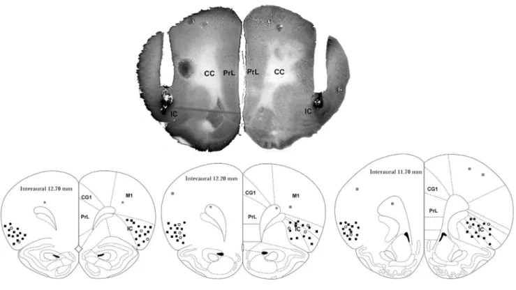
A representative photomicrograph of a coronal brain section depicting bilateral microinjection sites in the IC of one represen-tative rat is presented in Figure 1. A diagrammatic representation showing microinjection sites of vehicle, WB4101 and RX821002 into the IC and WB4101 and RX821002 into structures surrounding the IC is also presented in Figure 1.
Effect of IC pretreatment with different doses of the selectivea1-adrenoceptor antagonist WB4101 on cardiovascular changes evoked by acute restraint stress
Bilateral microinjection of different doses (0.3, 5, 10 and 15 nmol/100 nL, n = 5/group) of the selective a1-adrenoceptor
antagonist WB4101 into the IC did not affect either MAP or HR baseline values (Table 1). Representative experimental recordings showing effects of local microinjection of WB4101 into the IC on cardiovascular responses elicited by acute restraint stress are presented in Figure 2. Time-course analysis of restraint-evoked cardiovascular responses indicated that IC treatment with WB4101 (at doses of 5, 10 and 15 nmol/100 nl for MAP response and at dose of 15 nmol/100 nl for HR response) reduced both MAP (F(4,660)= 336, P,0.0001) and HR (F(4,660)= 50, P,0.0001)
responses, when compared with ACSF-treated animals (n = 7)
(Figure 3). There was also a significant effect over time for MAP (F(29,660)= 70, P,0.0001) and HR (F(29,660)= 27, P,0.0001)
responses, as well as a treatment x time interaction for the pressor response (MAP: F(116,660)= 6, P,0.0001; HR: F(116,660)= 1,
P.0.05). Nonlinear regression analysis revealed that WB4101 effects on restraint-evoked cardiovascular responses were dose-dependent, showing a significant association between drug dose and MAP (df = 18, r2= 0.85, P,0.05) and HR (df = 13, r2= 0.60, P,0.05) increases (Figure 3). The injection of WB4101 (n = 2) into structures surrounding the IC did not affect both MAP (F(1,60)= 2,9, P.0.05) and HR (F(1,60)= 0,2, P.0.05) responses
to restraint stress.
Effect of IC pretreatment with different doses of the selectivea2-adrenoceptor antagonist RX821002 on cardiovascular changes evoked by acute restraint stress
Bilateral microinjection of different doses (0.3, 5, 10 and 15 nmol/100 nL, n = 5/group) of the selective a2-adrenoceptor
antagonist RX821002 into the IC did not affect either MAP or HR baseline values (Table 2). Representative experimental recordings showing effects of RX821002 microinjection into the IC on cardiovascular responses induced by acute restraint stress are presented in Figure 2. Time-course analysis of restraint-evoked cardiovascular responses indicated that IC treatment with RX821002 at doses of 5, 10 and 15 nmol/100 nl reduced MAP response (F(4,630)= 216, P,0.0001) without affecting
restraint-evoked tachycardia (F(4,630)= 2, P.0.05), when compared with
ACSF-treated animals (n = 6) (Figure 4). There was also a significant effect over time for MAP (F(29,630)= 54, P,0.0001)
and HR (F(29,630)= 42, P,0.0001) responses, as well as a treatment
Figure 1. A photomicrograph of a coronal brain section, from one representative rat, which shows bilateral injection sites in the insular cortex.Diagrammatic representation based on the rat brain atlas of Paxinos and Watson (1997) indicating the microinjection sites of vehicle (white circles), WB4101 (black circles) and RX821002 (black squares) into the IC as well as WB4101 (gray circles) and RX821002 (gray squares) into structures surrounding the IC. Cg1 – cingulate cortex, area; PrL – prelimbic cortex, M1 – primary motor cortex; insular cortex – insular cortex, cc – corpus callosum, forceps minor of the corpus callosum (fmi).
x time interaction for the pressor response (MAP: F(116,630)= 3,
P,0.0001; HR: F(116,630)= 0.6, P.0.05). Nonlinear regression
analysis revealed that RX821002 effect on restraint-evoked pressor response was dose-dependent, showing a significant association between drug doses and MAP increase (df = 18, r2= 0.65, P,0.05) (Figure 4). The injection of RX821102 into structures surrounding the IC did not affect both MAP (F(1,60)= 1,4, P.0.05) and HR
(F(1,60)= 0,005, P.0.05) responses to restraint stress.
Discussion
The results of the present work provide the first direct evidence for the involvement of IC adrenoceptors in cardiovascular responses observed during aversive threats. We have shown that bilateral microinjection of the selectivea1-adrenoceptor antagonist
WB4101 into the IC reduced restraint-evoked pressor and tachycardiac responses in a dose-dependent manner. Moreover, IC treatment with the selective a2-adrenoceptor antagonist
RX821002 dose-dependently reduced MAP increase observed during restraint stress without affecting tachycardiac response.
Restraint stress is well accepted in the literature as an unconditioned and unavoidable aversive stimulus that elicits neuroendocrine and cardiovascular responses, the latter being characterized by sustained elevation of blood pressure, HR and the sympathetic activity that last through the restraint period [34– 37]. The IC receives an organized representation of visceral information and is also highly interconnected with subcortical limbic and autonomic-related regions. Based on this combination of sensory input and limbic connectivity it has been descript as an important cortical center for the integration of autonomic and behavioral responses during aversive threats [13]. Conversely, we have demonstrated that CoCl2-induced acute bilateral inhibition
of IC neurotransmission greatly attenuated both pressor and tachycardiac responses evoked by acute restraint stress [20]. However, due to the nonselective blockade of local neurotrans-mission caused by CoCl2[21,22], the possible neurotransmitter
involved was not identified.
It has been showed that diverse array of physical (e.g., immune challenge, hypoglycemia, hypotension, and cold exposure) and emotional (e.g., immobilization, electric shock, loud noise, and restraint stress) stressors activate brain noradrenergic mechanisms [1,23,26,38–42]. Noradrenergic neural terminals have been
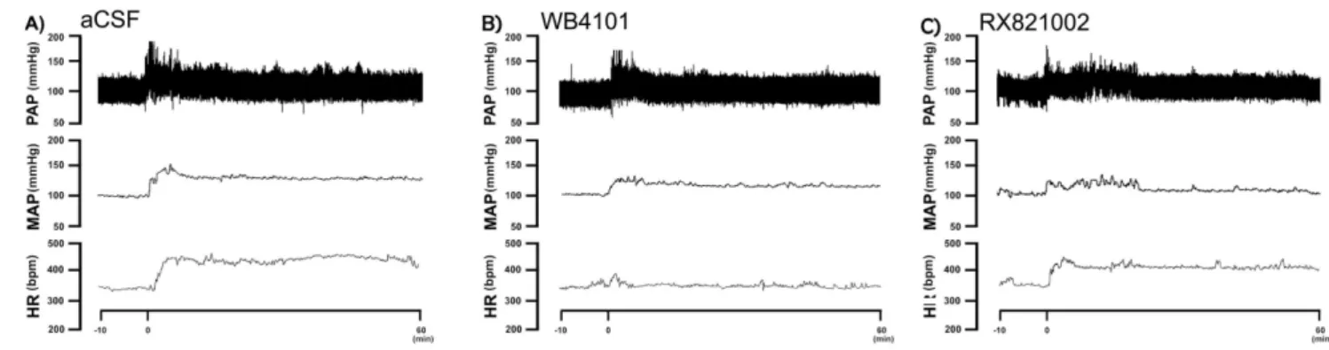
Figure 2. Representative recordings of mean arterial pressure (MAP), pulsatile arterial pressure (PAP) and heart rate (HR) of representative rats submitted to acute restraint stress. A)Recordings of MAP, PAP and HR of one representative rat treated with vehicle (ACSF) into the insular cortex and submitted to acute restraint stress. The onset of restraint is at t = 0.B)Recordings of MAP, PAP and HR of one representative rat treated with the selectivea1-adrenoceptor antagonist WB4101 into the insular cortex and submitted to acute restraint stress. The onset of restraint is at t = 0. Note the decrease in the MAP, PAP and HR responses to restraint stress in animals treated with WB4101 into the insular cortex. C) Recordings of MAP, PAP and HR of one representative rat treated with the selectivea2-adrenoceptor antagonist RX821002 into the insular cortex and submitted to acute restraint stress. The onset of restraint is at t = 0. Note the decrease in the MAP and PAP responses to restraint stress in animals treated with RX821002 into the insular cortex.
doi:10.1371/journal.pone.0083900.g002
Figure 3. Changes in mean arterial pressure (DMAP) and heart
rate (DHR) evoked by acute restraint stress in animals treated with different doses of the selectivea
1-adrenoceptor antago-nist WB4101 into the insular cortex. (Left)Time-course ofDMAP andDHR during acute restraint stress in rats treated with vehicle (ACSF, 100 nl, n = 7) or different doses (0.3, 5, 10 and 15 nmol/100 nl, n = 5/ group) of WB4101 into the insular cortex. The onset of exercise was at t = 0. Circles represent the mean and bars the S.E.M.#P,0.05, indicates a significant difference over the whole restraint stress period compared to vehicle treated animals; ANOVA followed by Bonferroni’s post test.
(Right)DMAP andDHR during acute restraint stress in rats treated with increasing doses of WB4101 (0.3, 5, 10 and 15 nmol/100 nl). V: vehicle (ACSF, 100 nl). Dose–effect curves were generated by nonlinear regression analysis. Data shown represent the means6S.E.M. of the variation of MAP and HR during the 60 min of restraint.#P,0.05, significantly different from vehicle group; one-way ANOVA followed by Bonferroni’s post test.
identified in the IC [30]. This IC innervation is mainly originated from noradrenergic cells grouped in the locus coeruleus (norad-renergic cell group A6) [27,28,30]. The present work has demonstrated that blockade of locala1-adrenoceptor by bilateral
microinjection of WB4101 into the IC was able to reduce both pressor and tachycardiac responses evoked by restraint stress. These results corroborate with effects observed previously following CoCl2-induced acute bilateral inhibition of IC
neuro-transmission [20], thus suggesting that local a1-adrenoceptors
mediates, at least in part, the IC influence on cardiovascular responses to restraint stress. Interestingly, blockade of local a2
-adrenoceptors caused by microinjection of RX821002 into the IC also reduced restraint-evoked pressor response, but without affecting tachycardiac response. Therefore, present data suggest that control of cardiac function during restraint stress by IC noradrenergic neurotransmission is due a selective activation of locala1-adrenoceptors, whereas control of blood pressure during
seems to be mediated by coactivation of local a1- and a2
-adrenoceptors.
The presence of specific noradrenergic mechanisms within the IC controlling restraint-evoked pressor and tachycardiac responses indicates that different neuronal pathways originating in the IC are involved in control of vascular and cardiac functions during stress. The existence of specific central nervous system circuitries controlling autonomic activity to different organs provides the structural substrate for specific local IC noradrenergic neurotrans-mission mechanisms modulating cardiovascular adjustments dur-ing restraint [43]. Conversely, it has been demonstrated that several brain regions selectively modulate stress-evoked blood pressure and HR responses [44–47]. The presence of specific noradrenergic mechanisms in the central nervous system modu-lating vascular and cardiac responses to stress has also been reported [33,48]. Therefore, our results corroborate with previous evidence of selective neural substrates controlling vascular and cardiac function during aversive threats.
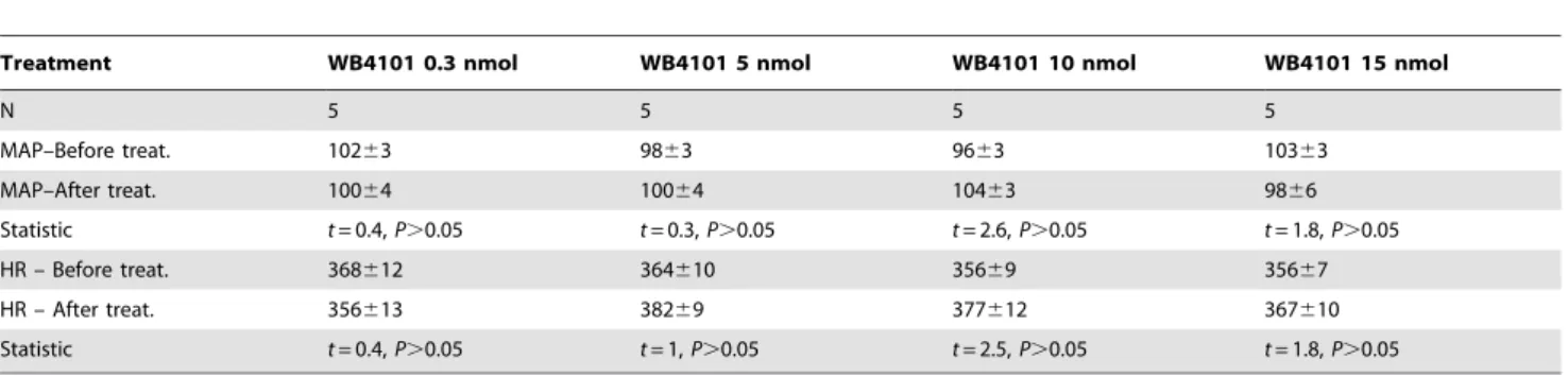
Noradrenaline is released in several central nervous system regions, including the prefrontal cortex [23,26], shortly after the onset of a stressful situation [1]. Since noradrenaline act through G protein-coupled receptors, which rapidly transfer their activation to downstream effectors, the rapid rise in their level is quickly translated into behavioral and physiological responses. This profile of fast release and action can explain why effects of local IC treatment with adrenoceptor antagonists are already observed during the early phase of restraint stress. However, it has been proposed that sustained and adaptive components of the stress responses (e.g., consolidation of the memory associated with the stressor) are mediated by mechanisms in the brain that affect gene expression and cell function [1]. A main mediator of these latter effects is corticosteroids acting through glucocorticoid receptors [1]. Since previous studies have demonstrated a role of the IC in Table 1.Effect of bilateral microinjections into the IC of crescent doses (0.3, 5, 10 and 15 nmol/100 nl) of the selectivea1 -adrenoceptor antagonist WB4101 on mean arterial pressure (MAP) and heart rate (HR) baseline.
Treatment WB4101 0.3 nmol WB4101 5 nmol WB4101 10 nmol WB4101 15 nmol
N 5 5 5 5
MAP–Before treat. 10263 9863 9663 10363
MAP–After treat. 10064 10064 10463 9866
Statistic t= 0.4,P.0.05 t= 0.3,P.0.05 t= 2.6,P.0.05 t= 1.8,P.0.05
HR – Before treat. 368612 364610 35669 35667
HR – After treat. 356613 38269 377612 367610
Statistic t= 0.4,P.0.05 t= 1,P.0.05 t= 2.5,P.0.05 t= 1.8,P.0.05
Student’st-test.
doi:10.1371/journal.pone.0083900.t001
Figure 4. Changes in mean arterial pressure (DMAP) and heart
rate (DHR) evoked by acute restraint stress in animals treated with different doses of the selectivea
2-adrenoceptor antago-nist RX821002 into the insular cortex.(Left) Time-course ofDMAP andDHR during acute restraint stress in rats treated with vehicle (ACSF, 100 nl, n = 6) or different doses (0.3, 5, 10 and 15 nmol/100 nl, n = 5/ group) of the selectivea2-adrenoceptor antagonist RX821002 into the insular cortex. The onset of exercise was at t = 0. Circles represent the mean and bars the S.E.M.#P,0.05, indicates a significant difference over the whole restraint stress period compared to vehicle treated animals; ANOVA followed by Bonferroni’s post test. (Right)DMAP and
the memory formation for aversive threat and hypothalamus-pituitary-adrenal axis control [2,4,9], further studies are necessary to investigate a possible role of IC in latter consequences of restraint stress.
Tachycardiac and pressor responses during stress are sympa-thetically mediated since they are abolished after the blockade of
b- anda-adrenoceptors, respectively [5,49,50]. Moreover, treat-ment with parasympathetic blocker increases the tachycardiac response evoked by psychological stress [33,51,52], thus suggesting the simultaneous activation of cardiac parasympathetic and sympathetic activity during psychological stress. It has been reported that the IC modulate the sympathetic nervous activity through a mandatory synapse in the ventrolateral medulla [53,54]. An IC control of cardiac parasympathetic activity has also been shown [31,55]. Therefore, activation of ICa1-adrenoceptors could
facilitate restraint-evoked tachycardiac response by stimulating facilitatory inputs to sympathetic medullary neurons and/or by stimulating inhibitory inputs to vagal neurons. Connections from the IC to sympathetic medullary neurons could also be the neural substrate for the facilitatory influence of IC a1- and a2
-adrenoceptors on the pressor response to stress.
Baroreflex stimulus–response curve resets toward higher blood pressure values during aversive threat [5,6]. It has been proposed that such changes on baroreflex activity play a facilitatory role in stress-evoked cardiovascular responses [3,7]. We have previously demonstrated that IC noradrenergic neurotransmission acting through activation of a1-adrenoceptor modulates the baroreflex
activity in a similar manner to that observed during stress [31]. Therefore, activation of IC a1-adrenoceptors could facilitate
cardiovascular responses to restraint stress through its modulation of baroreflex activity. However, once IC treatment with selective
a2-adrenoceptors does not affect baroreflex activity [31], it is
possible that IC control of restraint-evoked pressor response through this adrenoceptor occurs by mechanisms independent of the baroreflex.
An antero-posterior organization of IC control of cardiovascular function has been proposed. Predominantly depressor responses have been reported following stimulation of rostral regions of the IC [13]. However, stimulation of the posterior IC elicits either pressor response associated with tachycardia (rostral sites within the posterior IC) or depressor response followed by bradycardia (caudal sites within the posterior IC) [13]. Although these pieces of evidence, a possible regionalization in the IC control of cardiovascular adjustments to stress has never been reported. The injection sites within the IC in studies investigating the role of this cortical region in the cardiovascular control during stress
(including the present study) have centered within rostral regions of the IC [2,20]. Therefore, further studies are necessary in order to investigate a possible rostro-caudal organization of the IC control of cardiovascular function during aversive threat.
IC treatment with adrenoceptor antagonists did not affect either MAP or HR baseline values. Therefore, although the present study supports the hypothesis that IC noradrenergic neurotrans-mission plays an important role in modulating the cardiovascular responses to restraint stress, this neurotransmission is not involved in the tonic maintenance of cardiovascular function. These results corroborate with our previous data demonstrating no changes in cardiovascular parameters after blockade of either glutamatergic receptors or adrenoceptors into the IC [16,31]. However, present results contrast with data of other groups that observed increased arterial pressure and HR following microinjection of the neuronal blocker lidocaine into the IC of unanesthetized rats [53]. Lidocaine blocks both local synapses and passage fibers [56]. Therefore, since local IC pharmacological treatment with agents that selectively inhibits synapses without affecting passage fibers (e.g., CoCl2) does not affect cardiovascular basal parameters [20],
it is possible that effects observed previously after local lidocaine treatment is due the inhibition of fibers passing through the IC and targeting other brain regions. Furthermore, it is important to mention that other studies did not identify effects of local lidocaine microinjection or IC lesion on cardiovascular baseline parameters [17,18], thus supporting our results of absence of IC role in the tonic maintenance of cardiovascular function.
In conclusion, the present results show that noradrenergic neurotransmission in the IC modulates cardiovascular adjustments during restraint stress in a complex way. Our data provide evidence that IC noradrenergic neurotransmission acting through activation of both a1- and a2-adrenoceptors has a facilitatory
influence on pressor response during acute restraint stress. Moreover, IC a1-adrenoceptors also play a facilitatory role on
restraint-evoked tachycardiac response.
Acknowledgments
The authors wish to thank I.A.C. Fortunato and S.S. Guilhaume for technical help.
Author Contributions
Conceived and designed the experiments: FHFA CCC LBMR FMAC. Performed the experiments: FHFA CCC. Analyzed the data: FHFA CCC. Contributed reagents/materials/analysis tools: FHFA CCC LBMR FMAC. Wrote the paper: FHFA CCC. LBMR FMAC.
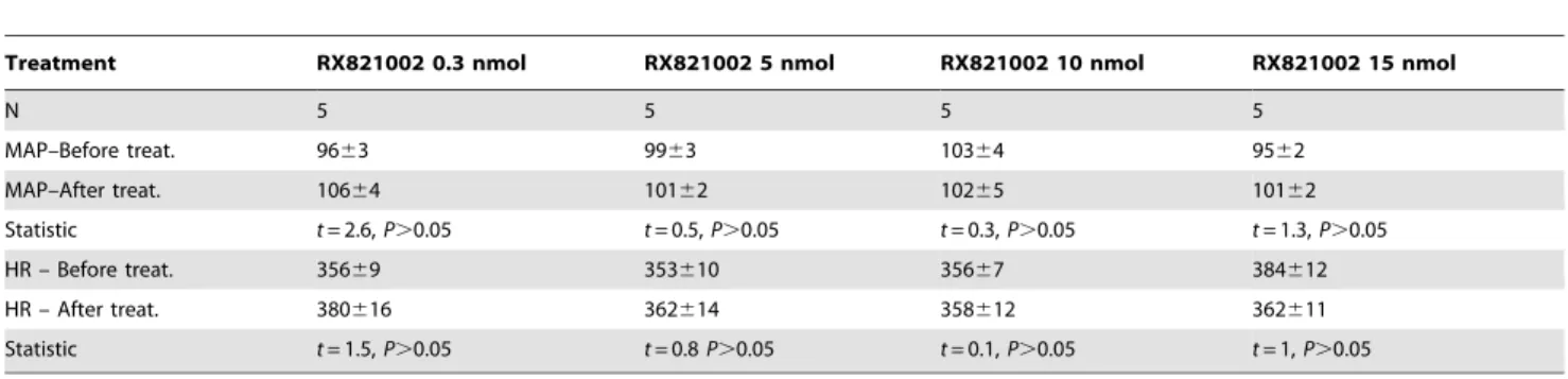
Table 2.Effect of bilateral microinjections into the IC of crescent doses (0.3, 5, 10 and 15 nmol/100 nl) of the selectivea2 -adrenoceptor antagonist RX821002 on mean arterial pressure (MAP) and heart rate (HR) baseline.
Treatment RX821002 0.3 nmol RX821002 5 nmol RX821002 10 nmol RX821002 15 nmol
N 5 5 5 5
MAP–Before treat. 9663 9963 10364 9562
MAP–After treat. 10664 10162 10265 10162
Statistic t= 2.6,P.0.05 t= 0.5,P.0.05 t= 0.3,P.0.05 t= 1.3,P.0.05
HR – Before treat. 35669 353610 35667 384612
HR – After treat. 380616 362614 358612 362611
Statistic t= 1.5,P.0.05 t= 0.8P.0.05 t= 0.1,P.0.05 t= 1,P.0.05
Student’st-test.
References
1. Joels M, Baram TZ (2009) The neuro-symphony of stress. Nat Rev Neurosci 10: 459–466.
2. Alves FH, Gomes FV, Reis DG, Crestani CC, Correa FM, et al. (2013) Involvement of the insular cortex in the consolidation and expression of contextual fear conditioning. Eur J Neurosci 38: 2300–2307.
3. Dampney RA, Horiuchi J, McDowall LM (2008) Hypothalamic mechanisms coordinating cardiorespiratory function during exercise and defensive behaviour. Auton Neurosci 142: 3–10.
4. Campeau S, Watson SJ (1997) Neuroendocrine and behavioral responses and brain pattern of c-fos induction associated with audiogenic stress. J Neuroendocrinol 9: 577–588.
5. Crestani CC, Tavares RF, Alves FH, Resstel LB, Correa FM (2010) Effect of acute restraint stress on the tachycardiac and bradycardiac responses of the baroreflex in rats. Stress 13: 61–72.
6. Hatton DC, Brooks V, Qi Y, McCarron DA (1997) Cardiovascular response to stress: baroreflex resetting and hemodynamics. Am J Physiol 272: R1588–1594. 7. Nosaka S (1996) Modifications of arterial baroreflexes: obligatory roles in cardiovascular regulation in stress and poststress recovery. Jpn J Physiol 46: 271– 288.
8. Schenberg LC, Vasquez EC, da Costa MB (1993) Cardiac baroreflex dynamics during the defence reaction in freely moving rats. Brain Res 621: 50–58. 9. Fornari RV, Wichmann R, Atucha E, Desprez T, Eggens-Meijer E, et al. (2012)
Involvement of the insular cortex in regulating glucocorticoid effects on memory consolidation of inhibitory avoidance training. Front Behav Neurosci 6: 10. 10. Resstel LB, Correa FM (2006) Involvement of the medial prefrontal cortex in
central cardiovascular modulation in the rat. Auton Neurosci 126–127: 130– 138.
11. Krettek JE, Price JL (1977) The cortical projections of the mediodorsal nucleus and adjacent thalamic nuclei in the rat. J Comp Neurol 171: 157–191. 12. Leonard CM (1969) The prefrontal cortex of the rat. I. Cortical projection of the
mediodorsal nucleus. II. Efferent connections. Brain Res 12: 321–343. 13. Verberne AJ, Owens NC (1998) Cortical modulation of the cardiovascular
system. Prog Neurobiol 54: 149–168.
14. Alves FH, Crestani CC, Resstel LB, Correa FM (2011) Cardiovascular effects of noradrenaline microinjected into the insular cortex of unanesthetized rats. Auton Neurosci 160: 90–98.
15. Nagai M, Hoshide S, Kario K (2010) The insular cortex and cardiovascular system: a new insight into the brain-heart axis. J Am Soc Hypertens 4: 174–182. 16. Alves FH, Crestani CC, Resstel LB, Correa FM (2009) N-methyl-D-aspartate receptors in the insular cortex modulate baroreflex in unanesthetized rats. Auton Neurosci 147: 56–63.
17. Saad MA, Huerta F, Trancard J, Elghozi JL (1989) Effects of middle cerebral artery occlusion on baroreceptor reflex control of heart rate in the rat. J Auton Nerv Syst 27: 165–172.
18. Saleh TM, Connell BJ (1998) Role of the insular cortex in the modulation of baroreflex sensitivity. Am J Physiol 274: R1417–1424.
19. Zhang ZH, Rashba S, Oppenheimer SM (1998) Insular cortex lesions alter baroreceptor sensitivity in the urethane-anesthetized rat. Brain Res 813: 73–81. 20. Alves FH, Crestani CC, Correa FM (2010) The insular cortex modulates cardiovascular responses to acute restraint stress in rats. Brain Res 1333: 57–63. 21. Kretz R (1984) Local cobalt injection: a method to discriminate presynaptic axonal from postsynaptic neuronal activity. J Neurosci Methods 11: 129–135. 22. Lomber SG (1999) The advantages and limitations of permanent or reversible
deactivation techniques in the assessment of neural function. J Neurosci Methods 86: 109–117.
23. Cecchi M, Khoshbouei H, Javors M, Morilak DA (2002) Modulatory effects of norepinephrine in the lateral bed nucleus of the stria terminalis on behavioral and neuroendocrine responses to acute stress. Neuroscience 112: 13–21. 24. Ma S, Morilak DA (2005) Chronic intermittent cold stress sensitises the
hypothalamic-pituitary-adrenal response to a novel acute stress by enhancing noradrenergic influence in the rat paraventricular nucleus. J Neuroendocrinol 17: 761–769.
25. Pardon MC, Gould GG, Garcia A, Phillips L, Cook MC, et al. (2002) Stress reactivity of the brain noradrenergic system in three rat strains differing in their neuroendocrine and behavioral responses to stress: implications for susceptibility to stress-related neuropsychiatric disorders. Neuroscience 115: 229–242. 26. Cecchi M, Khoshbouei H, Morilak DA (2002) Modulatory effects of
norepinephrine, acting on alpha 1 receptors in the central nucleus of the amygdala, on behavioral and neuroendocrine responses to acute immobilization stress. Neuropharmacology 43: 1139–1147.
27. Morrison JH, Grzanna R, Molliver ME, Coyle JT (1978) The distribution and orientation of noradrenergic fibers in neocortex of the rat: an immunofluores-cence study. J Comp Neurol 181: 17–39.
28. Morrison JH, Molliver ME, Grzanna R (1979) Noradrenergic innervation of cerebral cortex: widespread effects of local cortical lesions. Science 205: 313– 316.
29. Sara SJ, Segal M (1991) Plasticity of sensory responses of locus coeruleus neurons in the behaving rat: implications for cognition. Prog Brain Res 88: 571–585. 30. Ungerstedt U (1971) Stereotaxic mapping of the monoamine pathways in the rat
brain. Acta Physiol Scand Suppl 367: 1–48.
31. Alves FH, Crestani CC, Resstel LB, Correa FM (2009) Insular cortex alpha1-adrenoceptors modulate the parasympathetic component of the baroreflex in unanesthetized rats. Brain Res 1295: 119–126.
32. Crestani CC, Alves FH, Resstel LB, Correa FM (2008) Both alpha1 and alpha2-adrenoceptors mediate the cardiovascular responses to noradrenaline microin-jected into the bed nucleus of the stria terminal of rats. Br J Pharmacol 153: 583– 590.
33. Crestani CC, Alves FH, Tavares RF, Correa FM (2009) Role of the bed nucleus of the stria terminalis in the cardiovascular responses to acute restraint stress in rats. Stress 12: 268–278.
34. Barron BA, Van Loon GR (1989) Role of sympathoadrenomedullary system in cardiovascular response to stress in rats. J Auton Nerv Syst 28: 179–187. 35. Irvine RJ, White J, Chan R (1997) The influence of restraint on blood pressure
in the rat. J Pharmacol Toxicol Methods 38: 157–162.
36. Krieman MJ, Hershock DM, Greenberg IJ, Vogel WH (1992) Effects of adinazolam on plasma catecholamine, heart rate and blood pressure responses in stressed and non-stressed rats. Neuropharmacology 31: 33–38.
37. McDougall SJ, Paull JR, Widdop RE, Lawrence AJ (2000) Restraint stress : differential cardiovascular responses in Wistar-Kyoto and spontaneously hypertensive rats. Hypertension 35: 126–129.
38. Abercrombie ED, Jacobs BL (1987) Single-unit response of noradrenergic neurons in the locus coeruleus of freely moving cats. I. Acutely presented stressful and nonstressful stimuli. J Neurosci 7: 2837–2843.
39. Morilak DA, Fornal CA, Jacobs BL (1987) Effects of physiological manipulations on locus coeruleus neuronal activity in freely moving cats. III. Glucoregulatory challenge. Brain Res 422: 32–39.
40. Morilak DA, Fornal CA, Jacobs BL (1987) Effects of physiological manipulations on locus coeruleus neuronal activity in freely moving cats. II. Cardiovascular challenge. Brain Res 422: 24–31.
41. Page ME, Akaoka H, Aston-Jones G, Valentino RJ (1992) Bladder distention activates noradrenergic locus coeruleus neurons by an excitatory amino acid mechanism. Neuroscience 51: 555–563.
42. Valentino RJ, Foote SL, Page ME (1993) The locus coeruleus as a site for integrating corticotropin-releasing factor and noradrenergic mediation of stress responses. Ann N Y Acad Sci 697: 173–188.
43. Morrison SF (2001) Differential control of sympathetic outflow. Am J Physiol Regul Integr Comp Physiol 281: R683–698.
44. Mayorov DN, Head GA (2002) Ionotropic glutamate receptors in the rostral ventrolateral medulla mediate sympathetic responses to acute stress in conscious rabbits. Auton Neurosci 98: 20–23.
45. Busnardo C, Tavares RF, Resstel LB, Elias LL, Correa FM (2010) Paraventricular nucleus modulates autonomic and neuroendocrine responses to acute restraint stress in rats. Auton Neurosci 158: 51–57.
46. Deolindo MV, Reis DG, Crestani CC, Tavares RF, Resstel LB, et al. (2013) NMDA receptors in the lateral hypothalamus have an inhibitory influence on the tachycardiac response to acute restraint stress in rats. Eur J Neurosci 38: 2374–2381.
47. Tavares RF, Correa FM (2006) Role of the medial prefrontal cortex in cardiovascular responses to acute restraint in rats. Neuroscience 143: 231–240. 48. Daubert DL, McCowan M, Erdos B, Scheuer DA (2012) Nucleus of the solitary tract catecholaminergic neurons modulate the cardiovascular response to psychological stress in rats. J Physiol 590: 4881–4895.
49. Carrive P (2002) Cardiovascular and behavioural components of conditioned fear to context after ganglionic and alpha-adrenergic blockade. Auton Neurosci 98: 90–93.
50. Carrive P (2006) Dual activation of cardiac sympathetic and parasympathetic components during conditioned fear to context in the rat. Clin Exp Pharmacol Physiol 33: 1251–1254.
51. Baudrie V, Tulen JH, Blanc J, Elghozi JL (1997) Autonomic components of the cardiovascular responses to an acoustic startle stimulus in rats. J Auton Pharmacol 17: 303–309.
52. Iwata J, LeDoux JE (1988) Dissociation of associative and nonassociative concomitants of classical fear conditioning in the freely behaving rat. Behav Neurosci 102: 66–76.
53. Butcher KS, Cechetto DF (1995) Autonomic responses of the insular cortex in hypertensive and normotensive rats. Am J Physiol 268: R214–222.
54. Cechetto DF, Chen SJ (1992) Hypothalamic and cortical sympathetic responses relay in the medulla of the rat. Am J Physiol 263: R544–552.
55. Oppenheimer SM, Kedem G, Martin WM (1996) Left-insular cortex lesions perturb cardiac autonomic tone in humans. Clin Auton Res 6: 131–140. 56. Sandkuhler J, Maisch B, Zimmermann M (1987) The use of local anaesthetic