AISI 1005 Steel Plasma Treated by Diferent Thermochemical Surface Treatments
Keli Vanessa Salvador Damina,b*, Thiago de Souza Lamima, Francisco Cavilha Netoa, Cristiano Bindera,
Aloisio Nelmo Kleina, Ana Maria Maliskaa
Received: October 3, 2015; Revised: February 3, 2016; Accepted: July 13, 2016
To modify the surface structure of AISI 1005 steel and its properties without any dimensional loss,
diferent plasma surface treatments were performed at low temperatures (500 °C) in this study. The samples were subjected to single plasma treatments including: nitriding (N5% and N3%), carburizing (CE) and ferritic nitrocarburizing (NC) and to duplex treatments of nitriding followed by carburizing (N5%+CE and N3%+CE) and ferritic nitrocarburizing followed by carburizing (NC+CE). The gas mixture used for these treatments was varied as follows: nitriding (5%N2+95%H2 and 3%N2+97%H2),
carburizing (5%CH4+95%H2) and ferritic nitrocarburizing (5%N2+1.5%CH4+93.5%H2). A microstructural
characterization of the samples was carried out using optical and scanning electron microscopy in addition to XRD analysis. Microhardness testing was also performed. The XRD analysis showed a stabilization of the outermost cementite layer for all of the carburizing treatments. The results show
that a greater hardness increase was achieved for the nitriding treatment as well as a more regular
compound layer. However, a greater depth of hardening was obtained in samples with NC+CE and N5%+CE, which extended to the hardened depth to 800 μm.
Keywords: Plasma Carburizing, Plasma Nitriding, Plasma Ferritic Nitrocarburizing
* e-mail: [email protected]
1. Introduction
In recent decades, there have been constant advancements
in the techniques used for surface modiications of materials.
The importance of such techniques is linked to the fact that
modiications of the surface properties enable new applications
for materials due to changes in the chemical, physical, mechanical, metallurgical and tribological properties1-5. Among
these techniques is a plasma surface thermochemical treatment. Nitriding is a thermochemical treatment in which the
hardening of a surface is achieved by introducing nitrogen
through exposure in a nitriding atmosphere, which permits the formation of nitrides3,6,7. Nitriding is performed at
temperatures within the ferritic phase (500 – 570 °C) making
the subsequent quenching treatment used to increase the hardness unnecessary8. The same applies for the ferritic
nitrocarburizing process, which difers only in the composition of the atmosphere containing the carbon.
Carburizing is also a surface treatment utilizing carbon difusion. Generally, for carbon steels, this treatment occurs normally above 900 °C. However, for pieces that require
high dimensional and geometric controls, the use of the high
temperatures used in the carburizing treatment becomes a
problem, as it leads to a loss of dimensional control9.
Carburizing treatments at low temperatures has been
developed, as described in10-13, for stainless steels to avoid
sensitization above 450 °C in these materials, and also for
sintered pure iron and automobile gears, again, both to ensure dimensional control9,14. Furthermore, the literature contains
some studies describing plasma carburizing at low temperatures
as a method to produce surface layers of pure cementite15-18. This work evaluates the layers obtained in AISI 1005 steel
for diferent plasma surface treatments with regards to the structure, microstructure and hardness of the formed layers.
2. Material and Methods
The material used as a substrate in this study is AISI 1005 low carbon steel with a chemical composition shown
in Table 1. The samples were discs with a diameter of 25.4 mm and a height of 6 mm. All of the samples were ground,
polished and cleaned in an acetone ultrasonic bath prior to
the thermochemical treatments.
Seven diferent thermochemical treatments were carried out in a DC pulsed glow discharge reactor with the samples in the cathode coniguration. Single plasma treatments including nitriding, carburizing ferritic nitrocarburizing and double treatments of nitriding followed by carburizing and ferritic nitrocarburizing followed by carburizing were performed.
The parameters of these treatments and nomenclature used
are presented in Table 2. The working temperature was reached using auxiliary heating and the cooling of the samples performed in the plasma environment.
aMechanical Engineering Department, Federal University of Santa Catarina – UFSC, 88040-900,
Florianópolis, SC, Brazil
bTechnical Department in Mechanical, Federal Institute of Santa Catarina – IFSC, 89813-000,
Table 1: Chemical composition of AISI 1005 (wt.%).
C Si Mn P S Cr Fe
0,036 <0,002 0,173 0,036 0,014 0,01 balance
Table 2: Cycling and parameters of the treatments.
Nomenclature Treatment Gas Mixture Pressure (Torr) Time (h) DDP (V) (cmFlow 3/min)
Temperature
(oC)
N5% Nitriding 5%N2 / 95%H2 3 3 500 240 500
N3% Nitriding 3%N2 / 97%H2 3 3 500 240 500
NC Ferritic Nitrocarburizing 5%N2 / 1.5%CH4 / 93.5%H2 3 3 500 240 500
CE Carburizing 5%CH4 / 95%H2 5 3 500 240 500
N5%+CE Nitriding 5%N2 / 95%H2 3 3 500 240 500
Carburizing 5%CH4 / 95%H2 5 3 500 240 500
N3%+CE Nitriding 3%N2 / 97%H2 3 3 500 240 500
Carburizing 5%CH4 / 95%H2 5 3 500 240 500
NC+CE Ferritic Nitrocarburizing 5%N2 / 1.5%CH4 / 93.5%H2 3 3 500 240 500
Carburizing 5%CH4 / 95%H2 5 3 500 240 500
Conventional metallography procedures were performed to analyze the microstructure and the layers. The samples were etched with a Nital 2% solution and the obtained microstructures were examined using optical (Olympus BX60M) and scanning electron microscopy (Hitachi TM 3030).
A Philips X-ray difractometer (X’Pert MPD) with a copper Kα radiation source (λ = 1.5418 Å) was used to
determine the phases of the samples before and after the
plasma treatments. The numbers of JCPDS cards used to identify the phases were: 03-065-2412 (cementite), 03-065-4899 (α-Fe) 01-077-2006 (Fe4N) and
01-083-0879 (Fe3N).
The mechanical properties were evaluated for surface
microhardness using a Leco LM100AT with a Vickers indenter and a 0.01 kg load. For each treatment, a cut was made in the sample in the transverse direction to expose
a surface for microhardness measurements to be made at
diferent depths (25, 50, 75, 100, 125, 150, 200, 300, 400, 500, 650, 750 and 800 micrometers) to statistically determine the substrate hardness. Hardness measurements were also performed on the sample surface.
The large number of measurements used for the
microhardness tests were performed to ensure a maximum relative error of 10% and a reliability of 95%. Analysis test
hypotheses, estimation of variance and multiple comparisons
of means were also performed.
3. Results and discussion
3.1 Microstructure and X-ray difraction
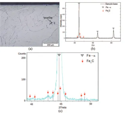
Figure 1(a) shows a cross section of the AISI 1005 steel sample surface without any thermochemical treatments. We
observe only the presence of the ferrite phase and a small
fraction of pearlite in the microstructure, which is conirmed by the XRD pattern presented in Figures 1 (b) and (c).
Figure 2 (a) shows the surface microstructure of the sample after plasma carburizing (CE). We observe a thin carburized layer, which appears to be homogeneous along the entire surface. This layer may be related to the increase
of the cementite peaks in the XRD patterns of this sample, as
seen in Figure 2 (b). Thus, the plasma carburizing treatment
promotes the formation of a thin Fe3C layer on the substrate. The same result was seen in9, which is attributed to the formation of this thin layer of cementite because of the
low difusivity of carbon at 500 °C, and the low solubility of these atoms in α-Fe, which is approximately 0.0025 wt.% at 500 °C. From this, the adsorbed carbon atoms on the surface do not have enough mobility to difuse into the
substrate, causing saturation of the element on the surface,
and therefore, the precipitation of cementite.
Figure 1: Sample base: (a) optical microscopy (b), XRD and (c) detailed XRD.
Figure 2: Sample CE: (a) scanning electron microscopy and (b) XRD.
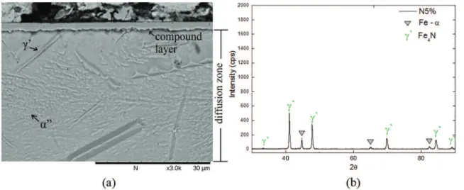
a difusion layer. The difusion zone is mainly formed by γ’-Fe4N in form of elongated needles, and also by α”-Fe16N2 nitrides in form of short needles, as suggested by19 and also reported in6,20. The compound layer is very regular and
consists of nitride γ’-Fe4N, as shown in the XRD spectrum
of Figure 3 (b).
The XRD analysis of sample N3% is shown in Figure 4 (b). The difractogram indicates the presence of Fe-α and discrete peaks corresponding to γ’-Fe4N and cementite.
Figure 4 (a) shows a micrograph of sample N3% obtained using SEM. Note that there was no formation of a compound layer, which was already expected according to authors who
claim that nitriding with a low nitrogen activity avoids the formation of the compound layer21-23.
Figure 5 (a) shows a micrograph of the cross section for the NC treatment. The formation of a compound layer and difusion zone was observed for this treatment.
It is possible to observe that the compound layer is
irregular and contained γ’-Fe4N and ε-Fe3N nitrides. Based on how the Fe3C carbide was conirmed in the XRD analysis, we may assume that the formation of Fe3C occurred. The cementite formation may have been promoted because of
the presence of 1.5 vol.% methane in the gas mixture that
Figure 3: Sample N5%: (a) scanning electron microscopy image and (b) XRD.
Figure 4: Sample N3%: (a) scanning electron microscopy and (b) XRD.
The presence of carbon is responsible for the stabilization and increase in the ε phase in the Fe-C-N phase diagram and a decrease in the phase γ’. According to Figueiredo24 the
γ’- Fe4N phase is unstable and tends to evolve into cementite
by releasing nitrogen and carbon enrichment.
The difusion zone of the NC sample is also shown in Figure 5 (a) and consists predominantly of nitride γ’-Fe4N
and a small fraction of α”-Fe16N2.
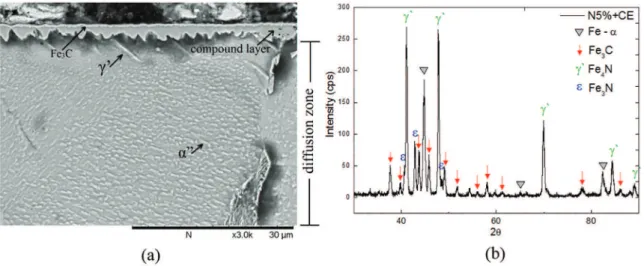
Figure 6 (a) shows a cross section of sample N5% + CE.
It is evident that there are three regions: a very thin outer
layer, a compound layer and a difusion zone.
The outer layer exhibits similar thin and regular characteristics as those observed in the CE treatment, as shown in Figure 2 (a). This layer is easily detached during the metallographic preparation procedure.
It is believed that the outer layer is composed of cementite
from the plasma carburizing performed after the nitriding treatment. Furthermore, in this treatment, an increase is
observed in the number and intensity of Fe3C peaks in the
XRD pattern, shown in Figure 6 (b), than those in samples not receiving the carburizing treatment. This may conirm
the assumption that a thin layer of cementite formed above
the compound layer in the N5%+CE samples.
The compound layer, shown in detail in Figure 6 (a), is visually the thicker layer among the diferent treatments performed in this study. Moreover, the layer contains γ’-Fe4N
and ԑ-Fe3N nitrides, as observed in the XRD pattern in Figure
6 (b), which also exhibits the presence of γ’-Fe4N and α”-Fe16N2 nitrides, which are associated with the difusion zone.
Sample NC + CE shows the same three layers observed in the treatment N5% + CE (Figure 7 (a)). The only diference is that the cementite layer is more discreet.
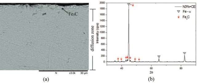
Micrographic analysis of the cross section of sample N3%+CE (Figure 8 (a)) revealed the formation of two regions: the outermost layer and the difusion zone. It is
assumed that the layer consists of cementite due to the
increased peaks of this compound shown in the sample’s
Figure 6: Sample N5%+CE: (a) scanning electron microscopy and (b) XRD.
XRD. Still, we observe that the formed layer exhibits some microcracking and has a thickness exceeding that obtained in the CE sample (Figure 2 (a)). Thus, we assume that the N3% treatment promoted an increase in the thickness of the hardened layer.
The sample difusion zone in sample N3%+CE is similar to that formed in sample N3%, and consists of nitrides and γ’-Fe4N e α”-Fe16N2.
3.2 Microhardness
Table 3 shows the results for the surface hardness and
the hardening depth obtained for each treatment.
Table 4 shows the layer thickness results obtained
using SEM. For the N3% treatment, no layer thickness was measured, as the sample exhibited no compound layer, which was previously discussed. It is important to note that there were some diiculties in the metallographic preparation of the N3%+CE and NC+CE samples. The cementite layer was
found to peel away from the sample during the polishing
step. For this reason, Table 4 shows the layer thicknesses for the N3%+CE and NC+ CE samples determined using 2000 grit sandpaper without any polishing. Because of this, we
avoided making comparisons between the layer thicknesses obtained using only sanded samples and those from polished
samples. This is because the imaged layer for only a sanded
sample is larger than the imaged layer for a sample that has
been polished.
Hypothesis testing and estimation of the variances
between the top hardness of the sample base and sample
CE shows that both are statistically identical with a 95% probability. This was already observed by Lamim9, who
attributed this result to the thin layer (in this case 0.45 µm)
of cementite, which is a very hard compound, on a ductile substrate which leads to the breakdown of the hardened layer and the concealment of its hardness by the hardness of the
Figure 7: Sample NC+CE: (a) scanning electron microscopy and (b) XRD.
Figure 8: Sample N3%+CE: (a) scanning electron microscopy and (b) XRD.
Table 3: Microhardnesses on the top and hardened depth of the studied samples.
Treatment Microhardness top (HV) Hardening depth (µm)
Base 192.4 ± 5.5 ─
CE 206.5 ± 14.9 100
N3%+CE 214.6 ± 12.9 500
N3% 215.7 ± 10.0 500
NC+CE 224.6 ± 20.6 800
NC 331.1 ± 22.5 650
N5%+CE 362.7 ± 59.7 800
N5% 443.9 ± 20.9 500
Table 4: Thickness of the layers obtained from the treatments.
Treatment Layer formed Layer thickness (µm)
CE Cementite 0.45 ± 0.02
N5% Compound layer 2.81 ± 0.26
NC Compound layer 1.92 ± 0.43
N5%+CE Cementite 0.58 ± 0.10
Compound layer 1.73 ± 1.03
NC+CE Cementite* 0.95 ± 0.10
Compound layer* 3.13 ± 0.48
N3%+CE Cementite* 1.73 ± 0.41
*Thickness measurements performed with only the layer sanded
(without polishing).
hardness measurement, Jönsson25 determined that the ratio of the indentation depth and sample thickness of the layer
must be within a critical range, which varies from 0.07 to 0.2. For the carburized sample, the relation is higher than the critical value, conirming the inluence of the substrate hardness values.
Although the presence of precipitates was not observed
in the CE samples, the proiles of the microhardness in the
The sample which exhibited the higher surface hardness was the one subjected to the nitriding treatment (N5%). This may be a result of the signiicant thickness (2.81 µm ± 0.26 µm) and regularity of the formed compound layer.
The results of the top microhardnesses of the sample
NC+CE (362.7 HV ± 59.7 HV) are similar to that presented
by Doré26, who used the same conditions as in this study and
obtained 360.7 HV ± 43.3 HV. The same was not observed for sample N5%+CE, which exhibited a hardness obtained of 149.0 HV ± 28.6 HV26, and difered signiicantly from
the value found in this work: 224.6 HV ± 20.6 HV. It is evident that when comparing sample N5% to samples NC, N5%+CE and NC+CE, sample N5% possesses the lowest hardness depth (500 μm), but has the highest surface hardness
This is attributed to the increased thickness of the compound layer that formed for this treatment compared to the others,
conirming the assumption that the greater the thickness of the compound layer, the smaller the difusion zone27.
The sample N3%+CE exhibited a microhardness of 214.6 HV ± 12.9 HV, which is statistically identical to sample N3% (215.7 HV ± 10.0 HV), despite the irst sample
having undergone the nitriding process to promote a greater
hardness. This result is due to the cementite layer formed during the N3%+CE treatment, leading to a very thin layer
upon a ductile substrate, resulting in the breakage of the
carburized layer and the concealment of hardness by the
hardness of the substrate25. This situation was mentioned
above for sample CE.
The treatments that had the highest hardening depth were
the NC+CE and the N5%+CE, which exhibited a hardened depth of 800 μm. This greater depth of hardening is attributed
to the longer hold time that the samples were subjected to the two successive treatments, which provided more time
to difuse nitrogen throughout the sample.
4. Conclusions
The highest surface hardness was obtained from the
nitriding treatment (N5%).
For the double treatments of N5%+CE, N3%+CE and NC+CE a ine layer of cementite was observed.
The nitrocarburized depth is 650 μm. A greater hardening depth was obtained in the samples that were nitrocarburized + carburizing and nitrided + carburized, which achieved a hardened depth of 800 μm.
5. Acknowledgements
The authors would like to thank the Materials Laboratory (LabMat) and Microstructural Characterization (LCM) of the Mechanical Engineering Department, Federal University of Santa Catarina.
6. References
1. Çavuşlu F, Usta M. Kinetics and mechanical study of plasma electrolytic carburizing for pure iron. Applied Surface Science. 2011;257(9):4014-4020.
2. Çetin A, Tek Z, Öztarhan A, Artunç N. A comparative study of
single and duplex treatment of martensitic AISI 420 stainless
steel using plasma nitriding and plasma nitriding-plus-nitrogen
ion implantation techniques. Surface and Coatings Technology. 2007;20(19-20):8127-8130.
3. Bendo T, Maliska AM, Acuña JJS, Binder C, Demetrio KB, Klein AN. Nitriding of surface Mo-enriched sintered iron: Structure
and morphology of compound layer. Surface and Coatings
Technology. 2014;258:368-373.
4. Klein AN, Cardoso RP, Pavanati HC, Binder C, Maliska AM, Hammes G, et al. DC Plasma Technology Applied to Powder Metallurgy:
an Overview. Plasma Science and Technology. 2013;15(1):70-81.
5. De Mello JDB, Binder C, Binder R, Klein AN. Efect of nature of nitride phases on microabrasion of plasma nitrided sintered iron. Tribology - Materials, Surfaces & Interfaces.2010;4(4):191-196.
6. Bendo T, Pavanati HC, Klein AN, Martinelli AE, Maliska AM.
Plasma Nitriding of Surface Mo-Enriched Sintered Iron. ISRN
Materials Science. 2011;2011:8p.
7. Hosseini SR, Ashraizadeh F. Accurate measurement and evaluation of the nitrogen depth proile in plasma nitrided iron. Vacuum. 2009;83(9):1174-1178.
8. Maliska AM. Inluência de elementos de liga e do oxigênio
no processo de nitretação por plasma em aços sinterizados. [Doctoral thesis]. Florianópolis: Federal University of Santa Catarina; 1995.
9. Lamim TS, Benardelli EA, Binder C, Klein AN, Maliska AM. Plasma carburizing of sintered pure iron at low temperature. Materials Research. 2015;18(2):320-327.
10. Sun Y. Kinetics of low temperature plasma carburizing of
austenitic stainless steels. Journal of Materials Processing
Technology. 2005;168(2):189-194.
11. Sun Y. Tribocorrosion behavior of low temperature plasma
carburized stainless steel. Surface and Coatings Technology.
2013;228(Suppl 1):s342-s348.
12. Scheuer CJ, Cardoso RP, Zanetti FI, Amaral T, Brunatto SF. Low-temperature plasma carburizing of AISI 420 martensitic stainless steel: Inluence of gas mixture and gas low rate. Surface and Coatings Technology.2012;206(24):5085-5090.
13. Souza RM, Ignat M, Pinedo CE, Tschiptschin AP. Structure and properties of low temperature plasma carburized austenitic stainless steels. Surface and Coatings Technology. 2009;204(6-7):1102-1105.
14. Baek JM, Cho YR, Kim DJ, Lee KH. Plasma carburizing process for the low distortion of automobile gears. Surface and Coatings Technology. 2000;131(1-3):568-573.
15. Siriwardane H, Pringle OA, Newkirk JW, James WJ. Microstructure and physical properties of iron carbide ilms
formed by plasma enhanced chemical vapor deposition. Thin
16. Li JL, O’Keefe TJ, James WJ. Iron carbide ilms formed by
plasma deposition and plasma carburizing. Materials Science
and Engineering: B. 1990;7(1-2):15-23.
17. Carpene E, Schaaf P. Formation of Fe3C surface layers by laser
plasma cementation. Applied Physics Letters. 2002;80:891-893.
18. Schaaf P, Kahle M, Carpene E. Reactive laser plasma coating
formation. Surface and Coatings Technology.
2005;200(1-4):608-611.
19. Metin E, Inal OT. Formation and growth of iron nitrides during ion-nitriding. Journal of Materials Science. 1987;22(8):2783-2788. 20. Gontijo LC, Machado R, Miola EJ, Casteletti LC, Nascente PAP.
Characterization of plasma-nitrided iron by XRD, SEM and
XPS. Surface and Coatings Technology. 2004;183(1):10-17.
21. Mittemeijer EJ, Slycke JT. Chemical potentials and activities of
nitrogen and carbon imposed by gaseous nitriding and carburising
atmospheres. Surface Engineering. 1996;12(2):152-162.
22. Mittemeijer EJ, Somers MAJ. Thermodynamics, kinetics, and process
control of nitriding. Surface Engineering. 1997;13(6):483-497.
23. Jack DH, Jack KH. Carbides and nitrides in steel. Materials
Science and Engineering. 1973;11(1):1-27.
24. Figueiredo RS. Análise da superfície em aços nitretados por
espectroscopia Mössbauer. [Dissertation]. Florianópolis: Federal University of Santa Catarina; 1991.
25. Jönsson B, Hogmark S. Hardness measurement of thin ilms. Thin Solid Films. 1984;114(3):257-269.
26. Doré M. Nitretação-cementação e carbonitretação-cementação
a plasma em baixas temperaturas de ferro puro sinterizado. [Final paper]. Florianópolis: Federal University of Santa Catarina; 2013.