DO:
D 10.1590/S1516-14392011005000072
*e-mail: [email protected]
Effects of Silica Addition on the Chemical, Mechanical and Biological Properties of
a New
α
-Tricalcium Phosphate/Tricalcium Silicate Cement
Loreley Morejón-Alonsoa*, Raúl García Carrodeguasb, Luis Alberto dos Santosa
a
Departamento de Materiais, Escola de Engenharia,
Universidade Federal do Rio Grande do Sul – UFRGS, Porto Alegre, RS, Brazil
bDepartamento de Cerámica, Instituto de Cerámica y Vidrio – CSIC, Madrid, Spain
Received: January 28, 2011; Revised: August 14, 2011
The addition of tricalcium silicate (C3S) to apatite cements results in an increase of bioactivity and improvement in the mechanical properties. However, adding large amounts raises the local pH at early stages, which retards the precipitation of hydroxyapatite and produces a loss of mechanical strength. The introduction of Pozzolanic materials in cement pastes could be an effective way to reduces basicity and enhance their mechanical resistance; thus, the effect of adding silica on the chemical, mechanical and biological properties of α-tricalcium phosphate/C3S cement was studied. Adding silica produces a reduction in the early pH and a decrease in setting times; nevertheless, the presence of more calcium silicate hydrate (C-S-H) delays the growth of hydroxyapatite crystals and consequently, reduces early compressive strength. The new formulations show a good bioactivity, but higher cytotoxicity than traditional cements and additions higher than 2.5% of SiD2 cause a lack of mechanical strength and an elevated degradability.
Keywords: calcium phosphate cements, hydroxyapatite, tricalcium silicate, pozzolan
1. Introduction
Calcium phosphate cements (CPCs) are a clinical alternative to traditional bioceramics because they are easy to handle and shape, mold themselves well to the contours of defective surfaces, and set in situ in the bone cavity to form a solid restoration1. Since their
development in the mid-80’s, CPCs have also attracted great interest due to their chemical similarity to the mineral phase of bone tissue and their good osteoconductivity2.
Dne of the most important formulations is based on α-tricalcium phosphate [α-Ca3(PD4)2; α-TCP], which sets in situ and forms a
calcium-deicient hydroxyapatite [Ca9(HPD4)(PD4)5(DH); CDHA] when hydrated3. However, it is not very strong under compression4 and
its mechanical strength is low when compared to that of cortical bone5,
limiting its application to areas subjected to low mechanical loads6.
On view of the excellent bioresorbability of CDHA, researchers have focused their efforts on overcoming the mechanical weakness
of calcium phosphate cements by using different illers, ibers and
reinforcing additives that lead to the formation of various multiphase
composites, based on the idea that the iller in the matrix may
eliminate crack propagation7. Nevertheless, the presence of illers
prevents bone ingrowths into pores and produces a denser cement with a slower resorption rate and hence a slower bone substitution8.
Therefore, it is dificult to increase the strength of these cements
without negatively affecting other properties.
Due to their spontaneous development of strength (spontaneous consolidation) towards water9, the addition of Ca
3SiD5 (C3S) to
the α-TCP-based cement could be an effective way to increase
its mechanical strength10 and improve the bioactivity and
biocompatibility of traditional CPCs11,12. However, the pH increase
during the initial stages delays setting times and prevents apatite formation in larger concentrations.
On the chemistry of ordinary Portland cement (DCP), synthetic colloidal silica (silica fume) is a highly reactive siliceous material
which reacts with the calcium hydroxide formed in hydrated cement paste (pozzolan). Although the mechanisms by which silica fume operates are unclear, the presence of small amounts of silica can accelerate the hydration of C3S by forming colloidal calcium silicate hydrate (C-S-H), thereby increasing the rate of early strength gain13.
Thus the aim of this work was to study the effects of adding silica on the chemical, mechanical and biological properties of α-TCP/C3S
cement after ageing in simulated body luid (SBF).
2. Experimental
2.1. Materials
To prepare the α-TCP/C3S/SiD2 cement, all chemicals of analytical grade were used.
α-TCP was prepared through solid state reaction, heating the appropriate mixture of γ-Ca2P2D7 (Extra Pure, Dyne) and CaCD
3
(Extra Pure, Nuclear) at 1300 °C for 5 hours followed by quenching in air14. After calcination, the product was wet milled for 4 hours in
a polyethylene jar with alumina balls using an alcoholic medium (anhydrous ethanol, 99.5%, Cromoline) to an average particle size inferior to 10 µm.
Tricalcium silicate powders were synthesized by sol-gel route, using Ca(ND3)2.4H2D and Si(DC2H5)4 (TEDS)15. Briely, suitable
2.2. Preparation of composite samples
Synthesized C3S (7.11 µm) was mixed with α-TCP (10.71 µm) in powder ratios of 0, 5.0 and 10.0 mass % and precipitated silica
WL180 (Auriquímica Ltda - Brazil) (surface area 133 m2.g–1 and mean
diameter 19.54 µm) was added in a molar ratio SiD2:C3S=2. The liquid phase was a sodium phosphate buffer prepared from NaH2PD4 and Na2HPD4.12H2D, and the liquid-to-powder ratio (L/P) was dependent of the content of C3S added ranging from 0.4 to 0.44 mL.g–1. Each
powder sample was carefully weighed and mixed with the liquid phase in appropriate powder-to-liquid ratio, packed into silicon molds and aged at 36.5 °C with controlled humidity for 24 hours. The samples
were identiied as α-TCP, 5SiD2 and 10SiD2 being the number the quantity of C3S added in mass percent.
2.3. Setting time measurement
Setting time of samples was measured according to ASTM C266-89 using a Gillmore Needles method16. Three specimens for each
formulation were tested and standard deviation was used as a measure of the standard uncertainty. Onitial setting time was determined as the end of moldability without serious damage to the cement structure
and the inal setting time as the time beyond which it is possible to
touch the cement without causing serious damage.
2.4. In vitro tests
To assess in vitro bioactivity, the 24 hours-set pastes were soaking
in simulated SBF at 36.5 °C SBF17 for 14 days and afterward, gently
rinsed with deionized water followed by ethanol dehydration and drying in atmospheric temperature.
The degradation behavior was characterized by monitoring
changes in weight loss in SBF18. For degradability test, the disks were
accurately weighed before and after immersion in SBF. The weight
loss (WL) was calculated according to Equation 1 being W0 the initial weight of the specimen and Wd the weight of the specimen dried after different degradation times (7, 14 and 21 days). Each measurement was performed three times and the average value was calculated.
(
0)
0 % 100 d W W WL W − = × (1)
2.5. Cytotoxicity test for cements
The cell viability assay was performed by direct contact test according to the OSD 10993-5(Biological evaluation of medical Part 5: Tests for in vitro cytotoxicity) using peripheral blood mononuclear
cells (PBMCs) and the MTT assay.
The PBMCs were assembled cultured in a Dulbecco’s modiied
Eagle’s Medium (DMEM) (Sigma) with HEPES (Sigma) (free acid, 2.5-3.7 g.L–1) supplemented with 10% fetal bovine serum (FBS)
(Cultilab, Sao Paulo, Brazil), at 37 °C in a humidiied atmosphere of
5% CD2 for one 24 and 48 hours. The dissolution extracts were prepared
adding the culture medium to cement discs previously incubated in SBF
for 7 days, placed in a 24-well plates and sterilized by ethylene oxide. The cell suspension was adjusted to a density of 105 cells in 0.5 mL of
HDMEM and was added to each well of a 24-well plate. As negative control, conventional α-TCP, also incubated in SBF for 7 days was used19.
The number of viable cells was quantitatively assessed by MTT test. After incubating at 37 °C and 5% CD2 for 24 and 48 hours, 150 µL of 3 mg.mL–1 3-(4,5-dimethylthiazol-2-yl)-2,5-diphenyl tetrazolium
bromide (MTT) solution was added to extract/cell constructs and cultured for 3 hours at 37 °C. Then 100 µL of dimethyl sulfoxide (DMSD) was added to each well, and the product was colorimetrically assessed with a Model Multiskan EX Microplate Reader (Labsystems, USA). Absorbances were read at a wavelength of 540 nm.
Experimental values were analyzed via one-way ANDVA test follow by Tukey’s Multiple Comparison Test.
2.6. Characterization techniques
Phase composition of the samples was determined by X-Ray Diffraction (XRD) in a PHOLLOPS diffractometer (X´Pert MPD) and Cu-target. Diffractograms were recorded employing Ni-iltered
radiation (λ = 1.5406 Å) with a step size of 0.05° and a time/step ratio of 1 second.
Morphological variations before and after soaking in SBF were
characterized by Scanning Electron Microscopy (SEM) using a JEDL microscope (JSM-6060) on gold-coated samples.
Compressive strength (CS) was measured in servohydraulic Universal Testing Machine (MTS 810) with a load measuring cell of 10 kN and a loading rate of 1 mm/min. The number of replicas was n = 10 and Student Multiple Comparison Test was used to compare mean values.
pH measures were carried out during soaking in SBF and lectures
were made in an µPA-210 pHmeter at 36,5 °C.
3. Results and Discussion
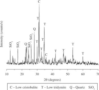
Figure 1 shows the X-ray of silica powder used in different
formulations. The results showed a mixture of different polymorphs in addition to amorphous form; even though that the only stable form under normal conditions is α-quartz and this is the form in which crystalline silicon dioxide is usually encountered. The phases found were: Cristobalite Low (JCPDS 76-0936); Tridymite M Low (JCPDS 76-0894); Quartz (JCPDS 79-1913); SiD2 (JCPDS 16-0980). The characterization of raw materials α-TCP and C3S was
described in previous works. For α-TCP powder, the presence of approximately 18% β-Ca3PD4 (β-TCP), determined by quantitative analysis19,20, was found in addition to α-TCP phase, due to the
presence of Mg2+ in raw materials. On the case of C
3S, the most
intense characteristic peaks of CaD due to the decomposition of Ca3SiD5 were observed21.
The initial and inal setting times of α-TCP and the different
composites are shown in Figure 2. The setting times of α-TCP were higher than those reported in the literature for similar materials22 due
Figure 2. Initial and inal setting time of the paste samples with different SiO2 contents (L/P) = 0.4 mL.g–1).
Figure 3. XRD patterns of cements 24 hours set.
Figure 4. XRD patterns of cements after 7 days in SBF.
Figure 5. XRD patterns of cements after 14 days in SBF. to the presence of β-TCP in the starting powder that delays -TCP
hydrolysis (Equation 2). When C3S is added, a signiicant increase in setting times is observed in relation to traditional -TCP cement (more than 200 minutes for initial setting time and 400 minutes
for inal setting time) this being directly related to the content of
C3S added12. The presence of SiD
2 reduces the setting time of the α-TCP/C3S compositions with a 5% weight content of C3S (5SiD2); however, the values obtained are still higher than those for α-TCP cements. Higher additions of SiD2 increased the setting time and are not suitable for immediate clinical applications23.
(
)
( )(
)(
)
( )3 4 2 s 2 9 4 4 5 s
3α −Ca PO +H O=Ca HPO PO OH (2)
surface of α-TCP particles preventing dissolution of the grains and precipitation of CDHA and increasing the setting times as a result.
( ) ( )
(
)
( )3 5 s 2 2 2 gel 2 s
Ca SiO +3H O=CaO.SiO .H O +2Ca OH (3)
(
)
2 s( ) 2 s( ) 2 2 ( )gelCa OH +SiO =CaO.SiO .H O (4)
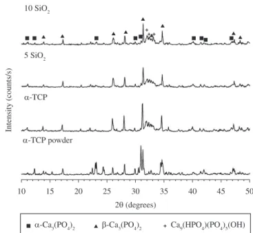
The x-ray diffraction patterns of different composites before
and after soaking in SBF are displayed in Figures 3-5. For α-TCP, mainly peaks of CDHA (JCPDS 46-0905) were observed in addition to β-TCP (JCPDS 09-0169) peaks at all soaking times. The β-TCP is considered an impurity in the starting powders as it not involved in the hydration reaction and remains unreacted; this is why their most intense peaks appear with great intensity in all diffraction patterns.
After 24 hours setting (Figure 3), in composites containing SiO2, the X-ray diffraction peaks of CHDA appeared less intense, and unreacted -TCP (JCPDS 09-0348) were also detected as a result of the delay in the dissolution of the α-TCP particles. The greater the amount of C3S/SiD2 added, the greater the intensity of the unreacted
α-TCP peaks and the lower the CDHA formed.
A few differences were observed in X-ray patterns of composites
after 7 and 14 days of SBF soaking (Figures 4-5). The X-ray spectra
of all formulations were very similar and the presence of CDHA, in addition to β-TCP and α-TCP, was observed in all cases. However, owing to the limitations of the technique, it was not possible to differentiate between the hydroxyapatite precipitated from the α-TCP
and that obtained by precipitation in SBF, and observations of the
surfaces and fracture surfaces by SEM were necessary to clarify this. Dn the other hand, the presence of calcium-silicate-hydrate (C-S-H) was not detected, probably due to the low content on the sample and the amorphous character of these hydrates.
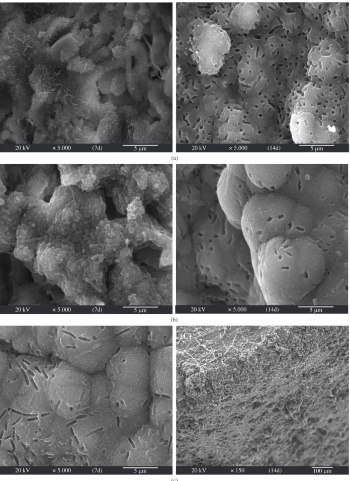
The observation by SEM is an effective way to estimate the bioactivity of materials as apatite grains and layers formed have differentiated features24; while several authors have considered the
materials to be biocompatible judging from the presence or absence
of a supericial layer of hydroxyapatite formed after immersion in simulated physiological solutions. Figure 6 shows the SEM images of α-TCP, 5SiD2 and 10SiD2 after soaking in SBF for 7 and 14 days. For
α-TCP cement (Figure 6a) a network of entangled needle-like crystals
or petal-like plates, typical for set α-TCP-based calcium phosphate cements, was observed after 7 days of immersion, whereas a layer of bone-like apatite, precipitated under physiological conditions,
was detected within 14 days of SBF soaking. Although measures
were taken to maintain aseptic conditions, bacterial contamination
by Bacillis and Cocci colonies was observed on samples surface25.
Adding small amounts of SiD2 produces a bumpy and amorphous
appearance after 7 days (Figure 6b) and a homogeneous layer of
CDHA with globular shape morphology, typical of bioactive materials after 14 days26 immersed in SBF. The higher the silicon content in
the composite, the shorter the time required for the formation of the
surface layer (Figure 6c). No typical features of type I C-S-H27 were
observed for silica additions maybe due to the large quantity of quartz present in the sample, since the SiD2 modiication used has a decisive inluence on the processes of formation of calcium silicate hydrates28.
Different microstructural features can be seen in the fracture
surface of different composites (Figure 7). Since early stages, α-TCP shows the typical petal-like plates covering the biggest grains and the growth of these plates with time, at the expense of smaller grains,
within the interstices of fracture surface (Figure 7a). With low
contents of SiD2 (Figure 7b) incipient precipitation of some CDHA crystals can be seen; crystals that grow with soaking time enclosing the larger α-TCP grains. For further additions of SiO2 (Figure 7c),
no signiicant differences with soaking time were found and the cross
section appeared covered or speckled with small distinct features that
could be identiied as small crystals of dry C-S-H29. No needle-like
crystals or petal-like plates were observed ascribed to the lack of time
for CDHA growth to occur besides insuficient time and conditions
for the material to react completely.
With the presence of C3S and SiD2, the HSiD3– ions are released
during the hydration of the composite paste, acting as sites for nucleation of apatite crystals and hence accelerating the deposition of apatite on the surface30.
Even though it was possible to reduce pH values with the addition of SiD2 (Figure 8), the compressive strength did not improve. After addition of SiD2, the values of compressive strength were inferior
to 1MPa, even after 7 days of soaking in SBF (Figure 9), and only
for 5SiD2, similar values to those of α-TCP cement were achieved after 14 days.
The increase in mechanical strength of the cement occurs as a result of the formation of the interlocking of hydroxyapatite crystals precipitated after α-TCP solubilization and in general, increases with
immersion time in SBF31. However, since the particles of SiD 2 act as
nucleation sites for C3S hydration and can cause blockage of the pores,
which densiies the hydrating gel structure, α-TCP dissolution and precipitation are delayed owing to the formation of a dense calcium silicate on the surface of α-TCP particles, hence causing the initial
low compressive strength. Moreover, although less signiicant, the
increase of porosity caused by the degradation of the materials in
early stages, also contributes to the decrease observed. For 5SiO2, with the increment in soaking time, and the advancement of the hydration reaction and crystal growth of CDHA, the compressive strength augmented.
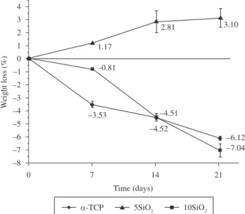
The implantation of materials into living tissue causes wound and foreign body reactions. To predict the possible harmful effects of materials on the surrounding host tissues, it is important to gain information on the degradability of the implanted materials32. The
relationship between the weight loss rate of the composites and the
immersion time in SBF (Figure 10) shows that the weight of α-TCP and 5SiD2 increased with time, whereas the weight of the composite cement decreased over time when the SiD2 content in the composite was 5% (10SiD2). Moreover, adding SiD2 produces an increase in the degradation rates of composites; however, the degradation rate of 5SiD2 sample was lower than those of the composites containing only C3S12 and the behavior observed was very similar to those of
traditional CPCs.
Degradability of some CPCs is very slow both in vivo and in vitro33 and conventional CPCs usually often experience a mass gain
after long soaking periods as a result of hydrolysis of α-TCP or due to the formation of apatite on the surface of the samples. Considering the fact that the degradability is primarily governed by the chemical composition, the reason for the higher degradation rate of the α-TCP/ C3S/SiD2 composites could be the higher solubility of the C-S-H as compared with CDHA. On addition, the dissolution of C-S-H and/or other phases could be larger than the amount of apatite deposited on the cement surface and formed due to the hydrolysis of α-TCP, so the weight of samples decreased.
Ot is generally accepted that the in vitro cell-material interaction
is a useful criterion in the evaluation of new biomaterials. Figure 11
shows the result of direct cytotoxicity test for composites against the
PBMCs after incubation for 24 and 48 hours. Although differences were no statistical signiicant, composites containing SiO2 seem to be more cytotoxic than traditional α-TCP. Furthermore, it was found that the viability of PBMCs showed a tendency towards a decrease
Figure 9. Compressive strength of the α-TCP α-TCP/C3S/SiD2 composites
after soaking in SBF.
Figure 10. Weight loss of the α-TCP and α-TCP/C3S/SiD2 composites after
soaking in SBF.
Figure 11. Cell viability of PBMCs on the different pastes after culturing for 24 and 48 hours.
some chemical transformations of the material in culture medium since after 7 days in aqueous media the pastes continues hydrating.
When only C3S is added, compositions are less cytotoxic and more compatible than pure α-TCP-based cement as a result of the dissolution of silicate ions present in α-TCP/C3S pastes that stimulate cell proliferation34,35. With the presence of SiD
2, more C-S-H is
formed and more HSiD3- is released during hydration which can
accelerate the hydroxyapatite deposition on the surface and enhance biocompatibility. However, contrary to our expectations, the results showed a fall in cell viability maybe due to the higher solubility of compositions and the poor cohesion of the cement in the presence of SiD2 which may have masked the actual results.
4. Conclusions
The addition of SiD2 reduces local pH during C3S hydrolysis and accelerates the hydration of C3S; however, it does not produce a noteworthy increase in compressive strength compared to conventional CPCs in the early stages as a result of the suppression of α-TCP dissolution. Furthermore, the presence of SiO2 increases the
degradability of cements and does not improve the biocompatibility of materials by reducing the cytotoxicity. Nevertheless, the α-TCP/ C3S/SiD2 composites possess excellent bioactivity, as indicated by
the early formation of bone-like apatite in SBF, being attributed to
the presence of C3S and not of SiD2.
Acknowledgements
This work was conducted with support from CAPES, the Brazilian
Government entity dedicated to the training of human resources.
References
1. Brown WE and Chow LC. A new calcium phosphate water-setting cement.
Cements Research Progress. 1986; 352-379.
2. LeGeros RZ, Chohayeb A and Shulman A.Apatitic calcium phosphates: possible dental restorative materials. Journal of Dental Research. 1982; 61:343.
3. Monma H. The hydration of alpha-tricalcium phosphate.
Yogo-kyokai-shi. 1976; 84: 209-213.
4. Ginebra MP, Boltong MG, Fernández E, Planell JA and Driessens
FCM. Effect of various additives and temperature on some properties
of an apatitic calcium phosphate cement. Journal of Material Science:
Materials in Medicine. 1995; 6:612-616. http://dx.doi.org/10.1007/
BF00121286
5. Santos LA, Cristina de Dliveira L, Cristina da Silva E, Garcia R,
Ortega A and Arruda A. Fiber Reinforced Calcium Phosphate. Artificial Organs. 2000; 24(3):212-216. http://dx.doi.org/10.1046/j.1525-1594.2000.06541.x
6. Yamamoto H, Niwa S, Hori M, Hattori T, Sawai K, Aoki S et al. Mechanical strength of calcium phosphate cement in vivo and in vitro.
Biomaterials 1998; 19:1587-1591. http://dx.doi.org/10.1016/S0142-9612(97)00121-X
7. Dorozhkin S. Calcium orthophosphate cements and concretes. Materials.
2009; 2:221-291. http://dx.doi.org/10.3390/ma2010221
8. Oshikawa K and Asaoka K. Estimation of ideal mechanical strength and critical porosity of calcium phosphate cement. Journal of Biomedical Material Research. 1995; 29:1537-1543. http://dx.doi.org/10.1002/ jbm.820291210
9. Lea FM. The Chemistry of Cement and concrete. London: Edward Arnold Ltd.; 1970.
10. Cárdenas LJ, Takeuchi A, Matsuya S and Ishikawa K. Effects of tricalcium
silicate addition on basic properties of α-tricalcium phosphate cement.
Journal of the Ceramic Society of Japan.2008; 116(1):83-7. http://dx.doi. org/10.2109/jcersj2.116.83
11. Morejon-Alonso L, Bareiro O, García Carrodeguas R and Santos LA.
Development and characterization of new dual setting calcium phosphate cement. On vitro behavior. On: Anais do 54th Congresso Brasileiro de Cerâmicas; 2010; Foz de Iguaçu, Brazil. Foz de Iguaçu; 2010.
12. Morejon-Alonso L, Ferreira OJB, Garcia Carrodeguas R and Santos LA. Bioactive composite bone cement based on α-tricalcium phosphate/ tricalcium silicate. Journal of Biomedical Material Research Part B. 2011. http://dx.doi.org/10.1002/jbm.b.31926
13. Zhao-Qi W and Young JF. The hydration of tricalcium silicate in
thepresence of colloidal silica. Journal of Materials Science.1984;
19:3477-3486. http://dx.doi.org/10.1007/BF02396922
14. Monma H, Goto M and Kohmura T. Effect of additives on hydration and hardness of tricalcium phosphate. Gypsum Lime. 1984; 188:11-6. 15. Zhao W and Chang J. Sol-gel synthesis and in vitro bioactivity of
tricalcium silicate powders. Material Letters. 2004; 58:2350-2353. http://dx.doi.org/10.1016/j.matlet.2004.02.045
16. American Society for Testing and Materials - ASTM. ASTM C266-89 A: Standart test method for time of setting of hydraulic-cement paste by Gillmore needles. 1995.
17. Kim HM, Miyazaki T, Kokubo T and Nakamura T. Revised Simulated
Body Fluid. Key Engineering Material. 2001; 192-195:47-50.
http://dx.doi.org/10.4028/www.scientiic.net/KEM.192-195.47
18. Wang Ch, Yan M, Chang H and Ding S. Degradation behavior of porous calcium phosphates. Journal of Medical and Biological Engineering. 2003; 23(3):159-164.
19. Morejón-Alonso L, Ferrari MB, Camassola M, Garcia R and Santos
LA. On vitro citotoxicity of a calcium phosphate-silicate composite bone cement. On: Proceedings of the 6th Congresso Latinoamericano de Órgãos Artificiais e Biomateriais; 2010; Gramado, Brazil. ABCM; 2010.
20. Morejón-Alonso L. Avaliação de cimentos ósseos de Fosfatos de Cálcio com adições de Aluminato e Silicato de Cálcio. [tese].Porto Alegre:
Universidade Federal do Rio Grande do Sul; 2011.
21. Morejon-Alonso L, Bareiro O, Garcia Carrodeguas R and Santos LA. In vitro bioactivity of a tricalcium silicate cement. On: Proceedings of the 53th Congresso Brasileiro de Cerâmicas; 2009; Guarujá, Brazil. São Paulo : Associação Brasileira de Cerâmica; 2009.
22. Ambard A and Mueninghoff L. Calcium Phosphate Cement: Review of mechanical and biological properties. Journal of Prosthodontics. 2006; 15:321-328. http://dx.doi.org/10.1111/j.1532-849X.2006.00129.x
23. Driessens FMC, Planell JA and Gil FG. Calcium phosphate bone cements.
On: Wise DL et al.,editors. Enciclopedic handbook of biomaterials and
bioengineering. New York: Ed. Marcel Decker; 1996. part B: Aplications.
p. 855-77.
24. International Organization for Standardization - ISO. ISO/FDIS 23317: Omplants for surgery. On vitro evaluation for apatite-forming ability of implant materials. OSD; 2007.
25. Gil FJ, Padro’s A, Manero JM, Aparicio C, Nilsson M and Planell JA. Growth of bioactive surfaces on titanium and its alloys for orthopaedic and dental implants. Materials Science and Engineering. 2002; C22:53-60. 26. Kokubo T and Takadama H. How useful is SBF in predicting in vivo bone
bioactivity? Biomaterials. 2006; 27: 2907-2915.
27. Kokubo T. Bioactive glass-ceramics: properties and applications. Ceramic Society ofJapan. 1991; 99:965. http://dx.doi.org/10.2109/jcersj.99.965
28. Diamond S. Cement paste microstructure: An overview at several levels. On: Proceedings of the conference on Hydraulic Cement Pastes: Their
Structure and Properties; 1977; Shefield. Shefield: University of Shefield; 1977. p. 2-29.
29. Baltaky K, Jauberthie R, Siauciunas R and Kaminskas R. Inluence
of modiication of SiO2 on the formation of calcium silicate hydrate.
Materials Science-Poland. 2007; 25(3):663-670.
30. Fonseca PC and Jennings HM. The effect of drying on early-age morphology of C-S-H as observed in environmental SEM. Cement and Concrete Research. 2010; 40:1673-1680. http://dx.doi.org/10.1016/j. cemconres.2010.08.007
31. Kokubo T. Bioactive glass-ceramics: properties and applications. Ceramic Society ofJapan. 1991; 99:965. http://dx.doi.org/10.2109/jcersj.99.965
32. Gruninger SE, Siew C, Chow LC, O’Young A, Ts’ao K and Brown WE. Evaluation of the biocompatibility of a new calcium-phosphate setting cement. Journal of Dental Research. 1984, 63:200.
33. Koerten HK and Van der Meulen J. Degradation of calcium phosphate ceramics. Journal of Biomedical Material Research. 1999; 44:78-86.
http://dx.doi.org/10.1002/(SOCO)1097-4636(199901)44:1%3C78::AOD-JBM9%3E3.0.CO;2-6
34. Hollinger JO, Brekke J, Gruskin E and Lee D. Role of bone
substitute. Clinical. Orthopedics. 1996; 324:55-66. http://dx.doi. org/10.1097/00003086-199603000-00008
35. Hench LL and West JK. Biological applications of bioactive glasses. Life Chemistry Reports. 1996; 13:187-241.