INTRODUCTION
The ordered double perovskite oxides, whose general formula is A2B’B’’O6, were irst proposed by Longo and Ward
[1] in 1961. According to those authors, A is an alkaline-earth divalent cation, B’ and B’’ are transition-metals and present an octahedral coordination with the anion O2- [2]. This type
of compound exhibits magnetic and electronic properties related to the strong interplay between structure, charge and spin ordering [3], which is the subject of nano-spintronic
studies. Spintronics is an emerging ield of science and technology that will most likely have a signiicant impact on the future of all aspects of electronics [4-6]. Moreover, spintronics is the next step towards the new technology of spin-based quantum computing and quantum information processing [7-9], voltage controlled spintronic devices for logical applications [10] and semiconductor devices [11,12].
The early discovery of large low-ield room-temperature magnetoresistance in these compounds (mainly in half-metallic Sr2FeMoO6, described by Kobayashi in 1998
Synthesis and structural characterization of the
Ca
2MnReO
6double perovskite
(Síntese e caracterização estrutural da dupla perovsquita Ca
2MnReO
6)
H. P. S. Corrêa1, I. P. Cavalcante1, D. O. Souza2, E. Z. Santos2, M. T. D. Orlando2, H. Belich2, F. J. Silva2, E. F.
Medeiro2, J. M. Pires2, J. L. Passamai2, L. G. Martinez3, J. L. Rossi3
1Universidade Federal de Mato Grosso do Sul, Campo Grande, MS
2Universidade Federal do Espírito Santo, Av. Fernando Ferrari 514, Vitória, ES 29075-910
3Instituto de Pesquisas Energéticas e Nucleares, IPEN-CNEN, Av. Prof. Lineu Prestes 2242, Cidade
Universitária, S. Paulo, SP 05508-000 [email protected]
Abstract
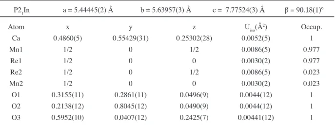
The Ca2Mn1Re1O6 double perovskite has been prepared in polycrystalline form by using the encapsulated quartz tube method. The partial oxygen pressure inside the quartz tube revealed this to be a crucial synthesis parameter for the production of a single structural phase sample. This parameter was controlled using the ratio between ReO2 and ReO3 content and the illing factor parameter (ratio between mass and total inner volume of the quartz tube). The morphology and chemical composition was investigated by scanning electron microscopy and energy dispersive X-ray spectroscopy. The crystal structure parameters were determined by analysis of the synchrotron high-resolution X-ray powder diffraction pattern. The analysis indicates that the sample is an ideal single-phase compound with a monoclinic crystal structure (space group P21/n) with a = 5.44445(2) Å;
b = 5.63957(3) Å; c = 7.77524(3) Å; and β = 90.18(1)º.Computer simulations were performed considering two cation valence conigurations, namely, (i) Mn2+Re6+ or (ii) Mn3+Re5+, for the Ca
2Mn1Re1O6 compound. XANES analysis measurements indicated +2.3 for the average valence of Mn (a mixture of Mn2+ and Mn3+) and +5.7 for the effective valence of Re (an intermediate valence between Re4+ (ReO
2) and Re6+ (ReO3)). As a summary, we concluded there is a mixed valence coniguration for Mn and Re in Ca2Mn1Re1O6 , taken into account the oxygen content of 6.0±0.1 .
Keywords:double perovskite, X-ray diffraction, lattice parameters.
Resumo
A dupla perovsquita Ca2MnReO6 na forma policristalina foi preparada utilizando o método do tubo de quartzo encapsulado. A
pressão parcial de oxigênio dentro do tubo de quartzo mostrou-se ser um parâmetro crucial para a produção de uma amostra
estrutural monofásica.Esse parâmetro foi controlado usando a relação entre o conteúdo dos precursores ReO2 e ReO3 e o
parâmetro fator de preenchimento (razão entre a massa e o volume interno total do tubo de quartzo). A morfologia e a composição química foi investigada através da microscopia eletrônica de varredura e espectroscopia de energia dispersiva de raios X . Os parâmetros de estrutura cristalina foram determinados através da análise do padrão de difração tomado com luz síncrotron de alta resolução. Finalmente, os parâmetros cristalinos foram comparados com os resultados oriundos dos cálculos de Bond-Valence Method oriundos do programa SPuDS. Como resumo, a análise indicou que a amostra é um composto monofásico com uma estrutura cristalina monoclínica (grupo espacial P21/c) com a = 5,44445(2) Å; b = 5,63957(3) Å; c = 7,77524(3) Å; and
b = 90,18(1)º.
[13]) stimulated interest in the study of the properties of ordered double perovskites, in the context of their potential application in the ield of spin electronics [14-17]. The focus of these studies was to characterize their magnetic and electronic properties as well as their crystallographic structures. Among them, the A2MReO6 series (namely
Re-based ordered double perovskites), with A = Ba, Sr, Ca and M = Cr, Fe, Mn, shows a wide variety of magnetic and electronic properties. Concerning the magnetic state, the majority of the compounds reveal ferromagnetic behavior with the coupling of the divalent magnetic M ion to Re [18].
The ideal structure of the double perovskites is based on the adapted tolerance factor t of the single perovskite [19]. In general, for double perovskites A2B’B’’O6, the tolerance
factor can be written as
t =
+ +
rA + r0 rB’
2 rB”
2 rO (A)
where rA, rB and rB’’ are the ionic radii of the respective ions and rO is the ionic radius of oxygen. The closer to t = 1, the more the structure corresponds to ideal cubic. Therefore, except in rare cases, one can consider the following rule for the double perovskite family: for 1.05 > t > 1.00 a cubic structure is adopted within the space group; for 1.00 > t > 0.97 the most likely structure corresponds to the I 4/m tetragonal space group and if t < 0.97 the compound becomes either monoclinic (P21/n) or orthorombic [20]. Philipp et al. [18] reached a similar conclusion by studying the CrW-based series. Lufaso et al. [19] reported that roughly 70% of all ordered double perovskites undergo octahedral tilting distortions. By considering 11 possible distinct types of octahedral tilting, it was shown that ive tilted systems accounted for ~97% of the reported structures. The ive dominant tilted systems reported, namely (aºaºaº), I 4/m (aºaºc-), (a-a-a-), I 2/m (aºb-b-) and P 2
1/n (a-a-b+), as
well as two additional tilted systems, (a+a+a+) and P
4/mnc (aºaºc+), can be simulated using the SPuDS (Structure
Prediction Diagnostic Software) [21].
Several reports like the ones by Philipp et al. [16] and Popov et al. [22] have studied the correlation between the A-site cation size and the properties of the double perovskites. Granado et al. [23] have concentrated on the spin-orbital manifestation of Re ion and its inluence on the electronic properties of the Ca2FeReO6 compound.
Herrero-Martín et al. [24] studied the X-ray absorption of the FeRe-base double perovskites series employing the most recent theoretical calculations in order to explain the magnetic and electronic results. Sikora et al. [25], employing X-ray magnetic circular dichroism at the Re L2,3 edges, observed
a considerable orbital magnetic moment, which implies an unquenched Re orbital moment, despite its octahedral coordination [26], in the similar series of FeRe-based double perovskites made by Herrero-Martín [24]. Finally, Serrate et al. [20] published a large topical review showing the importance of these materials for spintronic devices and that
the physics involved in these compounds is more complex and rich than expected.
This work was proposed by taking into account a scenario which the magnetic and electronics properties of the Ca2Mn1Re1O6 double perovskite present a strong correlation with structural order. The main goal was to investigate the synthesis and crystal structure of the monophasic compound Ca2Mn1Re1O6.
EXPERIMENTAL DETAILS
Ca2Mn1Re1O6 double perovskite oxide was synthesized
from CaO, MnO2, ReO2 and ReO3. The calcium oxide CaO
was prepared by decomposition of CaCO3 (Alfa Aesar
reagent, 99.9965%) irst at 950 °C for 24 h in dynamic vacuum and then at 1100 °C for 3 h under a lux of oxygen. The CaO removed from the furnace was immediately placed into a dry box. The MnO2 was used as purchased (Alfa Aesar,
puratronic, 99.999%). Both ReO2 (Aldrich, 99.9%) and
ReO3 (Aldrich, 99.9%) powders were also used as purchased.
The mixture of CaO, MnO2, ReO2 and ReO3 powders in the
2:1:0.9:0.1 stoichiometric ratio was ground and pelletized inside a dry glove box illed with Argon gas. This mixture of starting materials was wrapped in gold foil and sealed in an evacuated (10-2 torr) quartz tube. Care was taken not to
overheat the samples while sealing the quartz tubes under vacuum, due to the high vapor pressure of ReO3 at relatively
low temperatures. The 0.9:0.1 ratio between ReO2 and ReO3
was used in order to obtain a partial oxygen pressure inside the quartz tube at high temperatures during the irst thermal treatment. The ratio between sample mass and quartz tube inner volume was deined as the illing factor ff. The required illing factor was estimated to be ff@0.12g/cm3 in order
to obtain 3 bar of oxygen partial pressure during the irst thermal treatment at high temperature. Thispartial oxygen pressure inside the quartz tube revealed it to be a crucial synthesis parameter for the production of a single structural phase sample. The oxygen pressure inside the quartz tube after the irst thermal treatment at room temperature was 750 mmHg and it was measured by a home made pressure setup with accuracy of 20 mmHg. The oxygen stoichiometry of the sample was evaluated by taking into account the mass variation of the oxide mixture before and after the thermal treatment by an analytical balance (Sartorius, Model TE2145, 0.0001g accuracy). The quartz tube was placed inside a gas pressure furnace illed with argon gas at 20 bar pressure in order to avoid quartz tube leakage during the thermal treatment. The sample was sintered for a total time of 154 h at 985 °C with two intermediate grinding steps then inally cooled down slowly in the furnace.
Ca2Mn1Re1O6 and the regions of EDS microanalysis.
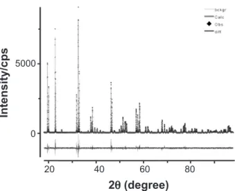
The high-resolution X-ray powder diffraction measurement was performed at the D10b-XPD beamline of the Brazilian Synchrotron Light Laboratory (LNLS), at Campinas city, using a wavelength λ = 1.54044 Å, at ambient pressure. A Ge(111) analyzer crystal was placed in a goniometer attached to the 2θ arm, and a scintillation
detector was used. The Rietveld reinement was performed using the GSAS+EXPGUI suite [27, 28]. The sample of double perovskite studied showed 100% of the desired phase Ca2MnReO6. Observed, calculated, and difference proiles are shown in Fig. 2, and the structure is drawn in Fig. 3 (a) and (b). The structure reported by Kato et al. [17] was used as the initial model for the reinement. The peak proile function was modeled using a convolution of the Thompson-Cox-Hastings pseudo-Voigt (pV-TCH) function [29] with the asymmetry correction described by Finger et al. [30], to account for the asymmetry due to the beam axial divergence. In order to account for the anisotropy in the half width of the relections, the bi-dimensional model described by Larson and Von Dreele [27] was used to account for the crystallite size and, for anisotropic strain, the model described by Stephens [31]. The value of the tolerance factor t was calculated using the bond valence parameters as obtained using the SPuDS simulation software [21]. SPuDS generates structures using computational tools for hypothetical compositions of those compounds where structural data are not available.
The local ReO6 and MnO6 octahedral oxygen coordination was investigated by X-ray absorption near edge (XANES) using the D08B – XAFS2 beamline. The measurements were recorded three times at Re LIII-edge (10.535 keV) and at Mn K-edge (6.540 keV) in transmission mode at room temperature. Si (111) channel-cut (2d = 6.271 Å) crystal produced a monochromatic beam in focus with 500 µm
diameter at 0.005 mrad (0.3 eV) resolution @ 7keV. The energy calibration was carried out using the irst inlection point of the XANES spectrum of Mn (K-edge = 6.540 keV) and Pt (LIII-edge = 11.564 keV) foils as references.
20
2q (degree)
Intensity/cps
5000
0
60
40 80
Figure 2: Observed (symbols - u), calculated (red line), background itted (green line) and difference (bottom blue line) X-ray powder diffraction proile of sample Ca2MnReO6, taken with energy 8048 eV. The goodness-of-it factors are Rwp = 0.1135, Rp = 0.0914,
c2 = 2.568 and R
F2 = 0.0549.
[Figura 2: Observado (símbolo -u), calculado (linha vermelha),
linha de fundo ajustada (linha verde) e diferença (linha de fundo
azul). Padrão de difração de raios X de pó da amostra Ca2MnReO6
tomado na energia 8048 eV. Os fatores da qualidade do ajuste
foram Rwp = 0,1135, Rp = 0,0914,c2 = 2,568 e R
F 2 = 0,0549.]
Figure 3: Schematic structure of Ca2MnReO6 (a) View of the unit cell along the crystallographic (110) direction corresponding to a pseudocubic a or b axis. The dark grey octahedra represent ReO6 and the light grey octahedra represent MnO6 ; opposite rotations of the octahedra along the viewing direction can be seen. (b) View along the crystallographic (001) direction showing in-phase rotations.
[Figura 3: Estrutura esquemática do Ca2MnReO6. (a) Vista de
célula unitária ao longo da direção cristalográica (110) que corresponde a um pseudocubico eixo a ou b. O octaedro cinza
escuro representa o ReO6 e o octaedro cinza claro representa o
MnO6; rotações opostas dos octaedros ao longo da direção podem
ser vistas. (b) Vista ao longo da direção cristalográica (001) mostrando as rotações em fase.]
Figure 1: Back scattered electron image of a Ca2Mn1Re1O6 sample carried out using a Zeiss Evo 40. The numbers (1, 2, 3 and 4) represent regions where the energy dispersive X-ray spectroscopy (EDS) microanalysis was performed.
[Figura 1: Imagem da amostra Ca2MnReO6 utilizando elétrons
retroespalhados usando um Zeiss EVO 40. Os números (1, 2, 3, e 4) representam regiões onde a microanálise por espectroscopia de raios X dispersiva (EDS) foi executada.]
3 2
1
RESULTS AND DISCUSSIONS
Oxygen stoichiometry
The CaO (99.99%), MnO2 (99.999%), ReO2 (Aldrich,
99.9%) and ReO3 (Aldrich, 99.9%) powders were mixed
in the 2:1:0.9:0.1 stoichiometric ratio. The stoichiometric equation is described below.
2.00x(CaO) + 1.00x(MnO2) + 0.90x(ReO2) +
0.10x(ReO3) Ca2Mn1Re1O6 + 0.05x(O2)
This mixture of starting materials was wrapped in gold foil and sealed in an evacuated (10-2 torr) quartz tube. The total
mass of oxide mixture and gold foil together was measured by an analytical balance (0.0001 g accuracy) before and after the thermal treatment. The oxygen stoichiometry was determined by taking into account the mass variation and the residual oxygen pressure measured in the quartz tube at room temparature after each thermal treatment, and also considering the analytical balance accuracy (0.0001 g) and the pressure measurement setup accuracy (20 mmHg). The inal oxygen stoichiometry was 6.0±0.1.
Scanning electron microscopy
A visual inspection of the SEM image (Fig. 1) indicates that the particles are almost equiaxial with sizes varying between 1 and 3 μm. The EDS elemental analysis of the particle center (number 1 in the image) detected the elements Ca, Mn, Re and O. The same elements were found at the other points (2, 3 and 4). No other elements were found at any of the points (1, 2, 3 and 4), taking into account the accuracy of EDS microanalysis system. The Ca2Mn1Re1 stoichiometry was conirmed by EDS.
The backscattered electron image indicates a chemically
homogeneus composition present at the center and on the periphery of the particles.
X-ray powder diffraction
A monoclinic structure (space group P 21/n) was used
to analyze the diffraction pattern. No secondary phases or impurities were observed in the diffractogram. The sample appeared to be homogeneous, with high crystalline quality, with no sign of anisotropic strain. The oxygen anions were constrained in order that they all have the same isotropic atomic displacement parameter. Reinements of occupancy at B and B’ sites indicate that the ordering of Mn and Re atoms is 98%, therefore cationic disorder into the Re/ Mn sites is @ 2%. Therefore, for the present discussion of
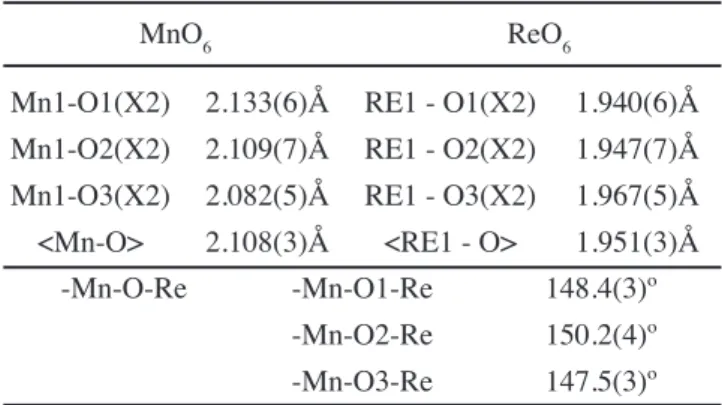
crystal structure, the B-site disorder will not be considered. The monoclinic structure is the preferred structure for rock-salt type ordering of double perovskites with t values lower than 0.97. A major consequence of the distortion is that the Mn—O—Re angle is considerably lower than 180º (in the present case equal ~149°. The overall itting quality is satisfactory. The reined structural parameters are given in Table I and the relevant bond-lengths and angles are given in Table II.
The Rietveld it results of the atomic positions in Table I show that Mn cations occupy the 2d position, Re the 2c position and the Ca cation and three oxygen anions occupy different 4e positions. The monoclinic structure with tilts of the octahedra is drawn in Figs. 3a and 3b. According to Glazer’s notation, there is an a-a-b+ coniguration along
the pseudo-cubic axes [32]. A positive superscript would denote the neighboring octahedra tilt in the same direction (in-phase) and a negative superscript implies in tilts of neighboring octahedral in opposite directions (out of phase). The view in Fig. 3a along the pseudo-cubic a (or b) axis shows octahedra rotations with opposite sign and Fig. 3b
Table I - Rietveld it results of the lattice and atomic parameters of sample Ca2MnReO6. The errors represent the
standard deviation (statistical only).
[Tabela I - Resultados de reinamentos de Rietveld da rede e dos parâmetros atômicos da amostra Ca2MnReO6. Os erros representam o desvio padrão (somente estatístico).]
P21In a = 5.44445(2) Å b = 5.63957(3) Å c = 7.77524(3) Å b = 90.18(1)º
Atom x y z Uiso(Å2) Occup.
Ca 0.4860(5) 0.55429(31) 0.25302(28) 0.0052(5) 1
Mn1 1/2 0 1/2 0.0086(5) 0.977
Re1 1/2 0 0 0.0030(2) 0.977
Re2 1/2 0 1/2 0.0086(5) 0.023
Mn2 1/2 0 0 0.0030(2) 0.023
O1 0.3155(11) 0.2861(11) 0.0496(9) 0.0044(12) 1
O2 0.2138(12) 0.8045(12) 0.0490(9) 0.0044(12) 1
shows the view along the crystallographic c axis with the in-phase rotation of the octahedra
The prediction of crystal structure and the value of t
calculated using the bond valence parameters were obtained using the SPuDS simulation software. The SPuDS output ile contains a complete crystallographic description of the
compound, including the space group, lattice parameters, atomic coordinates, bond-valence sums, individual bond valences and distances, tolerance factor, unit-cell volume, octahedral tilt angles, B—X—B’ bond angles and global instability. To calculate bond-valence sums the program uses the Brown model [33, 34]. This model assumes that the valence of a given cation is shared between the chemical bonds of the irst coordination sphere. It deines a phenomenological relationship between the formal valence of a bond and the corresponding bond lengths.
In this case, the metals Mn and Re can have two possible conigurations: (i) Mn2+Re6+ and (ii) Mn3+Re5+. Therefore, two
estimates of crystal structure by SPuDs for Ca2Mn2+Re6+O6
and Ca2Mn3+Re5+O6 perovskites (space group P 21/n) having
tilt system a-a-b+ are given in Table III.
Comparing the crystal parameters carried out from the Rietveld reinement (Table I) with those evaluated by SPuDs (Table III), one observes that the Rietveld reinement results are between the high value represented by Ca2Mn2+Re6+O6
and the low value presented by Ca2Mn3+Re5+O6. The
tolerance factor t evaluated for Ca2Mn3+Re5+O6 is higher
than that for Ca2Mn2+Re6+O6. However, both are lower than
0.97, which is in agreement with the P 21/n monoclinic
symmetry. The evaluated atomic positions of the Ca and O 4e sites are closer to the Rietveld reinement results. Table II - Length and bond angles of Ca2MnReO6 The errors
represent one standard deviation and are statistical only. [Tabela II - Comprimentos e ângulos de ligação do Ca2MnReO6. Os erros representam o desvio padrão e são estatísticos somente.]
MnO6 ReO6
Mn1-O1(X2) 2.133(6)Å RE1 - O1(X2) 1.940(6)Å Mn1-O2(X2) 2.109(7)Å RE1 - O2(X2) 1.947(7)Å Mn1-O3(X2) 2.082(5)Å RE1 - O3(X2) 1.967(5)Å <Mn-O> 2.108(3)Å <RE1 - O> 1.951(3)Å
-Mn-O-Re -Mn-O1-Re 148.4(3)º -Mn-O2-Re 150.2(4)º -Mn-O3-Re 147.5(3)º
Table III - SPuDS-predicte lattice parameters, atomic positions with tolerance factors t, lengths and angles bond for Ca2Mn2+Re6+O6 and Ca2Mn3+Re5+O6 perovskites (space group) having tilt system a-a-b+.
[Tabela III - SPuDS-parâmetros previstos de rede, posições atômicas com fator de tolerância t, comprimentos e ângulos de ligação para as perovsquitas Ca2Mn2+Re6+O
6 e Ca2Mn3+Re5+O6 (grupo espacial P21/n) apresentando um sistema torcido
a-a-b+.]
Formula a(Å) b(Å) c(Å) b(º) t
Ca2Mn2+Re6+O6 5.4714 5.7352 7.9095 89.95 0.897
Ca2Mn3+Re5+O
6 5.3850 5.5141 7.7031 89.99 0.943
Formula Ca x Ca y Ca z O1 x O1 y O1 z
Ca2Mn2+Re6+O6 0.4740 0.5735 0.2480 0.3226 0.2847 0.0555
Ca2Mn3+Re5+O
6 0.4883 0.5372 0.2495 0.2950 0.2814 0.0389
Formula O2 x O2 y O2 z O3 x O3 y O3 z
Ca2Mn2+Re6+O6 0.2117 0.8161 0.0554 0.6109 -0.0209 0.2336
Ca2Mn3+Re5+O
6 0.2171 0.7929 0.0389 0.5779 -0.3113 0.2441
Formula Mn1-O1 Mn1-O2 Mn1-O3 Re1-O1 Re1-O2 Re1-O3
Ca2Mn2+Re6+O
6 2.1978 2.1952 2.1964 1.9501 1.9479 1.9479
Ca2Mn3+Re5+O
6 2.0166 2.0163 2.0165 1.9276 1.9273 1.9274
Formula Mn-O1-Re Mn-O2-Re Mn-O3-Re
Ca2Mn2+Re6+O
6 145.61 146.06 145.14
Ca2Mn3+Re5+O
6 155.39 155.48 155.16
Moreover, by comparing Table II and Table III we observe that the angle Mn-O-Re and the bond distances Mn-O and Re-O have values between the evaluated limits deined by Ca2Mn2+Re6+O6 and Ca2Mn3+Re5+O6.
For each valence case (Mn2+ and Mn3+ conigurations)
there are two possibilities of spin state conigurations for the cations: the low-spin state and the high-spin state. These states arise from the split of crystalline ield in transition metal coordination complexes due to the competition between exchange force and crystalline electric ield, just like the octahedral ield in double perovskite compounds. The main implication for the whole system concerns magnetic properties, but there are some structural consequences. From Shannon’s table [35] we can see that the difference between the ionic radii in Mn3+, for example, lies in 10% (64.5 pm
for the high spin coniguration and 58 pm for the low one). In double perokskite compounds, the B’ site is usually occupied by a magnetic atom, for B’ = Mn2+ the exchange
split is about 4 eV [36]. The high spin coniguration is due to the crystalline ield energy ~ 1 eV, and occurs in most of the cases. On the contrary, the B’’ site is occupied by a non-magnetic atom and the exchange coupling is negligible, thus the low spin state prevails.
In summary, taking into account the comparative analysis of the parameters shown in Tables I, II and III, it is not possible to determine the exact cation valence coniguration (Mn2+Re6+ or Mn3+Re5+) for this Ca
2Mn1Re1O6 structural
monophasic compound. Magnetic measurements can bring more information about this double perovskite. However, in our point of view, the XANES analysis can complete description of local Mn-O and Re-O coniguration. In order to investigate the valence of Mn and Re in Ca2Mn1Re1O6,
X-ray absorption experiments were done. X ray absorption spectroscopy
Information about the average oxidation state of Mn and Re and the local structural distortion around these ions are provided by X-ray absorption spectroscopic investigation. The valence coniguration of the atoms is commonly determined by the chemical shift of the atomic absorption edge to high energy. Fig. 4 shows the Mn-K edge of the ordered double perovskite compound Ca2Mn1Re1O6 along
with the MnO (Mn2+), Mn
2O3 (Mn3+), MnO2 (Mn4+) and
KMnO4 (Mn6+) standards. Clearly, the proximity of the main
Mn edge of the Ca2Mn1Re1O6 perovskite to that of MnO
standard near the absorption coeficient µ = 1 (the box in Fig. 4) is consistent with a Mn+2.3 assignment. In addition,
the features indicated by the arrows suggest that the coordination of the Mn atom in double perovskite is rather similar to that of MnO standard.
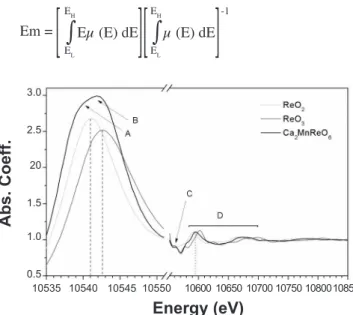
Fig. 5 shows the Re-LIII edge for the Ca2Mn1Re1O6
perovskite along with the ReO3 (Re6+) and ReO2 (Re5+)
standards. One may notice the splitting of the white-line feature in transition metals due to transitions into inal d states, namely eg/t2g states (A-B features in Fig. 5). The
coincidence of position of the Re-LIII edge of the double
perovskite and that of ReO3 suggests an approximate Re6+
state. A quantitative analysis may be taken if one uses the irst moment (Em) of the white-line feature (deined below)
in order to estimate the chemical shift:
Em = Eµ (E) dE µ (E) dE EH EH -1
EL EL
This method was used for the irst time by Alp et al. [37] to deine de Cu valence and charge transfer effect. Popov et al. [22] also have applied such method to study the valence of Mn and Re ions in a MnRe-based double perovskite series, just like in our case. The point to be considered here is the choice of the high-low energy limits in the integrals. The low-energy limit EL is insensitive to the cutoff since the Figure 4: Mn-K edge of compound Ca2Mn1Re1O6 with the standards. The box near the µ = 1 and the arrows indicate the
proximity of MnO coniguration.
[Figura 4: Borda K de absorção do Mn no composto Ca2Mn1Re1O6 com os padrões. A caixa próxima de µ = 1
e as setas indicam a semelhança do Ca2Mn1Re1O6 com a coniguração MnO.]
1,5
1,0
0,5
0,0
6540
Energy (eV)
A
bs
. C
oe
ff.
6565
655065556560 657565806585 6590
6545 6570
Figure 5: Re-L3 edge of the compound Ca2Mn1Re1O6 ploted
with the standards.
[Figura 5: Borda LIII do Rênio no composto Ca2Mn1Re1O6. desenhada em conjunto com os padrões de comparação.]
3.0
20
1.0 2.5
1.5
0.5 10535
Energy (eV)
Abs. Coeff.
background pre-edge subtraction process results in µ ~ 0 in this range, so it was chosen to be 100 eV below the edge in order to encompass the entire white-line feature. The high-energy cutoff was chosen to take into account the majority of the white-line feature weight but not so high to capture the EXAFS range (µ ~ 1.0). Accordingly, we have ixed the high-energy limit to have µ (EH) = 1.4. Taking as a reference
the Em values of the standards, it was found a itting point
+5.7 to a valence of the Re ions in Ca2Mn1Re1O6 double
perovskite compound. The coordination feature indicated by the C features in Fig. 5 agrees with the similar symmetry of Re ion (D4h) in ReO3 standard. The region D of Fig. 5 may be
regarded as a region of local environment information, and it is well known that the prominent peak moves to higher energies with decreasing the Re-O bond lengths. Thereby, in D-feature of Fig. 5 the position of perovskite peak is consistent with the result of intermediate valence between Re4+ (ReO
2) and Re6+ (ReO3) for Re ion. CONCLUSIONS
A structural monophasic Ca2MnReO6 double perovskite
oxide was synthesized from a mixture of CaO, MnO2, ReO2
and ReO3 powders in the nominal stoichiometric ratio
2:1:0.9:0.1 using the encapsulated quartz tube technique. The 0.9:0.1 ratio between ReO2 and ReO3 was used to guarantee
a partial pressure of oxygen inside the quartz tube at high temperatures. This partial pressure of oxygen was revealed to be a crucial synthesis parameter for the production of a single phase sample. The illing factor parameter was set to ff @ 0.12
g/cm3 in order to obtain a partial pressure of 3 bar inside the
tube at high temperatures for the irst thermal treatment. The EDS analysis conirmed that the product only contains the elements Ca, Mn, Re, and O, and the electron backscattered SEM images indicated an homogenous chemical compositon corresponding to the Ca2MnReO6 double perovskite oxide.
The Rietveld reinement indicated a monoclinic unit cell with rock-salt order of the Mn and Re ions, space group P 21/n,
with a = 5.44445(2) Å; b = 5.63957(3) Å; c = 7.77524(3) Å and β = 90.18(1)º, and show low Re/Mn cationic disorder (~ 3%). The Rietveld reinement also reveals there is only one structural phase. The SPuDS program simulation proposes two possible cation valence conigurations, namely, (i) Mn2+Re6+
or (ii) Mn3+Re5+, for the Ca
2Mn1Re1O6 compound. XANES
measurements indicated +2.3 for the average valence of Mn (a mixture of Mn2+ and Mn3+) and +5.7 for the effective valence
of Re (an intermediate valence between Re4+ (ReO
2) and Re6+
(ReO3)).
As a summary, based on our experiments, we concluded there is a mixed valence coniguration for Mn and Re in Ca2Mn1Re1O6, taking into account the oxygen content of
6.0±0.1. We suggest that some magnetic measurements are
necessary for a complete description of this behavior. ACKNOWLEDGEMENTS
We would like to thank the Brazilian agencies grants:
CNPq CT-Energ 504578/2004-9, CNPq 471536/2004-0, CNPq 480337/2007-1 and CAPES for inancial support. Thanks are also due to Companhia Siderurgica de Tubarão (ArcelorMittal). We gratefully acknowledge the Brazilian National Synchrotron Light Laboratory - LNLS (XPD and XAS measurements).
REFERENCES
[1] J. Longo, R. Ward, J. Am. Chem. Soc. 83 (1961) 2816. [2] A. W. Sleight, J. Longo, R. Ward, Inorg. Chem. 1 (1962) 245.
[3] J. M. D. Coey, M. Viret, S. von Molnar, Adv. Phys. 48 (1999) 167.
[4] S. A. Wolf, D. D. Awschalom, R. A. Buhram, J. M. Daughton, S. von Molnár, M. L. Roukes, A. Y. Chtchelkanova, D. M. Treger, Science 294 (2001) 1488. [5] S. A. Wolf, A. Y. Chtchelkanova, D. M. Treger, IBM J. Res. Dev. 50, 1 (2006).
[6] S. Parkin, X. Jiang, C. Kaiser, A. Panchula, K. Roche, M. Samant, Proc. IEEE 91, 5 (2003).
[7] P. Foldi, O. Kalman, M. G. Benedict, F. M. Peeters, Nano Lett. 8 (2008) 2556.
[8] M. N. Leuenberg, M. E. Flatté, D. D. Awschalom, Phys. Rev. Lett. 94 (2005) 107401.
[9] S. D. Sarma, J. Fabian, X. Hu, I. Zutic, Solid State Comm. 119 (2001) 207.
[10] C.-Y. You, S. D. Bader, J. Appl. Phys. 87 (2000) 5215. [11] H. Dery, L. J. Sham, Phys. Rev. Lett. 98 (2007) 046602. [12] A. Quesada, M. A. García, J. de La Venta, E. F. Pinel, J. M. Merino, A. Hernando, Eur. Phys. J. B 59 (2007) 457. [13] K.-I. Kobayashi, T. Kimura, H. Sawada, K. Terakura, Y. Tokura, Nature 395 (1998) 677.
[14] Z. Zeng, I. D. Fawcett, M. Greenblatt, M. Croft, Mater. Res. Bull. 36 (2001) 705.
[15] Z. Fang, K. Terakura, J. Kanamori, Phys. Rev. B 63 (2001) 180507(R).
[16] G. Popov, M. V. Lobanov, E. V. Tsiper, M. Greenblatt, E. N. Caspi, A. Borissov, V. Kiryukhin, J. W. Lynn, J. Phys.: Cond. Matt. 16 (2004) 135.
[17] H. Kato, T. Okuda, Y. Okimoto, Y. Tomioka, K. Oikawa, T. Kamiyama, Y. Tokura, Phys. Rev. B 69 (2004) 184412. [18] J. B. Philipp, P. Majewski, L. Alff, A. Erb, R. Gross, T. Graf, M. S. Brant, J. Simon, T. Walther, W. Mader, D. Topwal, D. D. Sarma, Phys. Rev. B 68 (2003) 144431. [19] M. W. Lufaso, P. W. Barnes, P. M. Woodward, Acta Cryst. B62 (2006) 397.
[20] D. Serrate, J. M. De Teresa, M. R. Ibarra, J. Phys.: Cond. Matt. 19 (2007) 023201.
[21] M. W. Lufaso, P. M. Woodward, Acta Crystallogr., Sect. B: Struct. Sci. 57 (2001) 725.
[22] G. Popov, M. Greenblatt, M. Croft, Phys. Rev. B 67 (2003) 024406.
[25] M. Sikora, Cz. Kapusta, M. Borowiec, C. J. Oates, V. Prochazka, D. Rybicki, D. Zajac, J. M. De Teresa, C. Marquina, M. R. Ibarra, Appl. Phys. Lett. 89 (2005) 062509. [26] J. M. Michalik, J. M. De Teresa, J. Blasco, P. A. Algarabel, M. R. Ibarra, Cz Kapusta, U. Zeitler J. Phys.: Condens. Matter 19 (2007) 506206.
[27] A. C. Larson, B. Von Dreele, General Structure Analysis System (GSAS), Report LAUR 86-748, Los Alamos, N. Mex., USA (2000).
[28] H. B. Toby, J. Appl. Crystallog. 4 (2001) 210.
[29] P. Thompson, D. E. Cox, J. B. Hastings, J. Appl. Crystallog. 20 (1987) 79.
[30] L. W. Finger, D. E. Cox, A. P. Jephcoat, J. Appl.
Crystallog. 27 (1994) 890.
[31] P. W. Stephens, J. Appl. Crystallog. 32 (1999) 281. [32] A. M. Glazer, Acta Crystallogr. B 28 (1972) 3384. [33] I. D. Brown, in M. O’Keefe, A. Navrotsky (Eds.): Structure and Bonding in Crystals, Vol 2, p. 1, Academic Press, New York (1981).
[34] N. E. Brese, M. O’Keefe, Acta Crystallogr. B 47 (1991) 192.
[35] R. D. Shannon, Acta Crystallogr. A 32 (1976) 751. [36] H. Wu, Phys. Rev. B 64 (2001) 125126.
[37] E. E. Alp, G. L. Goodman, L. Soderholm, S. M. Mini, M. Ramanathan, G. K. Shenoy, A. S. Bommannavar, J. Phys.: Condensed Matter. 1 (1989) 6463.