○ ○ ○ ○ ○ ○ ○ ○ ○ ○ ○ ○ A BSTRA CT○ ○ ○ ○ ○ ○ ○ ○ ○ ○ ○ ○ ○ ○ ○ ○ ○I N TRO D U CTI O N○ ○ ○ ○ ○ ○ ○ ○ ○ ○
The cell cycle, a complex process in which a large number of regulatory proteins is in-volved, can suffer alterations caused by differ-ent factors that can consequdiffer-ently transform a normal cell into a malignant one. These altera-tions are the result of the gradual accumula-tion of different genetic events, the main ones being the activation of oncogenes and the in-activation of tumor suppressor genes. These events, in turn, can lead to uncontrolled cell proliferation and the occurrence of neoplasia.1,3 The p16 tumor suppressor gene located on the short arm of chromosome 9 encodes a cyclin-dependent kinase 4 (cdk4) inhibitor that blocks cell division during the G1/S phase of the cell cycle by forming a complex that promotes the dissociation of cyclin from the kinase. The cyclin-dependent kinase 4 and cyclin-dependent kinase 6/cyclin D complexes play an important role in the phosphoryla-tion of the RB protein. Therefore, an increase in p16 expression in cells with a normal RB gene results in the blockade of the G1 phase, indicating the inhibitory function of the p16 gene in the cell cycle as well as its role in neo-plastic processes.4,5
Recently, greater importance has been at-tributed to the role of p16 gene alterations in human neoplasia, mainly as a result of the high frequency of such alterations observed for cell lines derived from different tumors and of the direct involvement of this gene in cell cycle regulation.4-8
In acute lymphoblastic leukemia (ALL), inactivation of the p15 and p16 genes repre-sents one of the most common genetic
mecha-nisms of tumorigenesis.9 Some authors have suggested that alterations in the p16 gene are associated with a higher frequency of relapse and lower survival in T-cell acute lymphob-lastic leukemias both in adults10 and chil-dren.11-13 Therefore, p16 inactivation might be of prognostic value in acute lymphoblastic leukemias, especially T-cell acute lymphoblas-tic leukemias, playing an important role in its genesis or evolution.6,11,14-24
The most frequent alterations in the p16 gene are total or partial homozygous deletions. These alterations are more frequently observed for T-cell acute lymphoblastic leukemias and are less likely in B-cell acute lymphoblastic leukemias. However, the reported frequency of these mutations is highly variable, ranging from 0 to 100% depending on the origin of the patient.12,17,18,23-31 In acute lymphoblastic leukemias, the frequency of nucleotide sub-stitutions in the p16 gene is estimated to be approximately 8%, ranging from 0 to 5% for B-cell acute lymphoblastic leukemias and from 0 to 13% for T-cell acute lymphoblastic leukemias.12,16,18,19,25,28,29
Based on the importance of the p16 gene in acute lymphoblastic leukemias, especially T-cell acute lymphoblastic leukemias, the aim of the present study was to determine prob-able alterations in the p16 gene in Brazilian children with acute lymphoblastic leukemias using the polymerase chain reaction-single stranded conformation polymorphism (PCR-SSCP) method and direct DNA sequencing and also to compare the event-free survival (EFS) using the Kaplan-Meier method and the log-rank test for patients carrying normal or altered p16 genes.
• Carlos Alberto Scrideli
• Luiz Gonzaga Tone
and deletions in childhood
acute lymphoblastic leukemias
Department of Pediatrics, Faculdade de Medicina de Ribeirão Preto,
Universidade de São Paulo, Ribeirão Preto, São Paulo, Brazil
Original Ar
ticle
CONTEXT: The p16 tumor suppressor gene encodes a cyclin-dependent kinase 4 inhibitor that blocks cell division during the G1 phase of the cell cycle. Al-terations in this gene have been reported for vari-ous neoplasia types, including acute lymphoblas-tic leukemias (ALL), especially T-cell acute lymphob-lastic leukemias (ALL).
OBJECTIVE: To determine probable alterations in the p16 gene in children with acute lymphoblastic leukemias using the polymerase chain reaction (PCR) and direct DNA sequencing and also to analyze event-free survival (EFS).
DESIGN: Retrospective study.
SETTING: Department of Child Care and Pediatrics, Faculty of Medicine of Ribeirão Preto, Universidade Federal de São Paulo.
PARTICIPANTS: Fifty-six children with ALL (mean age 4 years). Forty (71.43%) had B-cell and 12 (21.43%) had T-cell ALL; 4 (7.1%) were biphenotypic.
SAMPLE: DNA samples were extracted from bone marrow upon diagnosis and/or relapse. In 2 T-cell cases, DNA from cerebrospinal fluid (CSF) was analyzed.
MAIN MEASUREMENTS: Deletions or nucleotide sub-stitutions in exons 1, 2 and 3 of the p16 gene were determined by PCR and nucleotide sequencing. Event-free survival was determined by the Kaplan-Meyer and log-rank test for patients carrying normal and altered p16.
RESULTS: Deletions in exon 3 were observed in five cases. Abnormal migration in PCR was observed in seven cases for exon 1, six for exon 2, and five for exon 3. Mutations in exon 1 were confirmed by direct DNA sequencing in four cases and in exon 2 in two cases. The Kaplan-Meyer survival curves and the log-rank test showed no significant differences in 5-year EFS between children with normal or altered p16, or between patients with B-ALL carrying normal or altered p16 gene. Patients with T-ALL could not be evaluated via Kaplan-Meier due to the small number of cases.
CONCLUSIONS: Our results, particularly regarding de-letion frequency, agree with others suggesting that deletions in the p16 are initial events in leukemia genesis. The small number of samples did not al-low stablishment of correlation between childhood ALL and the p16 point mutations found in our study. Kaplan-Meier analysis revealed no significant cor-relation between EFS and alterations in ALL. The p16 alterations frequency observed for B and T-ALL agreed with reports from other centers.
○ ○ ○ ○ ○ ○ ○ ○ ○ ○ ○ ○ ○ ○M ETH O D S○ ○ ○ ○ ○ ○
DNA was extracted from the bone mar-row of 56 children with acute lymphoblastic leukemias treated at the Pediatric Oncology and Hematology Service, Hospital Univer-sitário, Faculdade de Medicina de Ribeirão Preto, Universidade de São Paulo (HCFMRP-USP), during the period from 1991 to 1997. Sixteen (28.57%) of the 56 children stud-ied were females and 40 (71.43%) were males. Immunophenotyping of the patients showed the following distribution: 40 children had B-cell acute lymphoblastic leukemias (71.43%), 12 had T-cell acute lymphoblastic leukemias (21.43%), and 4 had biphenotypic acute lym-phoblastic leukemias (7.14%). Sixty-two sam-ples of DNA from bone marrow collected at diagnosis and/or relapse and two samples from cerebrospinal fluid (CSF) from two central nervous system cases collected at relapse were analyzed via the polymerase chain reaction. DNA samples were obtained from 40 patients (25 B-acute lymphoblastic leukemias, 11 T-acute lymphoblastic leukemias and 4 bipheno-typic) at diagnosis only, from 8 patients (B-acute lymphoblastic leukemias) at diagnosis and relapse, and from 8 patients at relapse only (seven B-acute lymphoblastic leukemias and one T-acute lymphoblastic leukemia). Patient ages at diagnosis ranged from 6 months to 14 years, with a mean age of 4 years. Acute lym-phoblastic leukemia was classified according to immunophenotype criteria via cytofluo-rometric analysis using monoclonal antibod-ies carried out at the Hematology Laboratory. To verify the presence of probable dele-tions or base substitudele-tions in the p16 gene, the samples of DNA obtained from children with acute lymphoblastic leukemias at diagnosis and/or relapse were tested by the polymerase chain reaction-single stranded conformational polymorphism method. A control amplification was carried out using the β-globin gene (600-bp fragment) via multiplex polymerase chain reaction for those
polymerase chain reaction products that showed no amplification of the exon, in or-der to confirm deletion The samples show-ing abnormal migration upon sshow-ingle-stranded conformational polymorphism were submit-ted to nucleotide sequencing to confirm the probable mutation.
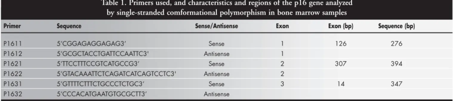
The polymerase chain reaction products of the bone marrow samples were analyzed by single-stranded conformational polymor-phism for possible point mutations in exons 1, 2 and 3 of the p16 gene using correspond-ing primer pairs (Table 1).17,30 For the polymerase chain reaction, each DNA sam-ple (0.1 µg/µl) was added to a mixture con-taining 2.5 mM buffer (0.2 M Tris-HCl, 0.5 M KCl, pH 8.4), 10 mM dNTPs (dATP,
dCTP, dGTP, dTTP), 50 mM MgCl2, 1%
DMSO, 10 µg/µl of the primer pair corre-sponding to the exon studied, and 5 U/µl Taq polymerase, in a final volume of 25 µl. All samples were submitted to the following am-plification conditions (for all exons): 35 suc-cessive denaturation cycles (1 minute at 94° C), annealing at 58° C for 1 minute, and ex-tension at 72° C for 1 minute.
For single-stranded conformational poly-morphism, the polymerase chain reaction products were diluted 1:10 in a solution con-taining 0.1% SDS (Sodium Dodecyl Sulfate) and 10 mM EDTA (Ethylene diamine tetraacetic acid) and an equal volume of dye solution containing 20 mM EDTA, 95% formamide, 0.05% bromophenol blue and 0.05% xylene cyanol were added. The sam-ples were denatured at 96° C for 10 minutes and a fraction was applied to non-denaturing 6% polyacrylamide gel and submitted to elec-trophoresis in 1X TBE buffer for 5 hours at 8 watts. The gel was stained with silver nitrate, developed, photographed and analyzed.
A 100-µl aliquot of each polymerase chain reaction product was used for DNA purifica-tion for direct sequencing of the samples that showed an abnormal electrophoretic migration pattern upon polymerase chain reaction-single
stranded conformational polymorphism. After DNA purification, samples were submitted to direct sequencing using the T7 kit (Pharmacia) and 6% non-denaturing polyacrylamide gel elec-trophoresis. The gel was then dried on filter pa-per and exposed to an X-ray film. The sequences obtained for each sample were compared to the normal SEG–HSPCDK[2182138] sequence consisting of U12818 (exon 1), U12820 (exon 2) and U12820 (exon 3).
Event-free survival was determined for patients carrying the normal or altered p16 gene by the Kaplan-Meier method. Curves were compared by the log-rank test. Statisti-cal analysis was carried out using the GraphPad Prism software, version 2.0 (GraphPad Soft-ware Inc., San Diego, CA).
The study was approved by the Ethics Committee of HCFMRP-USP (case no. 5800/2001).
○ ○ ○ ○ ○ ○ ○ ○ ○ ○ ○ ○ ○ ○ ○RESU LTS○ ○ ○ ○ ○
Sixty-two bone marrow DNA samples obtained at diagnosis and/or relapse and two cerebrospinal fluid samples obtained at relapse from 56 children with acute lymphoblastic leukemias were analyzed. In six DNA sam-ples from five patients that were not ampli-fied by polymerase chain reaction when tested with exon 3 primers, deletions were confirmed by multiplex reaction simultaneously with the ß-globin primers. The deletions detected were in four pre-B CALLA+ acute lymphoblastic leukemias and one T-acute lymphoblastic leukemia (Table 2). In three cases (two pre-B CALLA+ acute lymphoblastic leukemias and one T-acute lymphoblastic leukemia), the de-letions were observed only at diagnosis, in one they were observed at diagnosis and relapse, and in the last one only at the second relapse. Abnormal migration in the single-stranded conformational polymorphism method was observed in seven cases for exon 1, in six for exon 2 and in five for exon 3.
All samples showing abnormal behavior
Table 1. Primers used, and characteristics and regions of the p16 gene analyzed by single-stranded comformational polymorphism in bone marrow samples
Primer Sequence Sense/Antisense Exon Exon (bp) Sequence (bp)
P1611 5’CGGAGAGGAGAG3’ Sense 1 126 276
P1612 5’GCGCTACCTGATTCCAATTC3' Antisense 1
P1621 5’TTCCTTTCCGTCATGCCG3’ Sense 2 307 394
P1622 5’GTACAAATTCTCAGATCATCAGTCCTC3' Antisense 2
P1631 5’GTTTTCTTTCTGCCCTCTGC3’ Sense 3 14 347
upon single-stranded conformational poly-morphism were submitted to direct DNA sequencing. Base substitution in exon 1 was confirmed in four cases; two cases in exon 2 and none in exon 3 (Table 2).
Forty-eight patients with acute lymphob-lastic leukemias were analyzed regarding the event-free survival using Kaplan-Meier curves and the log-rank test, by comparing a group of children with a normal p16 gene (n = 41) with a group of children carrying the altered p16 gene (deletion or mutation) (n = 7). The 5-year event-free survival did not differ be-tween the two groups (p = 0.548). In addi-tion, no difference in the 5-year event-free survival between the two groups was observed for children with B-cell acute lymphoblastic leukemias (p = 0.73). Event-free survival could not be determined separately for the group with T-cell acute lymphoblastic leukemias, due to the small size of the sample.
○ ○ ○ ○ ○ ○ ○ ○ ○ ○ ○ ○ D I SCU SSI O N○ ○ ○ ○ ○ ○ ○ ○
Great advances have been made over the last few years in the understanding and treat-ment of childhood cancer,32 especially in stud-ies determining possible genetic alterations in tumor suppressor genes and oncogenes. These have allowed more precise diagnoses and the determination of important tumor markers for the evolutive analysis of neoplasia.
The p16 gene was first described by Serrano et al.5 in 1993, and since then numerous stud-ies on this gene have been published. In our work, 56 children with acute lymphoblastic leukemias were studied by polymerase chain reaction-single stranded conformational poly-morphism and direct DNA sequencing at di-agnosis and/or relapse in order to determine possible alterations in the p16 gene and their biological role in this disease.
Deletions were only observed in the exon 3 region of the DNA samples analyzed, and were identified in 10% (4/40) of B-cell acute lymphoblastic leukemia cases and 8% (1/12)
of T-cell acute lymphoblastic leukemia cases (Table 2). According to several authors, dele-tions are more frequent in T-cell acute phoblastic leukemias than in B-cell acute lym-phoblastic leukemias.12,17,18,,20,21,24-31 However, the frequency of p16 gene deletions in acute lymphoblastic leukemias is generally hetero-geneous, especially in T-cell acute lymphob-lastic leukemias, ranging from 0%33 to 100% for the latter,24 and from 4%34 to 50% for B-cell acute lymphoblastic leukemias.24 This heterogeneity seems to be related to the ori-gin of the patient, since frequencies vary ac-cording to the place where the studies are car-ried out. The results obtained in the present study for B-cell acute lymphoblastic leukemias are similar to those found in Australia (Perth),35 Germany,34 and France (Paris).25 The 8% incidence of deletions in T-cell acute lym-phoblastic leukemias observed in the present study can be considered to be low and is com-patible with frequencies obtained in studies carried out in Chicago (0%)32 and Japan (15%).29 According to Cayuela et al.,25 the reason for the variation in p16 gene deletions has yet to be established. Since Ribeirão Preto, Brazil, has been undergoing intense migration and has a population of many different eth-nic origins, we cannot conclude that our data are representative of the Ribeirão Preto region, especially considering that variables such as race and family origin were not analyzed.
Of the 18 cases for whom an abnormal migration pattern could be demonstrated upon single-stranded conformational poly-morphism, nucleotide substitutions were con-firmed in only six cases by direct nucleotide sequencing (four of exon 1 and two of exon 2). The occurrence of false abnormal bands might be due to three factors. First, electro-phoresis carried out at low temperatures pro-duces better quality bands but with a greater probability of false bands compared to elec-trophoresis carried out at room temperature.36 Second, it is possible that mutations occurred in the polymorphic regions of non-coding
introns adjacent to the exon. Finally, errors in single-stranded conformational polymor-phism analysis cannot be excluded, since bands that might be interpreted as subtle alterations are indeed normal bands.
Of the 56 patients studied, 11% presented nucleotide substitutions, with 5 out of 40 pa-tients (12%) having B-cell acute lymphoblas-tic leukemias and 1 out of 12 (8%) having T-cell acute lymphoblastic leukemias. Of the 6 cases presenting nucleotide substitutions, 4 were detected at diagnosis and 2 at relapse. Three patients (one early pre-B acute lym-phoblastic leukemias and two pre-B CALLA+ acute lymphoblastic leukemias) died and three (two pre-B CALLA+ acute lymphoblastic leukemias and one T-cell acute lymphoblastic leukemia) are currently in remission. Accord-ing to Dicciani et al.,12 it can be supposed that the mutations detected at diagnosis in the present study are probably related to early events in acute lymphoblastic leukemias. The following previously undescribed mutations were observed in four of our patients with nucleotide substitutions: GCAAla > GTAVal and GTAAla > TCASer, both in codon 32 (cases L325 and L339), AATAsn > ACTThr in codon 34 (case L126), and GCCAgr > TGCCys in codon 123 (case L147). Since data on mutations in child-hood acute lymphoblastic leukemias are scarce, it is difficult to establish whether these mutations play a role in the inactivation of the p16 gene or are polymorphisms. In the present study, TACTyr > TCCSer substitution was observed in codon 36 in a patient with pre-B CALLA+ acute lymphoblastic leukemias. In the literature, mutations in codon 36 have been described in 2 cases of adult lung cancer.37,38 In another case from our study (T-cell), a mutation was observed in a patient (L341) with central nervous system relapse whose bone marrow was normal and for whom DNA was isolated from the cer-ebrospinal fluid. Yoshida et al.39 observed nu-cleotide substitutions in the same codon, which produced a silent mutation in biliary
Table 2. Base substitutions compared to the SEG_HSPCDK [2182138] sequence
Patient Immunophenotype Mutation Position Codon Amino acid Phase Material Exon Base AA Wild type Mutated Wild type Mutated
L147 Pre-pre-B C > T 367 123 CGC TGC Arg Cys Diagnosis BM 2
L023 CALLA+ A > C 107 36 TAC TCC Tyr Ser 2nd relapse BM 1
L126 CALLA+ A > C 101 34 AAT ACT Asn Thr Diagnosis BM 1
L325 CALLA+ C > T 95 32 GCA GTA Ala Val Diagnosis BM 1
L339 CALLA+ G > T 94 32 GTA TCA Ala Ser Diagnosis BM 1
1. Grandér D. How do mutated oncogenes and tumor suppressor genes cause cancer? Med Oncol 1998;15(1):20-6. 2. Hunter T, Pines J. Cyclins and cancer. II: Cyclin D and CDK
inhibitors come of age. Cell 1994;79(4):573-82. 3. Schafer KA. The cell cycle: a review. Vet Pathol 1998;35(6):461-78. 4. Hannon GJ, Beach D. p15INK4B is a potential effector of TGF-beta-induced cell cycle arrest. Nature 1994;371(6494):257-61. 5. Serrano M, Hannon GJ, Beach D. A new regulatory motif in cell-cycle control causing specific inhibition of cyclin D/CDK4. Nature 1993;366(6456):704-7.
6. Caldas C, Hahn SA, da Costa LT, et al. Frequent somatic muta-tions and homozygous delemuta-tions of the p16 (MTS1) gene in pancreatic adenocarcinoma. Nat Genet 1994;8(1):27-32. 7. Cairns P, Mao L, Merlo A, et al. Rates of p16 (MTS1)
muta-tions in primary tumors with 9p loss. Science 1994;265(5170):415-7.
8. Nobori T, Miura K, Wu DJ, Lois A, Takabayashi K, Carson DA. Deletions of the cyclin-dependent kinase-4 inhibitor gene in multiple human cancers. Nature 1994;368(6473):753-6. 9. Guo SX, Taki T, Ohnishi H, et al. Hypermethylation of p16
and p15 genes and RB protein expression in acute leukemia. Leuk Res 2000;24(1):39-46.
10. Yamada Y, Hatta Y, Murata K, et al. Deletions of p15 and/or p16 genes as a poor-prognosis factor in adult T-cell leukemia. J Clin Oncol 1997;15(5):1778-85.
11. Fizzotti M, Cimino G, Pisegna S, et al. Detection of homozygous deletions of the cyclin-dependent kinase 4 inhibitor (p16) gene in acute lymphoblastic leukemia and association with adverse prognostic features. Blood 1995;85(10):2685-90. 12. Diccianni MB, Batova A, Yu J, et al. Shortened survival after
relapse in T-cell acute lymphoblastic leukemia patients with p16/ p15 deletions. Leuk Res 1997;21(6):549-58.
13. Carter TL, Reaman GH, Kees UR. INK4A/ARF deletions are acquired at relapse in childhood acute lymphoblastic leukemia: a paired study on 25 patients using real-time polymerase chain reaction. Br J Haematol 2001;113(2):323-8.
14. Kees UR, Ranford PR, Hatzis M. Deletions of the p16 gene in pediatric leukemia and corresponding cell lines. Oncogene 1996;12(10):2235-9.
15. Maloney KW, McGavran L, Odom LF, Hunger SP. Acquisition of p16(INK4A) and p15(INK4B) gene abnormalities between initial diagnosis and relapse in children with acute lymphoblas-tic leukemia. Blood 1999;93(7):2380-5.
16. Ohnishi H, Hanada R, Horibe K, et al. Homozygous deletions
○ ○ ○ ○ ○ ○ ○ ○ ○ ○ ○ ○ ○ ○ ○ ○ ○ ○ ○ ○ ○ ○ ○ ○ ○ ○ ○ ○ ○ ○ ○ ○ ○ ○ ○ ○ ○ ○ ○ ○ ○ ○ ○ ○ ○ ○ ○ ○ ○ ○ ○ ○ ○ ○ ○ ○ REFEREN CES○ ○ ○ ○ ○ ○ ○ ○
of p16/MTS1 and p15/MTS2 genes are frequent in t (1;19)-negative but not in t (1;19)-positive B precursor acute lymphob-lastic leukemia in childhood. Leukemia 1996;10(7):1104-10. 17. Rasool O, Heyman M, Brandter LB, et al. p15ink4B and
p16ink4 gene inactivation in acute lymphocytic leukemia. Blood 1995;85(12):3431-6.
18. Otsuki T, Clark HM, Wellman A, Jaffe ES, Raffeld M. Involve-ment of CDKN2 (p16 INK4A/MTS1) and p15INK4B/MTS2 in human leukemias and lymphomas. Cancer Res 1995;55(7):1436-40.
19. Stranks G, Height SE, Mitchell P, et al. Deletions and rearrangements of CDKN2 in lymphoid malignancy. Blood 1995;85(4):893-901.
20. Okuda T, Shurtleff SA, Valentine MB, et al. Frequent deletion of p16INK4a/MTS1 and p15INK4b/MTS2 in pediatric acute lymphoblastic leukemia. Blood 1995;85(9):2321-30. 21. Haidar MA, Cao XB, Manshouri T, et al. p16INK4A and
p15INK4B gene deletions in primary leukemias. Blood 1995;86(1):311-5.
22. Takeuchi S, Bartram CR, Seriu T, et al. Analysis of a family of cyclin-dependent kinase inhibitors: p15/MTS2/INK4B, p16/ MTS1/INK4A, and p18 genes in acute lymphoblastic leukemia of childhood. Blood 1995;86(2):755-60.
23. Delmer A, Tang R, Senamaud-Beaufort C, Paterlini P, Brechot C, Zittoun R. Alterations of cyclin-dependent kinase 4 inhibi-tor (p16 INK4A/MTS1) gene structure and expression in acute lymphoblastic leukemias. Leukemia 1995;9(7):1240-5. 24. Iolascon A, Faienza MF, Coppola B, della Ragione F, Schettini F,
Biondi A. Homozygous deletions of cycldependent kinase in-hibitor genes, p16(INK4A) and p18, in childhood T cell lineage acute lymphoblastic leukemias. Leukemia 1996:10(2):255-60. 25. Cayuela JM, Hebert J, Sigaux F. Homozygous MTS1
(p16INK4A) deletion in primary tumor cells of 163 leukemic patients. Blood 1995;85(3):854.
26. Cayuela JM, Madani A, Sanhes L, Stern MH, Sigaux F. Multi-ple tumor-suppressor gene 1 inactivation is the most frequent genetic alteration in T-cell acute lymphoblastic leukemia. Blood 1996:87(6):2180-6.
27. Hayette S, Thomas X, Bertrand Y, et al. Molecular analysis of cyclin-dependent kinase inhibitors in human leukemias. Leukemia 1997;11(10):1696-9.
28. Nakao M, Yokota S, Kaneko H, et al. Alterations of CDKN2 gene structure in childhood acute lymphoblastic leukemia: mu-tations of CDKN2 are observed preferentially in T lineage.
Leukemia 1996;10(2):249-54.
29. Ogawa S, Hangaishi A, Miyawaki S, et al. Loss of the cyclin-dependent kinase 4-inhibitor (p16; MTS1) gene is frequent in and highly specific to lymphoid tumors in primary human hematopoietic malignancies. Blood 1995;86(4):1548-56. 30. Quesnel B, Preudhomme C, Philippe N, et al. p16 gene
ho-mozygous deletions in acute lymphoblastic leukemia. Blood 1995;85(3):657-63.
31. Rubnitz JE, Behm FG, Pui CH, et al. Genetic studies of child-hood acute lymphoblastic leukemia with emphasis on p16, MLL, and ETV6 gene abnormalities: results of St Jude Total Therapy Study XII. Leukemia 1997;11(8):1201-6. 32. Okuda T, Shurtleff SA, Valentine MB, et al. Frequent deletion
of p16INK4a/MTS1 and p15INK4b/MTS2 in pediatric acute lymphoblastic leukemia. Blood 1995;85(9):2321-30. 33. Brenner MK, Pinkel D. Cure of leukemia. Semin Hematol
1999;36(4 Suppl 7):73-83.
34. Schröder M, Mathieu U, Dreyling MH, et al. CDKN2 gene deletion is not found in chronic lymphoid leukaemias of B-and T-cell origin but is frequent in acute lymphoblastic leukae-mia. Br J Haematol 1995;91(4):865-70.
35. Kees UR, Burton PR, Lü C, Baker DL. Homozygous deletion of the p16/MTS1 gene in pediatric acute lymphoblastic leukemia is associated with unfavorable clinical outcome. Blood 1997;89(11):4161-6.
36. Michaelides K, Schwaab R, Lalloz MRA, Schmidt W, Tuddenham JL. Mutational analysis: New mutations. In: McPherson MJ, Hames BD, Taylor GR, editors. PCR 2: A practical approach. Oxford: Oxford University Press; 1995.p257-88. 37. Washimi O, Nagatake M, Osada H, et al. In vivo occurrence of
p16 (MTS1) and p15 (MTS2) alterations preferentially in non-small cell lung cancers. Cancer Res 1995;55(3):514-7. 38. Okamoto A, Hussain SP, Hagiwara K, et al. Mutations in the
p16INK4/MTS1/CDKN2, p15INK4B/MTS2, and p18 genes in primary and metastatic lung cancer. Cancer Res 1995;55(7):1448-51.
39. Yoshida S, Todoroki T, Ichikawa Y, et al. Mutations of p16Ink4/ CDKN2 and p15Ink4B/MTS2 genes in biliary tract cancers. Cancer Res 1995;55(13):2756-60.
40. Volm M, Koomägi R, Stammler G, Rittgen W, Zintl F, Sauerbrey A. Prognostic implications of cyclins (D1, E, A), cyclin-depend-ent kinases (CDK2, CDK4) and tumor-suppressor genes (pRB, p16INK4A) in childhood acute lymphoblastic leukemia. Int J Cancer 1997;74(5):508-12.
tract carcinoma. Authors such as Otsuki et al.18 and Nakao et al.28 found low frequencies of nucleotide substitutions in the p16 gene in T-cell acute lymphoblastic leukemias, while oth-ers17,26 did not observe any mutation in T-cell acute lymphoblastic leukemias.
In the present study, analysis of all immunophenotypes as a whole did not reveal any difference in the event-free survival of chil-dren with acute lymphoblastic leukemias who carried a normal or altered p16 gene, similar to the findings of Rubnitz et al.31 With respect to children with B-cell acute lymphoblastic leukemias, there was also no difference in event-free survival between the two groups (p = 0.73),
in agreement with other studies.11,12,33,40 We were unable to analyze T-cell acute lymphob-lastic leukemias due to the small number of patients with this immunophenotype. How-ever, 2 patients with alterations in the p16 gene showed early relapse.
According to several authors, p16 inacti-vation may be an important factor for the as-sessment of the recurrence risk, especially in children with T-cell acute lymphoblastic leukemias.11-14 In the present study, no corre-lation could be established between the nu-cleotide substitutions found in six patients and patient survival or prognosis, probably due to the low frequency of mutations in this gene
in childhood leukemias.
○ ○ ○ ○ ○ ○ ○ ○ ○ ○ ○ CO N CLU SI O N○ ○ ○ ○ ○ ○ ○ ○ ○
CONTEXTO: O gene supressor tumoral p16 codifica uma proteína inibidora da quinase ciclina dependente 4(cdk4) que bloqueia a divisão celular na fase G1 do ciclo celular. Alterações neste gene foram relatadas em vá-rias neoplasias, incluindo as leucemias linfoblásticas agudas, principalmente da li-nhagem T.
OBJETIVO: Verificar prováveis alterações do gene p16 em leucemias linfoblásticas agudas em crianças, utilizando o método de reação de polimerase em cadeia - polimorfismo confor-macional em fita simples e seqüenciamento direto de nucleotídeos e comparar a sobrevida livre de eventos com a presença ou não de al-terações (deleções ou substituições de bases) do gene p16, utilizando o método de Kaplan-Meir e teste log-rank.
TIPO DE ESTUDO: Estudo retrospectivo.
LOCAL: Departamento de Puericultura e Pedia-tria da Faculdade de Medicina de Ribeirão Preto da Universidade de São Paulo.
PARTICIPANTES: Foram estudadas 56 crian-ças (16 do sexo feminino e 40 do masculino) com leucemia aguda com idade média de quatro anos. Quarenta pacientes (71,43%) tinham leucemias linfoblásticas agudas de células B, 12 (21,43%) tinham leucemias linfoblásticas agudas de células T e 4 (7,1%) eram bifenotípicas.
AMOSTRA: Amostras de DNA foram extraídas de medula óssea obtidas ao diagnóstico e/ou na recaída. Em dois casos de leucemias linfoblásticas agudas de células T, o DNA obti-do obti-do líquiobti-do céfalo-raquidiano foi analisaobti-do.
VARIÁVEIS ESTUDADAS: Deleções ou subs-tituições de bases nos éxones 1, 2 e 3 do gene p16 foram determinadas pela reação de polimerase em cadeia - polimorfismo confor-macional em fita simples e seqüenciamento de nucleotídeos. A sobrevida livre de eventos foi determinada segundo o método de Kaplan-Meier e teste log-rank para pacientes com gene p16 normal e alterado.
RESULTADOS: Em cinco casos (quatro
leuce-○ ○ ○ ○ ○ ○ ○ ○ ○ ○ ○ ○ ○ ○ ○ ○ ○ ○ ○ ○ ○ ○ ○ ○ ○ ○ ○ ○ ○ ○ ○ ○ ○ ○ ○ ○RESU M O○ ○ ○ ○ ○ ○
José Alexandre Rodrigues Lemos, MD. Biologist, De-partment of Pediatrics, Faculdade de Medicina de Ribeirão Preto, Universidade de São Paulo, Ribeirão Preto, São Paulo, Brazil.
Ricardo Defavery, MD. Assistant physician, Department of Pediatrics, Faculdade de Medicina de Ribeirão Preto, Universidade de São Paulo, Ribeirão Preto, São Paulo, Brazil.
Carlos Alberto Scrideli, MD. Assistant physician, Depart-ment of Pediatrics, Faculdade de Medicina de Ribeirão Preto, Universidade de São Paulo, Ribeirão Preto, São Paulo, Brazil.
Luiz Gonzaga Tone, MD. Associate professor, Depart-ment of Pediatrics, Faculdade de Medicina de Ribeirão Preto, Universidade de São Paulo, Ribeirão Preto, São Paulo, Brazil.
Sources of funding: None
Conflict of interest: Not declared
Date of first submission: July 15, 2002
Last of received: October 10, 2002
Accepted: November 29, 2002
Address for correspondence
Luiz Gonzaga Tone
Departamento de Pediatria da Faculdade de Medicina de Ribeirão Preto da Universidade de São Paulo Av. Bandeirantes, 3900
Ribeirão Preto/SP – Brasil – CEP 14049-900 Tel./Fax (+55 16) 633-0136
E-mail: [email protected]
COPYRIGHT © 2003, Associação Paulista de Medicina
○ ○ ○ ○ ○ ○Pu b l i sh i n g i n f o r m a t i o n○ ○ ○ ○ ○ ○ ○ ○ ○ ○ ○ ○ ○ ○
mias linfoblásticas agudas pré B CALLA+ e uma leucemia linfoblástica aguda T) foram observadas deleções no éxon 3. Foram verificadas migrações anômalas no polimor-fismo conformacional em fita simples em sete casos no éxon 1, seis no éxon 2 e cinco no éxon 3. A confirmação da mutação pelo seqüenciamento direto do DNA ocorreu em quatro casos no éxon 1 e 2 no éxon 2. Fo-ram comparadas as curvas de sobrevida pelo método de Kaplan-Meier e teste log-rank e não se obteve diferença estatisticamente sig-nificativa da sobrevida livre de eventos em cinco anos entre os grupos de criança com leucemia linfoblática aguda com p16 nor-mal e alterado e também entre os pacientes com leucemia linfoblástica aguda B com p16 normal e alterado. Não foi possível avaliar pelo método de Kaplan-Meier os pacientes com leucemia linfoblástica aguda T devido ao pequeno número de casos.
CONCLUSÕES: Os resultados obtidos, princi-palmente em relação à freqüência de deleções, foram concordantes com vários autores que sugerem que deleções do gene p16 possam ser eventos iniciais da gênese leucêmica. Ten-do em vista o pequeno número de amostras estudadas, não foi possível realizar a correla-ção entre leucemia linfoblática aguda na in-fância e as mutações de ponto no gene p16 encontradas neste estudo. Não se obteve significância estatística entre sobrevida livre de eventos e alterações do p16 em leucemia linfoblática aguda pelo método de Kaplan-Meier. A freqüência de alterações do p16 em leucemia linfoblástica aguda B e T foram con-cordantes com os estudos realizados em al-guns centros mundiais. Sugerimos a realiza-ção de uma pesquisa multicêntrica com um maior número de casos, principalmente so-bre leucemia linfoblástica aguda T, a fim que seja possível traçar um perfil de alterações do gene p16 em crianças brasileiras.
![Table 2. Base substitutions compared to the SEG_HSPCDK [2182138] sequence](https://thumb-eu.123doks.com/thumbv2/123dok_br/19025681.473331/3.892.67.823.931.1096/table-base-substitutions-compared-seg-hspcdk-sequence.webp)
