Lymph node ratio predicts tumor recurrence in stage III colon
Lymph node ratio predicts tumor recurrence in stage III colon
Lymph node ratio predicts tumor recurrence in stage III colon
Lymph node ratio predicts tumor recurrence in stage III colon
Lymph node ratio predicts tumor recurrence in stage III colon
cancer
cancer
cancer
cancer
cancer
O índice de linfonodos comprometidos como um preditor para a ocorrência de
O índice de linfonodos comprometidos como um preditor para a ocorrência de
O índice de linfonodos comprometidos como um preditor para a ocorrência de
O índice de linfonodos comprometidos como um preditor para a ocorrência de
O índice de linfonodos comprometidos como um preditor para a ocorrência de
recidivas tumorais no câncer de cólon estádio III
recidivas tumorais no câncer de cólon estádio III
recidivas tumorais no câncer de cólon estádio III
recidivas tumorais no câncer de cólon estádio III
recidivas tumorais no câncer de cólon estádio III
TIAGO L. DEDAVIDE SILVA1; DANIEL C. DAMIN2
A B S T R A C T A B S T R A C T A B S T R A C T A B S T R A C T A B S T R A C T
Objective: Objective: Objective: Objective:
Objective: To evaluate the lymph node ratio as a predictor for tumor recurrence in stage III colon cancer patients. Methods:Methods:Methods:Methods:Methods: Patients with stage III colon cancer who underwent curative resection between January 2005 and December 2010 were retrospectively reviewed. The main outcomes were tumor recurrence and death. The impact of lymph node ratio and other clinicopathological factors on disease-free survival were evaluated by uni- and multivariate analysis. Receiver operator characteristic (ROC) analysis was conducted in order to identify the best cutoff value for lymph node ratio to predict tumor recurrence. Disease-free survival was estimated by the Kaplan-Meier method. Results:Results:Results:Results:Results: Seventy patients were included in the study (50% male). The mean age was 64 years. Univariate analysis identified four factors for tumor recurrence: carcinoembryonic antigen, N stage, number of positive lymph nodes and lymph node ratio. Lymph node ratio was the one with the greatest magnitude of association. Receiver operator characteristic analyzes identified 0.15 as the best cutoff value. Patients with a lymph node ratio < 0.15 had a disease-free survival of 90% in 3 years (versus 64%, p = 0.011). Conclusion:Conclusion:Conclusion:Conclusion:Conclusion: Lymph node ratio is a strong predictor for tumor recurrence in stage III colon cancer.
Key words: Key words: Key words: Key words:
Key words: Lymph nodes. Colonic neoplasms. Lymphatic metastasis. Neoplasm staging. Survival.
Study conducted at the Department of Coloproctology, Porto Alegre Clinics Hospital – HCPA, and at the Department of Surgery, Federal University of Rio Grande do Sul – UFRGS, Porto Alegre, Rio Grande do Sul State – RS, Brazil.
1. MSH, Post-Graduation Program in Surgical Sciences, UFRGS; 2. Professor, Head, Department of Coloproctology, Porto Alegre Clinics Hospital, Department of Surgery, UFRGS.
INTRODUCTION
INTRODUCTION
INTRODUCTION
INTRODUCTION
INTRODUCTION
C
olorectal cancer (CRC) is the third most frequent tumor diagnosed in Brazil, with an incidence estimated in 28,000 new cases in 2010 1. Worldwide, it has beenassessed that over 1.2 million new cancer cases were diagnosed and 608,700 deaths have occurred in 2008 2 .En
bloc resection of the primary tumor with removal of regio-nal lymph nodes is the cornerstone therapy for patients with curable disease3,4.
Prognosis in CRC is clearly related to the degree of tumor infiltration through the bowel wall and the presence or absence of lymph node involvement. These two factors are the basis of the staging system elaborated for this disease
5. Almost 80 years after Dukes published his pioneer
classification 6, all the attempts to improve his proposal
were mostly based on refinements of these two data, creating subgroups. Among the current staging systems, the AJCC (American Joint Committee on Cancer) and UICC (International Union Against Cancer) TNM Classification of Malignant Tumors is the most widely accepted 7.
In TNM classification, CRC patients with lymph node metastases are classified as stage III. This is a heterogeneous group 8. It is well known that not only the
mere presence of lymph node metastases determines prognosis, but also the number of positive ones 9, a fact
corroborated by recent studies10,11. More recently, the total
number of resected lymph nodes has also been shown to be an independent prognostic factor 12. Trying to associate
these two observations, in order to obtain a single better prognostic factor, the lymph node ratio (LNR), i.e., the ratio of positive nodes divided by the total number of examined nodes, has been proposed by different authors 13.
MATERIALS AND METHODS
MATERIALS AND METHODS
MATERIALS AND METHODS
MATERIALS AND METHODS
MATERIALS AND METHODS
Patients Patients Patients Patients Patients
The study included all the patients who underwent resection of a primary colorectal cancer with curative intent attended by the staff of the Division of Coloproctology at the Porto Alegre Clinics Hospital (HCPA) from January 2005 to December 2010, and who were on stage III disease according to the 7th edition of the AJCC/UICC TNM
Classification of Malignant Tumors. The inferior border of the tumor must have been above the peritoneal reflection. Patients were excluded if they presented: (1) an R1 or R2 resection (microscopic evidence of invasive tumor in the resection margin or within 1 mm from it, or gross residual tumor that was not resected); (2) synchronic colorectal tumors in different segments, demanding total or subtotal colectomy; (3) familial adenomatous polyposis; (4) inflammatory bowel disease; and (5) other primary malignant tumors.
Surgeries were carried out predominantly through open access under oncologic principles. All patients received routine adjuvant chemotherapy consulting. Different chemotherapy regimens were possible.
Follow-up Follow-up Follow-up Follow-up Follow-up
All patients were followed up every 3 months for the first 2 years, every 6 months for the following 3 years and then once a year. Additional visits occurred when necessary. Blood tests with carcinoembryonic antigen measurement, chest X ray and ultrasonography of the liver were carried out. Surveillance colonoscopy and abdominal computed tomography were performed one year after surgery in most cases. Additional imaging studies were performed when recurrences were suspected.
Pathology Pathology Pathology Pathology Pathology
Surgical specimens were fixed in 10% buffered neutral formalin. The traditional inspection and palpation method was applied for lymph node harvesting. In cases in which fewer than 12 lymph nodes were detected, additional fat clearance was performed with Carnoy‘s solution in order to help identifying missing lymph nodes. Tissues were paraffin-embedded and histological evaluation was performed using hematoxylin-eosin staining. Metastatic deposits were defined as lymph nodes if these structures resembled nodes but contained no visible lymphatic tissue.
Data extraction Data extraction Data extraction Data extraction Data extraction
The following demographic and clinicopathological parameters were retrieved from patients‘ charts: age, gender, cancer site (right and left colon, by using middle transverse colon as partition), baseline carcinoembryonic antigen (CEA) serum level, body mass index (BMI), type of surgery (right or left colectomy, sigmoidectomy, anterior resection), tumor-node-metastasis (TNM) stage, degree of histological differentiation (well,
moderate or poor), lymphatic or venous invasion, number of resected nodes, number of metastatic nodes, lymph node ratio, adjuvant chemotherapy, regimen and number of cycles and follow-up time.
Primary endpoints were tumor recurrence and death. Tumor recurrence was defined as unequivocal evidence of disease from imaging methods or biopsy. For purpose of disease-free survival calculation, the date from the exam that proved recurrence was used. Operative death was defined as any death, regardless of cause, occurring (1) within 30 days after surgery in or out of hospital or (2) after 30 days during the same hospitalization subsequent to the operation.
Statistics Statistics Statistics Statistics Statistics
The chi-square test was used to compare groups with respect to categorical variables. Continuous variables were compared with the Student‘s t test or the Mann-Whitney U test. Cox proportional hazards regression model was used to identify the variables that could independently influence outcomes. Only covariates with trend-significance (o < 0.20) in univariate analysis were entered in multivariate analysis. Survival curves were estimated by the Kaplan-Meier method and compared using the logrank test. ROC analysis was performed to find the best lymph node ratio value predicting outcome. IBM SPSS (Statistical Package for the Social Sciences) version 18.0 (IBM Corp. Armonk, NY, USA) was used for the whole statistical analysis. A p value less than 0.05 was considered statistically significant.
RESULTS
RESULTS
RESULTS
RESULTS
RESULTS
Patient population Patient population Patient population Patient population Patient population
During the six years of the study period, 236 patients were submitted to surgical resection of intraperitoneal colorectal tumors with curative intent. Of these, 70 had stage III cancer and were in accordance with the inclusion and exclusion criteria. The mean follow-up time was 31.89 months (median 33, range 1 72). Operative mortality was 2.9%. Tumor recurrence was identified in 13 patients. Only 1 patient showed tumor recurrence after 36 months of follow-up, and in 85% of the cases, recurrence was within the first 2 years after surgery. Seven deaths were registered in the study, including the operative ones.
According to the 7th edition of the AJCC/UICC
TNM Classification of Malignant Tumors, five patients were on stage IIIA, 57 on IIIB, and eight on IIIC. In 90% of the cases, at least a 12 lymph node harvest has been achieved (median 18.5, range 5 51).
Tumor recurrence analysis Tumor recurrence analysis Tumor recurrence analysis Tumor recurrence analysis Tumor recurrence analysis
lymph node number and lymph node ratio. The lymph node ratio was the one with the greatest magnitude of association. These data, as well as the demographic and clinicopathological characteristics of the patients are displayed in Table 1.
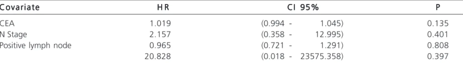
Two multivariate analysis models were developed. The first one considered all the covariates with p < 0.20 in univariate analysis. None of these was found to be an independent prognostic factor, but the tendency for a strong magnitude of association for lymph node ratio remained (Table 2). Considering the strong interdependency between N stage, positive lymph node number and lymph node ratio, a second model analyzed only CEA and lymph node ratio. The latter was the only independent factor for tumor recurrence (p = 0.029, hazard ratio 74.887, 95% confidence interval 1.550 3617.011).
ROC analysis identified a lymph node ratio value of 0.1491 (mnemonic 0.15) as the highest accuracy value to predict tumor recurrence.
Group analysis Group analysis Group analysis Group analysis Group analysis
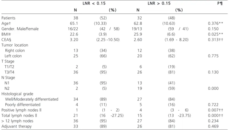
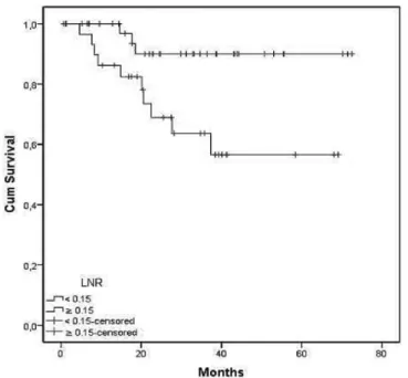
Based on ROC analysis, patients were grouped according to lymph node ratio. There were 38 patients with lymph node ratio < 0.15 and 32 with lymph node ratio e” 0.15. The characteristics of both groups are summarized in Table 3. Patients with lymph node ratio < 0.15 had a higher disease-free survival, estimated on 90% (versus 64%) at 36 month (Figure 1). This benefit was observed even when only patients in stage IIIB were considered (Figure 2). A lymph node
ratio < 0.15 was also associated with an impact on overall survival (Figure 3).
Table Table Table Table
1-Table 1- Demographic and clinicopathological characteristics and univariate analysis related to tumor recurrence.
All patients (N = 70)* All patients (N = 70)* All patients (N = 70)* All patients (N = 70)*
All patients (N = 70)* Univariate analysisUnivariate analysisUnivariate analysisUnivariate analysisUnivariate analysis N
N N N
N (%)(%)(%)(%)(%) R RR RR RR RR R IC 95%IC 95%IC 95%IC 95%IC 95% PPPPP
Age† 64 (38.9 - 82.6) 0.998 (0.948 - 1.051) 0.942
Gender. Male/Female 35/35 (50 / 50) 0.978 (0.328 - 2.914) 0.968
BMI‡ 23.7 (6.1) 0.980 (0.789 - 1.093) 0.717
CEA§ 3.10 (1.86 - 8.43) 1.026 (1.003 - 1.049) 0.024
Tumor location
Right colon 25 (36)
Left colon 45 (64) 1.236 (0.414 - 3.690) 0.704
T Stage
T1/T2 8 (11)
T3/T4 62 (89) 0.730 (0.161 - 3.304) 0.683
N Stage
N1 49 (70)
N2 21 (30) 3.981 (1.323 - 11.981) 0.014
Histological grade
Well/Moderately differentiated 61 (87)
Poorly differentiated 9 (13) 1.242 (0.275 - 5.618) 0.778
Positive lymph nodes || 2 (1 - 4) 1.172 (1.033 - 1.330) 0.014
Total lymph nodes || 18.5 (14 - 26) 0.999 (0.942 - 1.059) 0.966
LNR¶ 0.156 89.644 (2.425 - 3313.874) 0.015
Adjuvant therapy 59 (86) 0.569 (0.125 - 2.580) 0.465
* Data are number of patients (percentage), unless otherwise indicated. HR = hazard ratio. CI = confidence interval. † Age, mean (range), years. ‡ BMI = body mass index, mean (standard deviation), kg/m2. § CEA = carcinoembryonic antigen, median (interquartile range), ng/mL. || Number
of lymph nodes, median (interquartile range). ¶ LNR = lymph node ratio, mean.
Table 2 Table 2 Table 2 Table 2
Table 2 - Multivariate analysis related to tumor recurrence*.
Covariate Covariate Covariate Covariate
Covariate H RH RH RH RH R CI 95%CI 95%CI 95%CI 95%CI 95% PPPPP
CEA 1.019 (0.994 - 1.045) 0.135
N Stage 2.157 (0.358 - 12.995) 0.401
Positive lymph node 0.965 (0.721 - 1.291) 0.808
20.828 (0.018 - 23575.358) 0.397
DISCUSSION
DISCUSSION
DISCUSSION
DISCUSSION
DISCUSSION
The objectives of the TNM Classification of Malignant Tumors have been stated as: to aid the clinician in the planning of treatment; to give some indication of prognosis; to assist in evaluation of the results of treatment; to facilitate the exchange of information between treatment centers; to contribute to the continuing investigation of human cancer 7. Since its 6th edition, the AJCC and the
UICC have been working in the direction of rendering the stage III colorectal cancer group of patients less heterogeneous. The subdivision of patients in stages IIIA, IIIB and IIIC is an example of this development. Nevertheless, most patients were placed in subgroup IIIB, providing no solution for this important question.8,14,15. The 7th edition
added complexity to the system, with the creation of N1 and N2 subdivisions A and B, and redefined stage III subgroups, accounting for an even greater heterogeneity in subgroup IIIB 16, 17.
The LNR has been proposed as a better prognostic factor than the number of metastatic lymph nodes13. Our
study clearly demonstrates the magnitude of association of this factor with tumor recurrence. However, to incorporate this information in a staging system, it must be simplified
as a cutoff, not as a continuous variable. Different approaches have been used for this aim, mainly based on quartile division. It is our opinion that the better way do determinate a cutoff is trough the ROC analysis.
Two previous studies assessed this method on colon cancer based on different outcomes. Galizia et al. studied the impact of LNR on disease-specific survival, finding a cutoff of 0.18 18. Greenberg et al. applied ROC analysis
to overall survival, identifying 0.13 as the best cutoff 19. Our
study is the first to use this method in disease-free survival, resulting in a value of 0.15. The similar cutoffs to LNR found in three independent studies, each one based on different outcomes, reinforce the importance of considering incorporating this tool in future editions of the TNM classification.
Although frequently discussed together, some studies indicate differences in lymph node spread patterns between colon and rectum cancers. Wang et al. assessed patients with colorectal carcinoma and showed that rectum surgical specimens tend to present lower lymph node retrieval, but a higher number of metastatic ones, which results in a higher lymph node ratio, even after T stage consideration 20. Therefore, the cutoff values found in colon
cancer may not be the same for rectal cancer. In fact, studies
Table 3 Table 3 Table 3 Table 3
Table 3 - Group comparing according to LNR*.
LNR < 0.15 LNR < 0.15 LNR < 0.15 LNR < 0.15
LNR < 0.15 LNR LNR LNR LNR >LNR >>> 0.15> 0.15 0.15 0.15 0.15 P ¶P ¶P ¶P ¶P ¶ N
N N N
N (%)(%)(%)(%)(%) NNNNN (%)(%)(%)(%)(%)
Patients 38 (52) 32 (48)
Age† 65.1 (10.33) 62.8 (10.63) 0.376**
Gender. Male/Female 16/22 (42 / 58) 19/13 (59 / 41) 0.150
BMI‡ 22.6 (3.9) 25.9 (6.6) 0.025**
CEA§ 3.20 (2.25 -10.50) 2.60 (1.69 - 8.20) 0.313††
Tumor location
Right colon 13 (34) 12 (38)
Left colon 25 (66) 20 (62) 0.775
T Stage
T1/T2 2 (5) 6 (19)
T3/T4 36 (95) 26 (81) 0.130
N Stage
N1 36 (95) 13 (41)
N2 2 (5) 19 (59) 0.000
Histological grade
Well/Moderately differentiated 34 (89) 27 (84)
Poorly differentiated 4 (11) 5 (16) 0.722
Positive lymph nodes || 1 (1 - 2) 4 (3 - 6) 0.007††
Total lymph nodes || 21 (16 -27.25) 15 (13 -23.75) 0.000††
> 12 lymph nodes 36 (95) 27 (84) 0.234
Adjuvant therapy 33 (89) 26 (81) 0.469
* Data are number of patients (percentage), unless otherwise indicated. LNR = lymph node ratio. † Age, mean (standard deviation), years. ‡ BMI = body mass index, mean (standard deviation), kg/m2. § CEA = carcinoembryonic antigen, median (interquartile range), ng/mL. || Number of lymph
nodes, median (interquartile range).
on rectal cancer addressing this issue found diverse results, ranging from 0.21 to 0.60 21, 22. This variability can be largely
explained by the use of preoperative radiotherapy. It seems clear that the interpretation of the nodal status of patients previously submitted to radiotherapy must be done in separate, and it is possible that in the future colon and rectum staging systems become independent, as recently occurred to appendix cancer.
This study included only patients with intraperitoneal colorectal tumors. Although some patients
Figure 3 Figure 3 Figure 3 Figure 3
Figure 3 - Kaplan-Meier overall survival curves for patients with LNR < 0.15 (superior line) and > 0.15 (inferior line). Logrank test p = 0.024.
Figure 2 Figure 2 Figure 2 Figure 2
Figure 2 - Kaplan-Meier disease-free survival curves for stage IIIB patients with LNR < 0.15 (superior line) and > 0.15 (inferior line). Logrank test p = 0.016.
Figure 1 Figure 1 Figure 1 Figure 1
-Figure 1 - Kaplan-Meier disease-free survival curves for patients with LNR < 0.15 (superior line) and > 0.15 (inferior line). Logrank test p = 0.011.
with rectosigmoid junction tumors have been included, with tumors whose inferior borders could be located in the upper rectum, this procedure is in accordance with inclusion criteria of major clinical trials on colon cancer adjuvant therapy 23, 24, thus reflecting common clinical practice.
Stratification of the patients according to their lymph node ratio showed that patients with an LNR greater than or equal to 0.15 had both a higher number of positive nodes and a lower number of lymph nodes retrieved. This trend was found by other authors 25, 26. This observation
agrees with studies that analyzed the impact of lymph node harvest on colon cancer prognosis, which suggests that factors other than staging accuracy may account for the improvement in survival associated with the increased number of lymph nodes evaluated 12,27.
The explanation of the aforementioned trends could be in the immune system. It is very plausible that the number of nodes present in a given patient reflects, at least in part, the underlying interaction between tumor and host
28. Galon et al. 29 have found that type, density, and location
of immune cells within the colorectal cancer formed patterns of immune response that could predict the survival of some patients more accurately than standard histopathologic staging methods. George et al. 30, in a study
on tumor aggressiveness, but also on patient immune response.
The minimum number of lymph nodes resected for the LNR to be considered a reliable prognostic factor is still under debate. The American College of Surgeons, the American Society of Clinical Oncology and the National Quality Forum endorsed that at least 12 lymph nodes should be removed and pathologically examined in resectable primary colon cancer as the quality measure of hospital performance 31-33 . This recommendation is also present in
TNM classification(7). Four studies evaluated the influence
of lymph node harvest on LNR in colon cancer, with mixed results. The largest of these studies, published by Wang et al., demonstrated that the LNR remained an independent prognostic factor even in patients with less than 10 lymph nodes examined 34, a fact also confirmed by Vaccaro et
al.35 Controversially, Berger et al. found that the LNR lost
its discriminatory capacity in patients with less than 10 lymph nodes harvested, but these authors included patients with stage II colon cancer in their statistical analysis 36. In the
study by Park et al., the LNR had no prognostic significance
in patients with less than 12 resected lymph nodes, but interpretation of this observation is hampered by the choice of the LNR cutoffs being based on previous data from gastric cancer 37.
In our study, only 10% of patients had less than 12 resected lymph nodes. The limited size of our sample prevents an analysis of this subgroup of patients. In fact, the major limitations of our study reside in our sample size and its retrospective nature. The small number of deaths observed during follow-up also prevented us to perform a detailed analysis on the influence of the LNR in disease-specific survival or overall survival.
CONCLUSION
CONCLUSION
CONCLUSION
CONCLUSION
CONCLUSION
Lymph node ratio is a strong predictor for tumor recurrence on colon cancer. Future editions of TNM Classification of Malignant Tumors should consider the inclusion of this factor in the stratification of patients with stage III disease.
R E S U M O R E S U M O R E S U M O R E S U M O R E S U M O
Objetivo: Objetivo: Objetivo: Objetivo:
Objetivo: avaliar o índice de linfonodos comprometidos na ocorrência de recidivas tumorais em pacientes com câncer de cólon estádio III. Métodos:Métodos:Métodos:Métodos:Métodos: foram avaliados de maneira retrospectiva todos os pacientes com câncer de cólon estádio III submetidos à ressecção curativa do tumor primário entre janeiro de 2005 e dezembro de 2010. Os desfechos de interesse foram a ocorrência de recidivas tumorais e morte. O impacto do índice de linfonodos comprometidos e das demais variáveis clínico-patológicas na sobrevida livre de doença foi avaliado através de análise uni e multivariável. De modo a identificar-se o ponto de corte de maior acurácia para utilização do índice de linfonodos comprometidos como um preditor de recidivas tumorais realizou-se a análise da curva característica de operação do receptor. A sobrevida livre de doença foi avaliada através de curvas de Kaplan-Meier. Resultados:
Resultados: Resultados: Resultados:
Resultados: setenta pacientes foram incluídos no estudo (50% masculinos). A média de idade foi 64 anos. A análise univariável identificou quatro fatores determinantes para a ocorrência de recidivas tumorais: antígeno carcinoembrionário, estadiamento N, número de linfonodos positivos e índice de linfonodos comprometidos. O índice de linfonodos comprometidos foi o que demons-trou a maior magnitude de associação. A análise da curva característica de operação do receptor identificou 0,15 como o ponto de corte ideal. Pacientes com um índice de linfonodos comprometidos <0,15 apresentavam uma sobrevida livre de doença de 90% em três anos (versus 64%, P=0,011). Conclusão: Conclusão: Conclusão: Conclusão: Conclusão: o índice de linfonodos comprometidos é um forte preditor para recidivas tumorais no câncer de cólon estádio III.
Descritores: Descritores: Descritores: Descritores:
Descritores: Linfonodos. Neoplasias do colo. Metástase linfática. Estadiamento de neoplasias. Sobrevivência.
REFERENCES
REFERENCES
REFERENCES
REFERENCES
REFERENCES
1. Brasil. Ministério da Saúde. Instituto Nacional de Câncer José Alencar Gomes da Silva. Estimativa 2010: Incidência de câncer no Brasil [Internet]. Rio de Janeiro: INCA; 2009. Disponível em: http:// www2.inca.gov.br/wps/wcm/connect/agencianoticias/site/home/ noticias/2009/lancamento_estimativa_2010
2. Jemal A, Bray F, Center MM, Ferlay J, Ward E, Forman D. Global cancer statistics. CA Cancer J Clin. 2011;61(2):69-90. Erratum in: CA Cancer J Clin. 2011;61(2):134.
3. Engstrom PF, Arnoletti JP, Benson AB 3rd, Chen YJ, Choti MA, Cooper HS, et al. NCCN Clinical Practice Guidelines in Oncology: colon cancer. J Natl Compr Canc Netw. 2009;7(8):778-831. 4. Engstrom PF, Arnoletti JP, Benson AB 3rd, Chen YJ, Choti MA,
Cooper HS, et al. NCCN Clinical Practice Guidelines in Oncology: rectal cancer. J Natl Compr Canc Netw. 2009;7(8):838-81.
5. Puppa G, Sonzogni A, Colombari R, Pelosi G. TNM staging system of colorectal carcinoma: a critical appraisal of challenging issues. Arch Pathol Lab Med. 2010;134(6):837-52.
6. Dukes CE. The classification of cancer of the rectum. J Pathol Bacteriol. 1932;35(3):323-32.
7. Sobin L, Gospodarowicz M, Wittekind C, editors. TNM classification of malignant tumors. 7th ed. New York: Wiley-Blackwell; 2009. 8. Merkel S, Mansmann U, Papadopoulos T, Wittekind C,
Hohenberger W, Hermanek P. The prognostic inhomogeneity of colorectal carcinomas stage III: a proposal for subdivision of stage III. Cancer. 2001;92(11):2754-9.
10. Gunderson LL, Jessup JM, Sargent DJ, Greene FL, Stewart AK. Revised TN categorization for colon cancer based on national survival outcomes data. J Clin Oncol. 2010;28(2):264-71. 11. Gunderson LL, Jessup JM, Sargent DJ, Greene FL, Stewart A.
Revised tumor and node categorization for rectal cancer based on surveillance, epidemiology, and end results and rectal pooled analysis outcomes. J Clin Oncol. 2010;28(2):256-63.
12. Chang GJ, Rodriguez-Bigas MA, Skibber JM, Moyer VA. Lymph node evaluation and survival after curative resection of colon cancer: systematic review. J Natl Cancer Inst. 2007;99(6):433-41. 13. Ceelen W, Van Nieuwenhove Y, Pattyn P. Prognostic value of the lymph node ratio in stage III colorectal cancer: a systematic review. Ann Surg Oncol. 2010;17(11):2847-55.
14. Greene FL, Stewart AK, Norton HJ. A new TNM staging strategy for node-positive (stage III) colon cancer: an analysis of 50,042 patients. Ann Surg. 2002;236(4):416-21; discussion 421. 15. Greene FL, Stewart AK, Norton HJ. New tumor-node-metastasis
staging strategy for node-positive (stage III) rectal cancer: an analysis. J Clin Oncol. 2004;22(10):1778-84.
16. Nitsche U, Maak M, Schuster T, Künzli B, Langer R, Slotta-Huspenina J, et al. Prediction of prognosis is not improved by the seventh and latest edition of the TNM classification for colorectal cancer in a single-center collective. Ann Surg. 2011;254(5):793-800; discussion 800-1.
17. Tong LL, Gao P, Wang ZN, Song YX, Xu YY, Sun Z, et al. Is the seventh edition of the UICC/AJCC TNM staging system reasonable for patients with tumor deposits in colorectal cancer? Ann Surg. 2012;255(2):208-13.
18. Galizia G, Orditura M, Ferraraccio F, Castellano P, Pinto M, Zamboli A, et al. The lymph node ratio is a powerful prognostic factor of node-positive colon cancers undergoing potentially curative surgery. World J Surg. 2009;33(12):2704-13.
19. Greenberg R, Itah R, Ghinea R, Sacham-Shmueli E, Inbar R, Avital S. Metastatic lymph node ratio (LNR) as a prognostic variable in colorectal cancer patients undergoing laparoscopic resection. Tech Coloproctol. 2011;15(3):273-79.
20. Wang H. Patterns of lymph node metastasis are different in colon and rectal carcinomas. World J Gastroenterol. 2010;16(42):5375. 21. Shin JY, Hong KH. Prognostic significance of lymph node ratio in stage III rectal cancer. J Korean Soc Coloproctol. 2011;27(5):252-9.
22. Dekker JW, Peeters KC, Putter H, Vahrmeijer AL, van de Velde CJ. Metastatic lymph node ratio in stage III rectal cancer; prognostic significance in addition to the 7th edition of the TNM classification. Eur J Surg Oncol. 2010;36(12):1180-6.
23. Prandi M, Lionetto R, Bini A, Franciono G, Accarpio G, Anfossi A, et al. Prognostic evaluation of stage B colon cancer patients is improved by an adequate lymphadenectomy: results of a secondary analysis of a large scale adjuvant trial. Ann Surg. 2002;235(4):458-63.
24. Le Voyer TE, Sigurdson ER, Hanlon AL, Mayer RJ, Macdonald JS, Catalano PJ, et al. Colon cancer survival is associated with increasing number of lymph nodes analyzed: a secondary survey of intergroup trial INT-0089. J Clin Oncol. 2003;21(15):2912-9.
25. Kim YS, Kim JH, Yoon SM, Choi EK, Ahn SD, Lee SW, et al. Lymph node ratio as a prognostic factor in patients with stage III rectal cancer treated with total mesorectal excision followed by chemoradiotherapy. Int J Radiat Oncol Biol Phys. 2009;74(3):796-802.
26. Kobayashi H, Enomoto M, Higuchi T, Uetake H, Iida S, Ishikaea T, et al. Clinical significance of lymph node ratio and location of nodal involvement in patients with right colon cancer. Dig Surg. 2011;28(3):190-7.
27. Storli K, Søndenaa K, Furnes B, Leh S, Nesvik I, Bru T, et al. Improved lymph node harvest from resected colon cancer specimens did not cause upstaging from TNM stage II to III. World J Surg. 2011;35(12):2796-803.
28. Baxter NN. Is lymph node count an ideal quality indicator for cancer care? J Surg Oncol. 2009;99(4):265-8.
29. Galon J, Costes A, Sanchez-Cabo F, Kirilovsky A, Mlecnik B, Lagorce-Pagés C, et al. Type, density, and location of immune cells within human colorectal tumors predict clinical outcome. Science. 2006;313:1960-4.
30. George S, Primrose J, Talbot R, Smith J, Mullee M, Bailey D, et al. Will Rogers revisited: prospective observational study of survival of 3592 patients with colorectal cancer according to number of nodes examined by pathologists. Br J Cancer. 2006;95(7):841-7. 31. American College of Surgeons. CoC Quality of Care Measures.
[cited 2012 Feb 08]. Available from: http://www.facs.org/cancer/ qualitymeasures.html.
32. American Society of Clinical Oncology. ASCO/NCCN Quality Measures: Breast and Colorectal Cancers. [cited 2012 Feb 08]. Avaliable from: http://www.nccn.org/professionals/ quality_measures/.
33. National Quality Forum. At least 12 regional lymph nodes are removed and pathologically examined for resected colon cancer. [cited 2012 Feb 08]. Avaliable from: http://www.qualityforum.org/ MeasureDetails.aspx?actid=0&SubmissionId=455#k=colon+cancer. 34. Wang J, Hassett JM, Dayton MT, Kulaylat MN. Lymph node ratio: role in the staging of node-positive colon cancer. Ann Surg Oncol. 2008;15(6):1600-8.
35. Vaccaro CA, Im V, Rossi GL et al. Lymph node ratio as prognosis factor for colon cancer treated by colorectal surgeons. Dis Colon Rectum. 2009;52(7):1244-50.
36. Berger AC, Sigurdson ER, LeVoyer T, Hanion A, Mayer RJ, Macdonald JS, et al. Colon cancer survival is associated with decreasing ratio of metastatic to examined lymph nodes. J Clin Oncol. 2005;23(34):8706-12.
37. Park IJ, Choi GS, Jun SH. Nodal stage of stage III colon cancer: the impact of metastatic lymph node ratio. J Surg Oncol. 2009;100(3):240-3.
Received on 03/10/2012
Accepted for publication 05/12/2012 Conflict of interest: None.
Source of funding: None.
How to cite this article: How to cite this article: How to cite this article: How to cite this article: How to cite this article:
Silva TLD, Damin DC. Lymph node ratio predicts tumor recurrence in stage iii colon cancer. Rev Col Bras Cir. [periódico na Internet] 2013;40(6). Disponível em URL: http://www.scielo.br/rcbc
Address for correspondence: Address for correspondence: Address for correspondence: Address for correspondence: Address for correspondence: Dr. Daniel C. Damin