Guanylate cyclase inhibition by methylene blue in circulatory
Guanylate cyclase inhibition by methylene blue in circulatory
Guanylate cyclase inhibition by methylene blue in circulatory
Guanylate cyclase inhibition by methylene blue in circulatory
Guanylate cyclase inhibition by methylene blue in circulatory
shock caused by acute necrotizing pancreatitis: a word of caution
shock caused by acute necrotizing pancreatitis: a word of caution
shock caused by acute necrotizing pancreatitis: a word of caution
shock caused by acute necrotizing pancreatitis: a word of caution
shock caused by acute necrotizing pancreatitis: a word of caution
based on a porcine model
based on a porcine model
based on a porcine model
based on a porcine model
based on a porcine model
Inibição da guanilato ciclase pelo azul de metileno no choque circulatório
Inibição da guanilato ciclase pelo azul de metileno no choque circulatório
Inibição da guanilato ciclase pelo azul de metileno no choque circulatório
Inibição da guanilato ciclase pelo azul de metileno no choque circulatório
Inibição da guanilato ciclase pelo azul de metileno no choque circulatório
causado por pancreatite aguda necrosante: uma palavra de cuidado embasada
causado por pancreatite aguda necrosante: uma palavra de cuidado embasada
causado por pancreatite aguda necrosante: uma palavra de cuidado embasada
causado por pancreatite aguda necrosante: uma palavra de cuidado embasada
causado por pancreatite aguda necrosante: uma palavra de cuidado embasada
em modelo suíno
em modelo suíno
em modelo suíno
em modelo suíno
em modelo suíno
CAROLINE FLOREOTO BALDO1; VERENA KISE CAPELLINI2; ANDREA CARLA CELOTTO2; FABIANE SÔNEGO3; LUIS FERNANDO TIRAPELLI2;
MARCELO BATALHÃO4; EVELIN CAPELLARI CÁRNIO4; JOSÉ SEBASTIÃODOS SANTOS2; PAULO ROBERTO BARBOSA EVORA2
A B S T R A C T A B S T R A C T A B S T R A C T A B S T R A C T A B S T R A C T
Objective: Objective: Objective: Objective:
Objective: To study the therapeutic application of guanylate cyclase inhibition by methylene blue in an experimental model of acute pancreatitis in pigs. Methods:Methods:Methods:Methods: acute necrotizing pancreatitis was induced in anesthetized pigs by the retrograde infusion ofMethods: 1 ml/kg of 5% sodium taurocholate and 8 U/kg enterokinase in the pancreatic duct. Three groups were studied (n = 5): control (C), pancreatitis (AP), and MB bolus followed by pancreatitis (MB+P). The data included serum and abdominal fluid enzymes, hemodynamic variables, arterial hemogasometry, abdominal fluid volume, inflammatory markers, plasma nitrite/nitrate (NOx), plasma myeloperoxidase (MPO) and plasma malondialdehyde (MDA). One- and two-way analysis of variance (ANOVA) was performed, followed by the Bonferroni test (p < 0.05). Results:Results:Results:Results:Results: amylase and lipase were three and 10-fold higher in the AP group. Myeloperoxidase activity was 50% higher in the AP group. The hemodynamic data indicated early hypovolemic shock followed by cardiogenic shock. Severe fluid translocation to the peritoneal cavity was observed. Plasma NOx remained unchanged. The MB+P group had a five-fold increase in MDA compared with the C group. Conclusion: Conclusion: Conclusion: Conclusion: Conclusion: preemptive application of MB in pigs with AP demonstrated no significant effects on hemodynamic and inflammatory variables. The use of MB is inadequate in cases of exponential NO release, and extreme caution must be exercised, given the increase in lipid peroxidation based on the malondialdehyde dosage.
Key words: Key words: Key words: Key words:
Key words: Pancreatitis. Nitric oxide. Free radicals Methylene blue. Guanylate cyclase.
Work performed at the Laboratory of Cardiovascular and Endothelial Function of the Department of Surgery and Anatomy, Faculty of Medicine of Ribeirão Preto, University of São Paulo, Ribeirão Preto – USP-RP, São Paulo State – SP, Brazil.
1. Post-Graduate Student, Department of Surgery and Anatomy, Faculty of Medicine of Ribeirão Preto, USP-RP; 2. Post-Graduate Student, Department of Pharmacology, Faculty of Medicine of Ribeirão Preto, USP-RP; 3. Associate Professor, Department of Surgery and Anatomy, Faculty of Medicine of Ribeirão Preto, USP-RP; 4. Laboratory Technician, Department of General and Specialized Nursing, School of Nursing of Ribeirão Preto, USP-RP; 5. Associate Professor, Department of General and Specialized Nursing, School of Nursing of Ribeirão Preto, USP-RP; 6. Professor, Department of Surgery and Anatomy, Faculty of Medicine of Ribeirão Preto, USP-RP.
INTRODUCTION
INTRODUCTION
INTRODUCTION
INTRODUCTION
INTRODUCTION
S
ince the discovery and isolation of nitric oxide (NO) as an endothelium-derived relaxing factor in the early 1990s, several studies have been performed to better understand the implications of this autacoid in shock and inflammatory syndromes. A poor outcome associated with the exponential release of NO by inducible nitric oxide synthase (iNOS) implies that this pathway may be a potential target for numerous therapeutic interventions.The soluble guanylyl cyclase (sGC)–cyclic 3’,5’-guanosine monophosphate (cGMP)–NO pathway is associated with endothelial dysfunction, microcirculatory derangement, severe loss fluid, and critical hypotension1. Inhibition of the sGC–cGMP pathway by methylene blue
(MB), a thiazide die, has been shown to be remarkably effective in distributive shock that is unresponsive to exogenous catecholamine administration2-5. Methylene blue may be a rescue therapy option to treat circulatory shock caused by acute pancreatitis (AP).
in severe AP. This was the rationale for the present study, which investigated the preemptive use of MB for AP in pigs. The study design included hemodynamic shock parameters and inflammatory markers.
METHODS
METHODS
METHODS
METHODS
METHODS
Animals, Anesthesia, And Instrumentation Animals, Anesthesia, And Instrumentation Animals, Anesthesia, And Instrumentation Animals, Anesthesia, And Instrumentation Animals, Anesthesia, And Instrumentation Female Dalland pigs that weighed 16 to 22 kg were fasted the night prior to the study, but water was available ad libitum. All experimental procedures and protocols were approved by the Ethics Committee for Ani-mal Use of the University of São Paulo, Campus of Ribei-rão Preto, that followed the Ethical Principles on Animal Experimentation of the Brazilian College of Animal Experimentation (COBEA) (Protocol number: 119/2007, approved in 29/10/2007).
The animals were anesthetized with an intramuscular injection of 0.5 mg/kg midazolam (Dormid, Cristália Produtos Químicos Ltda, Itapira, SP, Brazil) combined with 10 mg/kg tiletamine/aolazepam (Telazol, Fort Dodge, IA, USA). Anesthesia was maintained by intravenous administration of 20 mg/kg/h fentanyl (Fastfan, Cristália Produtos Químicos Ltda, Itapira, SP, Brazil) and 15 mg/kg/h propofol (Propovan, Cristália Produtos Químicos Ltda, Itapira, SP, Brazil). All drugs were delivered by a syringe infusion pump (Syringe Infusion Pump, Harvard Apparatus, South Natick, MA, USA).
Tracheostomy was performed on all animals immediately after the induction of anesthesia. A Swan-Ganz CCOmbo CCO/SvO2 744HF75 catheter (Edwards Lifesciences, Irvine, CA, USA) and polyethylene catheter were threaded into the dissected right common jugular vein and left carotid artery, respectively. Once the hemodynamic catheters were in place, a medial laparotomy was performed, beginning 3 cm from the xyphoid and extending 5 cm caudally, and the pancreas was exposed. Careful manipulation of the pancreas enabled the identification, dissection, and catheterization of the main pancreatic duct, located distally to the third portion of the mesenteric border of the duodenal arch. Catheterization was achieved with an 18-gauge polypropylene catheter and fixed in place with a 3-0 cotton suture. The catheter was connected to an extension line, and a syringe primed with a solution of taurocholate and enterokinase was adjusted to the animal’s weight.
After instrumentation, 20 min elapsed to allow for anesthesia stabilization. Volemia support was achieved with an intravenous infusion of 5 ml/kg/h of 0.9% sodium chloride. Hemodynamic parameters were recorded every 30 min for a total of 6 h of observation. Venous and arterial blood samples were taken immediately before and every 2 h after the induction of AP. Abdominal fluid samples, volu-me assessvolu-ment and pancreatic tissue harvesting were performed at the end of the experimental period.
Groups Groups Groups Groups Groups
The animals were randomly assigned to one of the following three groups: Control (C group; n = 5; the animals were surgically instrumented, and the pancreas was gently manipulated; the pancreatic duct was not catheterized); Acute Pancreatitis (AP group; n = 5; the pancreatic duct was catheterized followed by AP induction); and Acute Pancreatitis Pretreated with Methylene Blue (MB+P group; n = 5; immediately prior to AP induction, the animals were given an intravenous bolus of 1% MB).
Induction of Acute Pancreatitis Induction of Acute Pancreatitis Induction of Acute Pancreatitis Induction of Acute Pancreatitis Induction of Acute Pancreatitis
The model used to induce AP consisted of an intra-ductal retrograde injection of 1 ml/kg of 5% sodium taurocholate (Sigma, St. Louis, MO, USA) combined with 8 U/kg enterokinase (Sigma), performed over 2 min with a syringe pump (Syringe Infusion Pump, Harvard Apparatus).
Methylene Blue Administration Methylene Blue Administration Methylene Blue Administration Methylene Blue Administration Methylene Blue Administration
Methylene blue (1%) was prepared by mixing 10 mg of MB powder with 1 ml of sterile water. The solution was prepared immediately before use, and all infusion lines were carefully wrapped with aluminum foil to protect them from light-induced degradation. Intravenous MB (3 mg/kg) was administered for 5 min after the stabilization period with a syringe pump (Syringe Infusion Pump, Harvard Apparatus).
Serum and Ascites Amylase and Lipase Serum and Ascites Amylase and Lipase Serum and Ascites Amylase and Lipase Serum and Ascites Amylase and Lipase Serum and Ascites Amylase and Lipase Amylase and lipase concentrations were measured using the fixed-time kinetic method (Amylase Ref. 11, Labtest Diagnóstica S.A., Lagoa Santa, Minas Gerais, Brazil) and enzymatic activity method (Lipase Liquiform Ref. 107, Labtest Diagnóstica S.A., Lagoa Santa, Minas Gerais, Brazil), respectively, according to the manufacturer’s instructions.
Plasma Nitrite and Nitrate (NOx) Plasma Nitrite and Nitrate (NOx) Plasma Nitrite and Nitrate (NOx) Plasma Nitrite and Nitrate (NOx) Plasma Nitrite and Nitrate (NOx)
Nitric oxide levels were obtained by measuring the plasma concentrations of its stable end-products nitrite (NO2-) and nitrate (NO
3
mM), yielding a standard curve. The values are expressed as micrometers. Hemodynamic Parameters Hemodynamic Parameters Hemodynamic Parameters Hemodynamic Parameters Hemodynamic Parameters
A Swan-Ganz CCOmbo CCO/SvO2 744HF75 catheter (Edwards Lifesciences) was placed in the right jugular vein and lumen of the main pulmonary artery. The left carotid was simultaneously catheterized. Mean blood pressure (MBP), mean pulmonary arterial pressure (MPAP), pulmonary wedged capillary pressure (PWCP), and heart rate (HR) were recorded using the MP System 100 A (BioPac System, Santa Barbara, CA, USA). The cardiac index (CI), venous hemoglobin oxygen saturation (SvO2) and core temperature were recorded using the Vigilance System (Edwards Lifesciences). As a complement to the hemodynamic study, arterial blood samples were subjected to hemogasometry with a previously calibrated portable hemogasometer (iSTAT Portable Clinical Analyzer, Abbott Laboratories, Abbott Park, IL, USA), Abbott Laboratories). Additionally, abdominal cavity fluid was carefully aspirated with a suction pump and transferred to a gauged burette to determine the ascites volume.
Inflammatory Markers Interleukin-1 Inflammatory Markers Interleukin-1 Inflammatory Markers Interleukin-1 Inflammatory Markers Interleukin-1 Inflammatory Markers Interleukin-1bbbbb and and and and and Interleukin-6, Leukocyte Exudates, and Interleukin-6, Leukocyte Exudates, andInterleukin-6, Leukocyte Exudates, and Interleukin-6, Leukocyte Exudates, and Interleukin-6, Leukocyte Exudates, and Myeloperoxidase
MyeloperoxidaseMyeloperoxidase Myeloperoxidase Myeloperoxidase
Inflammation was investigated by measuring interleukin-1b (IL-1b) and IL-6 concentrations in serum with the aid of a commercial porcine-specific kit and according to the manufacturer’s instructions (Porcine IL-6 Immunoassay, catalog no. P6000; Porcine IL-1b
Immunoassay, catalog no. PLB00; R&D Systems, Minneapolis, MN, USA).
To count neutrophils we used peritoneal tissue dyed using the Giemsa-modified Wolbach method (i.e., the main cells that constitute the leukocyte exudate and acute inflammatory response). All analyses were performed using a Zeiss Axioskop 2 plus microscope. The analyzed fields were photographed using an Axio Cam Hrc camera coupled to the microscope and saved using Axio Vision 4.6 software. Neutrophil activity was assessed by measuring myeloperoxidase (MPO) activity in pancreatic tissue using the tetramethylbenzidine (TMB) method. One milliliter of Buffer 1 (0.1 M NaCl, 0.015 M NaEDTA diluted in 0.02 M NaPO4, pH 4.7) was added to 0.5 mg of tissue and subjected to a Polytron at 13,000 rpm. This process was repeated three times. The solution was then centrifuged at 3000 rpm for 15 min, and the supernatant was discarded. The lysate was resuspended with 2.5 ml of Buffer 2 (1 g H-TAB in 200 ml of 0.05 M NaPO4, pH 5.4) and stored in a -70°C freezer. Before the final analysis, the sample was thawed and centrifuged at 10,000 rpm at 4°C for 15 min. Serial dilutions of the sample plus 25 ml TMB and 100 ml hydrogen peroxide (H2O2) were allowed to react for 5 min while protected from light. Sulfuric acid (H2SO4) was added to stop the reaction, and the samples were subjected to an
enzyme-linked immunosorbent assay (ELISA) to read light absorbance at 450 nm.
Malondialdehyde Malondialdehyde Malondialdehyde Malondialdehyde Malondialdehyde
Indirect free radical activity was estimated by measuring malondialdehyde (MDA) levels in pancreatic tissue using the thiobarbiturate technique. Briefly, 20 mg of the tissue sample was sonicated in 200 ìl of 1.15% chilled KCl and centrifuged at 10,000 rpm at 4°C for 10 min. Trichloroacetic acid (10%, 200 ml) was added to 100 ìl of the supernatant, allowing the new solution to incubate on ice for 15 min. The samples were then centrifuged at 2,200 rpm for 15 min, and 0.67% thiobarbiturate acid (TBA) was added to the new supernatant, allowing the new solution to stand in boiling water at 95°C for 50 min. After cooling, the samples were duplicated and subjected to an ELISA reader adjusted to wavelength absorbance at 532 nm.
Statistics Statistics Statistics Statistics Statistics
The present study followed the design of a controlled randomized prospective experimental study. Mean values recorded every 30 and 120 min were subjected to two-way repeated-measures analysis of variance (ANOVA), followed by the Bonferroni post hoc test with a 5% probability of type I errors (p < 0.05). Mean values obtained at the end of the 6 h period were subjected to one-way ANOVA, followed by the Bonferroni post hoc test (p < 0.05). The statistical analysis was performed using Prism 5.0 software (GraphPad, San Diego, CA, USA).
RESULTS
RESULTS
RESULTS
RESULTS
RESULTS
Experimental Model Experimental Model Experimental Model Experimental Model Experimental ModelSerum and Ascites Amylase and Lipase
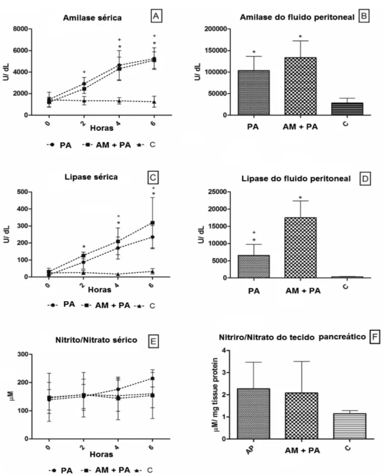
Pancreatitis induced significant increases in serum amylase and lipase concentrations. Methylene blue administration delayed the increase in amylase and lipase but did not decrease it (Figure 1A-D). Lipase assessment in abdominal fluid revealed greater concentrations of this enzyme when MB was administered (Figure1D).
Plasma Nitrite and Nitrate (NOx) Plasma Nitrite and Nitrate (NOx) Plasma Nitrite and Nitrate (NOx) Plasma Nitrite and Nitrate (NOx) Plasma Nitrite and Nitrate (NOx)
Although slight increases in NO levels were found in the AP group compared with the C and MB+AP groups, the means between groups remained statistically unchanged in plasma and pancreatic tissue (Figure 1E and F).
Hemodynamic Parameters Hemodynamic Parameters Hemodynamic Parameters Hemodynamic Parameters Hemodynamic Parameters
Figure 1 Figure 1 Figure 1 Figure 1
-Figure 1 - Mean values and standard deviation plotted for serum amylase (A), peritoneal fluid amylase (B), serum lypase (C), peritoneal fluid lypase (D), serum nitrite and nitrate (E) and peritoneal fluid nitrite and nitrate (F) of control pigs and pigs submitted to acute hemorrhagic necrotizing pancreatitis by retrograde pancreatic intra-ductal infusion of sodium taurocholate and entrokinase, pre-treated or not with methylene blue. +C differs from AP. *C differs from MB+AP. Significance for error type I was considered
Figure 2 -Figure 2 -Figure 2 Figure 2
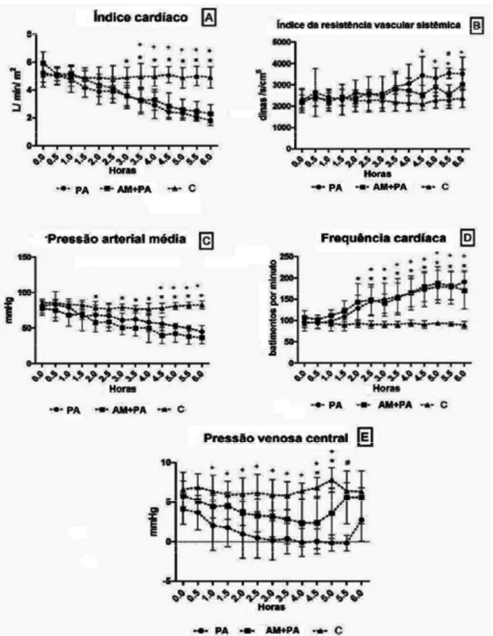
-Figure 2 - Mean values and standard deviation plotted for cardiac index (A), systemic vascular resistance index (B), mean arterial blood pressure (C), heart rate (D) and central venous pressure (E) of control pigs and pigs submitted to acute hemorrhagic necrotizing pancreatitis by retrograde pancreatic intra-ductal infusion of sodium taurocholate and entrokinase, pre-treated or not with methylene blue. +C differs from AP. *C differs from MB+AP. # AP differs from MB+AP. Significance for error type I was
decreased until the fifth hour and reached negative values (-0.16 ± 1 mmHg) that paralleled the decreases in CI and MBP and increases in HR and SVRI (Fig. 2). At the sixth hour, CVP increased (2.74 ± 2.65 mmHg). The administration of MB in the MB+P group generated means that followed the same pattern as the AP group. The means of SVRI, MPAP (Figure 2B and C), and PCWP were halfway between the means calculated for the C and AP groups and were not statistically different. Methylene blue delayed the reduction of CVP, revealing a significant decrease at hours 4.5 and 5 compared with the C group (Figure 2E).
Inflammatory Markers Inflammatory Markers Inflammatory Markers Inflammatory Markers Inflammatory Markers Interleukins-1â and -6
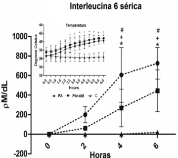
Surprisingly, IL-1â levels were zero and IL-6 levels significantly increased 2 h after the induction of AP compared with the C group (199.14 ± 82 pM/dl). Methylene blue decreased IL-6 to levels that were similar to controls. Hyperthermia coincided with increased IL-6 levels in all groups (Figure 3).
Myeloperoxidase Myeloperoxidase Myeloperoxidase Myeloperoxidase Myeloperoxidase
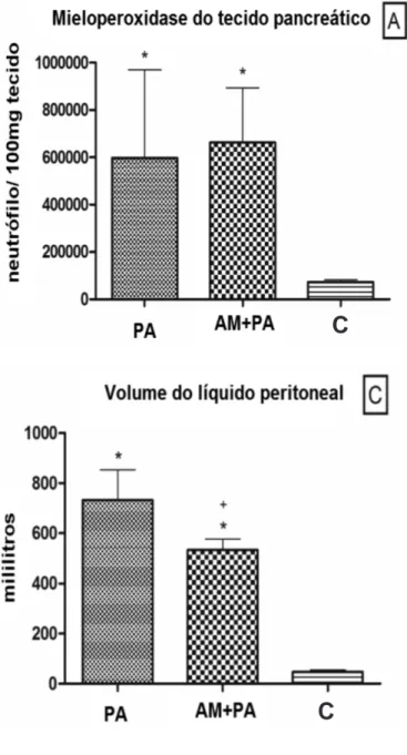
This measure of neutrophil activity increased after AP induction compared with controls (C group, 76,532 ± 11,360 neutrophils/100 mg tissue; AP group, 425,412 ± 123,021 neutrophils/100 mg tissue). Methylene blue administration did not result in significant differences compared with the AP group (MB+AP group, 663,926 ± 66,319; Figure 4A).
Lipid Peroxidation Lipid Peroxidation Lipid Peroxidation Lipid Peroxidation Lipid Peroxidation Malondialdehyde
Levels of this lipid peroxidation indicator increased in the AP and MB+AP groups compared with the C group. Methylene blue administration prior to AP induction worsened MDA levels compared with the AP group (MB+AP group, 41 ± 3 µM/mg protein; AP group, 25 ± 4 µM/mg protein; Figure 4B).
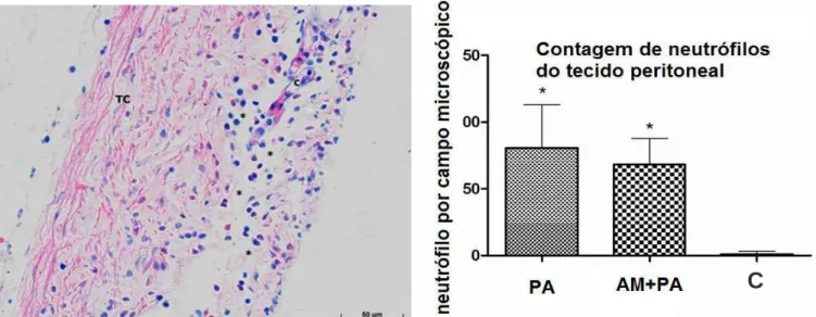
Peritoneal Tissue Neutrophil Count Peritoneal Tissue Neutrophil Count Peritoneal Tissue Neutrophil Count Peritoneal Tissue Neutrophil Count Peritoneal Tissue Neutrophil Count The mean numbers of neutrophils were higher in the AP and MB+AP groups compared with the C group, with no significant difference between the AP and MB+AP groups (Figure 5).
DISCUSSION
DISCUSSION
DISCUSSION
DISCUSSION
DISCUSSION
Experimental Model Experimental Model Experimental Model Experimental Model Experimental Model
Considering the experimental model, the retrograde infusion of taurocholate caused premature activation of trypsinogen, amylasemia, edema, inflammatory leukocyte migration, acinar necrosis, and the local generation of inflammatory mediators7.All of these features were observed in the present study, causing
Figure 3 Figure 3 Figure 3 Figure 3
-Figure 3 - Mean values and standard deviation plotted for Interleukin 6 and temperature of control pigs and pigs submitted to acute hemorrhagic necrotizing pancreatitis by retrograde pancreatic intra-ductal infusion of sodium taurocholate and entrokinase, pre-treated or not with methylene blue. +C differs from
AP. *C differs from MB+AP. # AP differs from MB+AP.
Significance for error type I was considered when p<0,05.
necrohemorragic pancreatitis associated with high concentrations of amylase and lipase in serum and in ascites. High concentrations are often used to characterize and quantify the severity of the disease8. Necrotizing hemorrhagic pancreatitis was also confirmed by macroscopic and microscopic evaluations (data not shown). The group treated with MB exhibited a delay in the increase in serum amylase but no effect on lipase. In abdominal fluid, lipase increased in the MB+AP group. The reduced levels of these enzymes in blood compared with abdominal fluid may be attributable to local release and compartmentalization syndrome9.
Hemodynamic Study Hemodynamic Study Hemodynamic Study Hemodynamic Study Hemodynamic Study
From a hemodynamic perspective, the present model presented an initial hyperdynamic stage of hypovolemic shock followed by the onset of cardiogenic shock, clinical signs of Systemic Inflammatory Response Syndrome and death. At the sixth hour of observation, the animals were on the verge of death. The first effect of AP was a significant decrease in CVP and PCWP, caused by volume depletion and reduced venous return initiated at the first hour, followed by increasing means toward the end of the study. The hemodynamic consequences of AP were devastating and resulted from intense vascular volu-me depletion. The initial findings resembled those seen during hypovolemic shock, associated with the release of vasoactive factors and inflammatory mediators and circulatory dysfunction. Retrograde infusion of NaT and EK in pigs promoted hypovolemia mainly 2 h after the
induction of AP, reflected by decreased CI, MBP, and CVP and increased HR and SVRI, similar to other reports13. Inspections of the CVP recordings revealed two patterns. First, the MB-mediated inhibition of sGC initially appeared to prevent severe reductions of CVP. Indeed, a reduction of accumulated fluid was found in the abdominal cavity, which might have contributed to more sustained venous return and thus CVP. Second, CVP trended upward toward the end of the experiment in all groups, which was attributable to the reduced myocardial function that resulted from the release of a depressor factor during AP14, and resulted in severe hyperkalemia/hypocalcemia, which was attributable to extensive necrosis. This observation, together with the direct recordings of decreased CI, may be the most significant finding of the cardiovascular parameters and may indicate the occurrence of cardiogenic shock that resulted from the depression of myocardial contractility. Previous studies have linked myocardial depression to the late response to proinflammatory mediators released during endotoxemia14, sepsis15, and hemorrhagic shock16. The etiology of myocardium depression is a complex and delayed response to cytokines released during these various forms of inflammation, the mechanisms of which are associated with a decreased â-adrenergic response17, decreased energy synthesis18, and decreased sensitivity of contractile
Figure 4 -Figure 4 -Figure 4
-Figure 4 -Figure 4 - Mean values and standard deviation plotted for pancreatic tissue myeloperoxidase (A), pancreatic tissue malondialdehyde (B) and peritoneal fluid volu-me (C) of control pigs and pigs submitted to acute hemorrhagic necrotizing pancreatitis by retrograde pancreatic intra-ductal infusion of sodium taurocholate and entrokinase, pre-treated or not with methylene blue. +Difference between AP and MB+AP. *Different
from C. Significance for error type I was considered when p<0,05.
C
proteins to calcium19. A brief period of sustained MBP and CI was responsive to reflex tachycardia but followed by an imminent reduction caused by severe hypovolemia and reduced preload and inotropy. In the AP group, an increase in PVRI was observed, possibly caused by hypoxic pulmonary vasoconstriction associated with reduced cardiac output. The occurrence of vasodilation was ruled out after calculating an increase in SVRI, which can be explained by an increase in catecholamine release resulting from volume depletion or a lack of an increase in NO associated with circulatory shock caused by AP. The lack of an increase in NO during shock may interfere with the beneficial effects of MB-mediated inhibition of sGC. After the initial stable phase, CI and HR declined, together with increased SVRI, PAP, PCWP, and CVP. Si-milar results have also been described elsewhere8,13.
Inflammation Inflammation Inflammation Inflammation Inflammation
An intense inflammatory reaction was confirmed by increased levels of IL-6, MPO, and MDA and fever. Serum concentrations of IL-1â, the main cytokine related to iNOS expression and leukocyte adhesion to endothelia20, were zero in all groups. Suggesting the absence of IL-1â would be impractical and unreasonable because, together with tumor necrosis factor-a, it is a main trigger and amplifier of inflammation21. Undetectable levels of IL-1â may be an element of non-NO experimen-tal models. The mean value of zero may reflect methodological problems that are difficult to evaluate, since we ensured the proper handling of the samples and appropriate assays. High serum concentrations of IL-6 in the AP group represented an appropriate prognostic tool. Preemptive administration of MB reduced IL-6 concentrations, but the values remained above control
levels, suggesting that a mild systemic antiinflammatory effect can be attributed to MB-induced inhibition of sGC. Hyperthermia is a reaction to the release of inflammatory mediators, especially IL-6, that promote the release of endogenous pyrogens22. This is the main feature of the inflammatory response observed in all groups subjected to AP, the onset of which coincided with increased MDA. Methylene blue did not prevent hyperthermia in the present study, despite its well-known antioxidant effects23.
Increased MPO activity, reflecting greater neutrophilia, was observed in the AP and MB+AP groups. Greater MPO activity has been associated with MB administration in an ischemia-reperfusion injury model24,25. However, we did not observe any stimulating effect of preemptive MB administration on MPO activity.
As mentioned above, the onset of AP is followed by intense pancreatic and inflammatory enzyme activation and the induction of mediators that increase vascular permeability with plasma leakage, edema, and third space fluid collection named pancreatic ascites. Amylase, a reliable indicator of membrane leakage, was markedly increased in this study. In the present model, post mortem observations indicated that the pancreas was the only source of leakage, with significant volume accumulation. The 28% reduction of accumulated volume seen in the MB+AP group may be attributable to MB-induced inhibition of sGC. Pancreatic ascites is an important source of toxic and vasoactive substances26 and is thought to be related to remote inflammation and pancreatic complications. The volume reduction observed in the present study was not associated with improved disease outcome. The peritoneal analysis revealed a possible correlation between AP severity and the spread of inflammation to extra-pancreatic tissues.
Figure 5 Figure 5 Figure 5 Figure 5
-Figure 5 - Peritoneal tissue neutrophil count in control pigs and pigs submitted to acute hemorrhagic necrotizing pancreatitis by retrograde pancreatic intra-ductal infusion of sodium taurocholate and entrokinase, pre-treated or not with methylene blue. The picture to the left shows the peritonium of an animal of the AP group showing large numbers of neutrophils (*)(*)(*)(*)(*) with blue nuclei and pink cytoplasm in the conective tissue (TC)(TC)(TC)(TC); capillary (C)(TC) (C)(C)(C)(C). Giemsa, 400x.
Lipid Peroxidation Lipid Peroxidation Lipid Peroxidation Lipid Peroxidation Lipid Peroxidation
Preemptive MB increased MDA, confirming our previous studies that used the same porcine model. Methylene blue prevents the formation of superoxide and its ability to function as a final acceptor of divergent electrons from oxidase sites 8. In fact, MB presents antioxidant effects but has redox characteristics. Plasma concentrations greater than 5 µM, which are easily obtained with clinical doses of MB of 1.5-2 mg/kg, increase oxygen capture, free radical generation and the consumption of antioxidant factors such as glutathione27. Other determinants of the redox activity of MB are the experimental model, dosage regimen and simple premise that MB is a prime promoter of lipid peroxidation.
In conclusion, the induction of this type of pancreatitis was acute and devastating. The progression of the disease brought the body to the verge of exhaustion. The adoption of a gradual and slow model of pancreatitis associated with increased NO release may allow the observation of the beneficial effects of MB. For now,
however, the use of MB is inadequate in cases of exponential non-NO release, and extreme caution must be taken given the increase in lipid peroxidation. Some of the beneficial effects of MB, including decreased ascites volume and delayed increases in amylase levels, do not justify the use of MB in fulminant pancreatitis. The constant physiological release of NO, a powerful modulator of vasotone, should be understood, and the inhibition of biological effects can have negative consequences, especially on microcirculation. Additionally, this study supports the concept that MB acts in situations of NO overproduction, similar to observations in sepsis, anaphylaxis and cardiopulmonary bypass vasoplegic syndrome.
Acknowledgements Acknowledgements Acknowledgements Acknowledgements Acknowledgements
Technical assistance from the Laboratory of Biochemistry - Department of Anatomy and Surgery, and from the Laboratory of Pain and Inflammation - Department of Pharmacology is gratefully acknowledged.
R E S U M O R E S U M O R E S U M O R E S U M O R E S U M O
Objetivo: Objetivo: Objetivo: Objetivo:
Objetivo: estudar o uso terapêutico do bloqueio da guanilato ciclase pelo azul de metileno em um modelo experimental de pancreatite aguda grave em suínos. MétodosMétodosMétodosMétodos: a pancreatite aguda necrotizante foi induzida em porcos anestesiados por infusãoMétodos ductal pancreática retrógrada de 1ml/kg de taurocolato de sódio a 5% e 8U/kg de enteroquinase. Três grupos foram estudados (n=5): controle (C), pancreatite (PA), “bolus” de azul seguido por pancreatite (AM+PA). Os dados incluíram enzimas séricas e do líquido abdominal, variáveis hemodinâmicas, hemogasometria arterial, volume de líquido abdominal, marcadores inflamatórios plasmáticos, nitrito/nitrato e mieloperoxidase e malondialdeído plasmático. Aplicou-se a análise de variância seguida do pós-teste de Bonferroni (p<0,05). ResultadosResultadosResultadosResultadosResultados: os valores de amilase e lipase foram três e dez vezes mais elevados no grupo PA. A atividade da mieloperoxidase foi 50% superior no grupo PA. Os dados hemodinâmicos indicaram choque hipovolêmico precoce seguido de choque cardiogênico. Observou-se grave translocação de líquidos para a cavidade peritoneal. A nitrito/nitrato plasmática permane-ceu inalterada. O grupo AM+PA teve aumento de cinco vezes do mieloperoxidase em comparação com o grupo C. Conclusões:Conclusões:Conclusões:Conclusões:Conclusões: a utilização de azul de metileno em suínos com pancreatite não demonstrou efeitos significativos sobre variáveis hemodinâmicas e inflamatórias. Seu uso terapêutico na pancreatite necro-hemorrágica pode ser inadequado e extremo cuidado deve ser tomado dado o aumento da peroxidação lipídica evidenciado pelo aumento dos valores do malondialdeído.
Descritores: Descritores: Descritores: Descritores:
Descritores: Pancreatite. Óxido nítrico. Radicais livres. Azul de metileno. Guanilato ciclase.
REFERENCES
REFERENCES
REFERENCES
REFERENCES
REFERENCES
1. Pastor CM, Suter PM. Evidence that humans produce less nitric oxide than experimental animals in septic shock. Crit Care Med. 1998;26(6):1135.
2. Evora PR, Ribeiro PJ, de Andrade JC. Methylene blue administration in SIRS after cardiac operations. Ann Thorac Surg. 1997;63(4):1212-3. 3. Evora PR. Should methylene blue be the drug of choice to treat
vasoplegias caused by cardiopulmonary bypass and anaphylactic shock? J Thorac Cardiovasc Surg. 2000;119(3):632-4.
4. Buzato MA, Viaro F, Piccinato CE, Evora PR. The use of methylene blue in the treatment of anaphylactic shock induced by compound 48/80: experimental studies in rabbits. Shock. 2005;23(6):582-7. 5. Evora PR, Simon MR. Role of nitric oxide production in naphylaxis and its relevance for the treatment of anaphylactic hypotension with methylene blue. Ann Allergy Asthma Immunol. 2007;99(4):306-13.
6. Meirelles RF Jr, Ceneviva R, Viaro F, Baldo CF, Evora PR. Methylene blue improves hemodynamic shock but increases lipoperoxidation
in severe acute pancreatitis pig model. Acta Cir Bras. 2008;23 Suppl 1:8-16; discussion 16.
7. Laukkarinen JM, Van Acker GJ, Weiss ER, Steer ML, Perides G. A mouse model of acute biliary pancreatitis induced by retrograde pancreatic duct infusion of Na-taurocholate. Gut. 2007;56(11):1590-8.
8. Li W, Yan X, Wang H, Zhang Z, Yu W, Ji D, et al. Effects of continuous high-volume hemofiltration on experimental severe acute pancreatitis in pigs. Pancreas. 2007;34(1):112-9.
9. Dugernier TL, Laterre PF, Wittebole X, Roeseler J, Latinne D, Reynaert MS, et al. Compartmentalization of the inflammatory response during acute pancreatitis: correlation with local and systemic complications. Am J Respir Crit Care Med. 2003;168(2):148-57.
11. Ozturk F, Gul M, Esrefoglu M, Ates B. The contradictory effects of nitric oxide in caerulein-induced acute pancreatitis in rats. Free Radic Res. 2008;42(4):289-96.
12. Bachetti T, Pasini E, Suzuki H, Ferrari R. Species-specific modulation of the nitric oxide pathway after acute experimentally induced endotoxemia. Crit Care Med. 2003;31(5):1509-14.
13. Yekebas EF, Strate T, Zolmajd S, Eisenberger CF, Erbersdobler A, Saalmüller A, et al. Impact of different modalities of continuous venovenous hemofiltration on sepsis-induced alterations in expe-rimental pancreatitis. Kidney Int. 2002;62(5):1806-18.
14. Lefer AM, Glenn TM, O’Neill TJ, Lovett WL, Geissinger WT, Wangensteen SL. Inotropic influence of endogenous peptides in experimental haemorrhagic pancreatitis. Surgery 1971;69(2):220-8.
15. Ao L, Song Y, Fullerton DA, Dinarello CA, Meng X. The interaction between myocardial depressant factors in endotoxemic cardiac dysfunction: role of TNF-alpha in TLR4-mediated ICAM-1 expression. Cytokine. 2007;38(3):124-9.
16. Joulin O, Petillot P, Labalette M, Lancel S, Neviere R. Cytokine profile of human septic shock serum inducing cardiomyocyte contractile dysfunction. Physiol Res. 2007;56(3):291-7.
17. Meng X, Ao L, Song Y, Raeburn CD, Fullerton DA, Harken AH. Signaling for myocardial depression in hemorrhagic shock: roles of Toll-like receptor 4 and p55 TNF-alpha receptor. Am J Physiol Regul Integr Comp Physiol. 2005;288(3):R600-6.
18. Kumar A, Paladugu B, Mensing J, Kumar A, Parrillo JE. Nitric oxide-dependent and -inoxide-dependent mechanisms are involved in TNF-alpha-induced depression of cardiac myocyte contractility. Am J Physiol Regul Integr Comp Physiol 2007;292(5):R1900-6. 19. Kelm M, Schäfer S, Dahmann R, Dolu B, Perings S, Decking UK, et
al. Nitric oxide induced contractile dysfunction is related to a reduction in myocardial energy generation. Cardiovasc Res. 1997;36(2):185-94.
20. Merx MW, Weber C. Sepsis and the heart. Circulation. 2007;116(7):793-802.
21. Leszczynski D, Josephs MD, Foegh ML. IL-1 beta-stimulated leucocyte-endothelial adhesion is regulated, in part, by the cyclic-GMP-dependent signal transduction pathway. Scand J Immunol. 1994;39(6):551-6.
22. Jambrik Z, Gyöngyösi M, Hegyi P, Czakó L, Takács T, Farkas A, et al. Plasma levels of IL-6 correlate with hemodynamic abnormalities in acute pancreatitis in rabbits. Intensive Care Med. 2002;28(12):1810-8.
23. Riedel W, Lang U, Oetjen U, Schlapp U, Shibata M. Inhibition of oxygen radical formation by methylene blue, aspirin, or alpha-lipoic acid, prevents bacterial-lipopolysaccharide-induced fever. Mol Cell Biochem. 2003;247(1-2):83-94.
24. Salaris SC, Babbs CF, Voorhees WD 3rd. Methylene blue as an inhibitor of superoxide generation by xanthine oxidase. A potential new drug for the attenuation of ischemia/reperfusion injury. Biochem Pharmacol. 1991;42(3):499-506.
25. Ilhan H, Alatas O, Tokar B, çOlak O, Paºao”lu O, Koku N. Effects of the anti-ICAM-1 monoclonal antibody, allopurinol, and methylene blue on intestinal reperfusion injury. J Pediatr Surg. 2003;38(11):1591-5.
26. Sugimoto M, Takada T, Yasuda H, Nagashima I, Amano H, Yoshida M, et al. The lethal toxicity of pancreatic ascites fluid in severe acute necrotizing pancreatitis. Hepatogastroenterology. 2006;53(69):442-6.
27. May JM, Qu ZC, Cobb CE. Reduction and uptake of methylene blue by human erythrocytes. Am J Physiol Cell Physiol. 2004;286(6):C1390-8.
Received on 10/10/2012
Accepted for publication 1312/2012 Conflict of interest: none.
Source of funding: The project was financially supported by FAEPA/ Clinical Hospital at the Ribeirão Preto Faculty of Medicine, University of Sao Paulo and CAPES.
How to cite this article: How to cite this article: How to cite this article: How to cite this article: How to cite this article:
Baldo CF, Capellini VK, Celotto AC, Sônego F, Tirapelli LF, Batalhão M, Cárnio EC, Santos JS, Évora PRB. Guanylate cyclase inhibition by methylene blue in circulatory shock caused by acute necrotizing pancreatitis: a word of caution based on a porcine model. Rev Col Bras Cir. [periódico na Internet] 2013;40(6). Disponível em URL: http:// www.scielo.br/rcbc