523
Rev Soc Bras Med Trop 49(4):523-526, July-August, 2016 doi:10.1590/0037-8682-0331-2015
Case Report
Nocardia nova
causing empyema necessitatis after
lung re-transplantation: a case report
Cecília Bittencourt Severo
[1],[2],
Letícia Beatriz Matter
[3],[4],
Flávio de Mattos Oliveira
[1],
Agueda Palmira Castagna Vargas
[4],
Sadi Marcelo Schio
[5],
José de Jesus Peixoto
Camargo
[5],
Bruno Hochhegger
[2],[6]and
Luiz Carlos Severo
[1],[7][1]. Laboratório de Micologia, Hospital Santa Rita, Irmandade Santa Casa de Misericórdia de Porto Alegre, Porto Alegre, Rio Grande do Sul, Brasil. [2]. Departamento de Ciências Básicas, Universidade Federal de Ciências da Saúde de Porto Alegre, Porto Alegre, Rio Grande do Sul, Brasil. [3]. Departamento de Ciências da Saúde, Universidade Regional Integrada do Alto Uruguai e das Missões, Santo Ângelo, Rio Grande do Sul, Brasil.
[4]. Departamento de Medicina Veterinária Preventiva, Universidade Federal de Santa Maria, Santa Maria, Rio Grande do Sul, Brasil. [5]. Serviço de Transplante Pulmonar, Irmandade Santa Casa de Misericórdia de Porto Alegre, Porto Alegre, Rio Grande do Sul, Brasil.
[6]. Serviço de Radiologia, Irmandade Santa Casa de Misericórdia de Porto Alegre, Porto Alegre, Rio Grande do Sul, Brasil. [7]. Departamento de Medicina Interna, Universidade Federal do Rio Grande do Sul, Porto Alegre, Rio Grande do Sul, Brasil.
Abstract
We report herein a case of thoracic infection due to Nocardia nova following lung re-transplantation performed for emphysema
related to alpha-1-antitrypsin defi ciency. The infection extended from the lung into the pleural space, thoracic wall, and mediastinum,
presenting as pericarditis and empyema necessitatis. Nocardia nova was identifi ed by 16S ribosomal deoxyribonucleic acid (rDNA) sequencing and phylogenetic analysis. According to a literature search of PubMed, LILACS and MEDLINE databases,
we describe herein the fi rst case of empyema necessitatis caused by N. nova species in a transplanted patient.
Keywords: Nocardia nova. Pericarditis. Empyema necessitatis.
Corresponding author: Dr. Luiz Carlos Severo. e-mail: [email protected]
Received 11 November 2015 Accepted 5 April 2016
INTRODUCTION
Empyema necessitatis refers to infl ammatory tissue that extends directly from the pleural cavity into the thoracic
wall, forming a mass (a cold abscess requiring drainage) in
the extra-pleural soft tissues. It is a very rare complication of
different infectious etiologies; tuberculosis and actinomycosis are the most common causative organisms(1). Recently, cases of nocardiosis with chest wall extension(2) and N. asteroides
empyema necessitatis have been observed(3). Herein, we report the fi rst case of empyema necessitatis caused by N. nova.
CASE REPORT
A 50-year-old woman underwent lung re-transplantation in July 2011, after developing chronic lung allograft dysfunction. The patient had a long history of emphysema secondary to
alpha-1-antitrypsin defi ciency. She was subjected to
double-lung volume-reduction surgery in 2003 and single-double-lung transplantation in 2006. The lung re-transplantation in 2011 was bilateral. Her immunosuppressive regimen consisted of prednisone, mycophenolate, and tacrolimus. Five months
after re-transplantation, the patient presented to the transplant
clinic with dyspnea. Bronchoscopy identifi ed stenosis of the
left bronchial anastomosis. Endoscopic attempts to manage the stenosis failed and a lower left lobectomy was performed.
One month later, she presented to the clinic with dyspnea and a painful draining mass located on her left lateral chest wall.
On examination, she was afebrile and an external fi stula was identifi ed in the left inframammary region. The patient received intravenous ceftriaxone as empiric antimicrobial therapy for
subcutaneous infection, with consequent clinical improvement. However, when the therapy was stopped, the problem worsened and the chest radiograph revealed a pleural effusion.
In the investigation, the pleural fl uid consisted of a purulent
exudate (Figure 1A). Gram-positive filamentous bacilli
(Figure 1B) were detected in the exudate. These bacilli were
decolorized by Ziehl-Neelsen staining. A contrast-enhanced
axial computed tomography (CT) scan with a mediastinal
window demonstrated atelectasis in the left lower lobe, associated with bilateral pleural and pericardial effusions. In addition, pleural and pericardial thickening was revealed, suggesting a complicated effusion. The clinical and cytological
fi ndings were suggestive of actinomycosis. The patient was
thus treated with penicillin, but failed to show any clinical improvement. Kinyoun staining was performed and revealed
branching, weakly acid-fast, fi laments (Figure 1C). Aerobic
524
Severo CB et al. - Empyema necessitatis caused by Nocardia nova
FIGURE 1. Nocardia nova pictures. (A) Purulent pleural fl uid obtained via aspiration of the pleural cavity. (B) Staining performed directly from the pleural effusion showing Gram-positive rod-shaped bacteria. (C) Partially acid-fast branching fi laments showed by Kinyoun Staining. Note: Figures B and C were
observed by microscopy at 1,000-times magnifi cation.
A B C
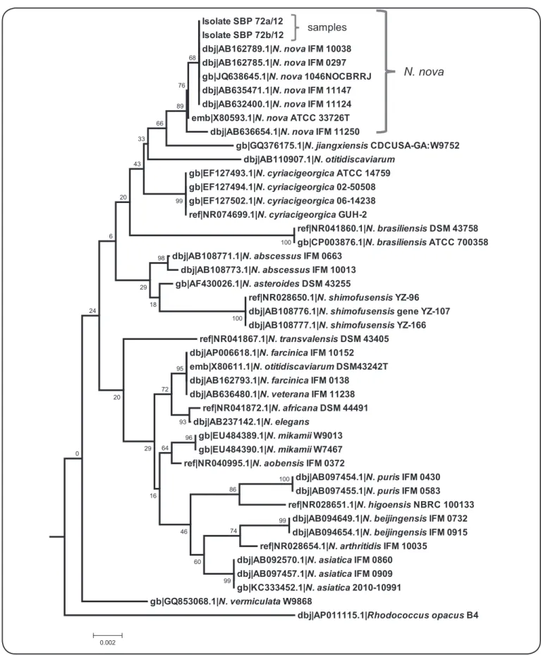
In the diagnosis, deoxyribonucleic acid (DNA) from isolated colonies (SBP72a/12: sampled from pleural fl uid;
and SBP72b/12 sampled from the pleural cavity secretion) was isolated and molecular diagnosis was performed by
analyzing the partial sequence (approximately 1,300 base pairs)
of 16S ribosomal ribonucleic acid (rRNA) gene amplifi ed with universal primers(4). Polymerase chain reaction (PCR)
products were sequenced by ACTGene Análises Moleculares Ltda [Centro de Biotecnologia, Universidade Federal do Rio Grande do Sul (UFRGS), Porto Alegre, RS, Brazil] using the automatic sequencer ABI-PRISM® 3100 Genetic Analyzer
(Applied Biosystems). Consensus sequences were assembled using Staden Package 4. Figure 2 shows the strong homology of the samples with others N. nova strains and the relationship with others species available in GenBank dataset. The partial 16S rRNA gene sequences of SBP72a/12 and SBP72b/12 were deposited in the GenBank database.
In thefollow-up, N. nova was identifi ed, the patient received
treatment with intravenous trimethoprim/sulfamethoxazole (TMP/SMX) 320/1,600mg every six hours for one month, with
success. Central nervous system disease was ruled out. Finally, the patient was discharged on oral TMP/SMX (160/800 BID). At discharge, she was asymptomatic, and the lesions had improved both clinically and radiologically. At the time of her last
follow-up visit, fi ve months after discontinuation of antimicrobial
therapy, she remained well.
DISCUSSION
Traditional identifi cation of Nocardia to species level by phenotyping is cumbersome and the results are sometimes
diffi cult to interpret. The recent introduction of molecular methods has had an enormous impact on the taxonomy of
Nocardia and has resulted in the recognition of numerous new species. Worldwide, respiratory and disseminated infections are
most often due to members of the previously broadly defi ned
N. asteroides complex, which contains three strains: Nocardia
asteroides sensu stricto, Nocardia farcinica, and N. nova
complex. The latter includes N. nova species(5).
In a search of PubMed/MEDLINE and LILACS databases, we found no previous report of empyema necessitatis caused by N. nova. The key words used for the search were Nocardia nova, nocardiosis, lung transplant, and empyema necessitatis, and it
was performed until September 2015. We describe here the fi rst
case of empyema necessitatis caused by N. nova species in a transplanted patient.
A contemporary case-control study found that the incidence of N. nova isolated from solid organ transplant recipients was higher than N. farcinica (49% vs. 28%, respectively)(6). In
a review of 17 patients with an underlying malignancy and Nocardia bacteremia, N. nova complex was demonstrated to be responsible for 35% of the cases. Bacterial adhesion to intravenous catheters may play a role in the incidence of intravascular N. nova infection(7) (8).
Infection of the lung parenchyma and pleural space by N. nova has previously been observed(9) (10) (11). However, direct extension from the lung into the pleural cavity, progressing to
penetration of the mediastinum, with consequent pericarditis and
empyema necessitates–specifi cally in a lung-transplant recipient
– has not previously been reported. Physicians should be aware of this possibility in order to obtain and submit appropriate thoracic specimens to the laboratory to optimize the recovery of this microorganism.
In conclusion, we advocate maintaining a high index
525 Rev Soc Bras Med Trop 49(4):523-526, July-August, 2016
Isolate SBP 72a/12 Isolate SBP 72b/12
dbj|AB162789.1|N. novaIFM 10038 dbj|AB162785.1|N. novaIFM 0297 gb|JQ638645.1|N. nova1046NOCBRRJ dbj|AB635471.1|N. novaIFM 11147 dbj|AB632400.1|N. novaIFM 11124 emb|X80593.1|N. novaATCC 33726T
dbj|AB636654.1|N. novaIFM 11250
gb|GQ376175.1|N. jiangxiensisCDCUSA-GA:W9752 dbj|AB110907.1|N. otitidiscaviarum
gb|EF127493.1|N. cyriacigeorgicaATCC 14759 gb|EF127494.1|N. cyriacigeorgica02-50508 gb|EF127502.1|N. cyriacigeorgica06-14238 ref|NR074699.1|N. cyriacigeorgicaGUH-2
ref|NR041860.1|N. brasiliensisDSM 43758 gb|CP003876.1|N. brasiliensisATCC 700358 dbj|AB108771.1|N. abscessusIFM 0663
dbj|AB108773.1|N. abscessusIFM 10013 gb|AF430026.1|N. asteroidesDSM 43255
ref|NR028650.1|N. shimofusensisYZ-96 dbj|AB108776.1|N. shimofusensisgene YZ-107 dbj|AB108777.1|N. shimofusensisYZ-166 ref|NR041867.1|N. transvalensisDSM 43405
dbj|AP006618.1|N. farcinicaIFM 10152 emb|X80611.1|N. otitidiscaviarumDSM43242T dbj|AB162793.1|N. farcinicaIFM 0138
dbj|AB636480.1|N. veteranaIFM 11238 ref|NR041872.1|N. africanaDSM 44491 dbj|AB237142.1|N. elegans
gb|EU484389.1|N. mikamiiW9013 gb|EU484390.1|N. mikamiiW7467 ref|NR040995.1|N. aobensisIFM 0372
dbj|AB097454.1|N. purisIFM 0430 dbj|AB097455.1|N. purisIFM 0583 ref|NR028651.1|N. higoensisNBRC 100133
dbj|AB094649.1|N. beijingensisIFM 0732 dbj|AB094654.1|N. beijingensisIFM 0915 ref|NR028654.1|N. arthritidisIFM 10035
dbj|AB092570.1|N. asiaticaIFM 0860 dbj|AB097457.1|N. asiaticaIFM 0909 gb|KC333452.1|N. asiatica2010-10991 gb|GQ853068.1|N. vermiculataW9868
dbj|AP011115.1|Rhodococcus opacusB4 68
76
89
66
33
99 43
100 20
98
100 18
29 6
95
93 72
96 64
100 86
99 74
99 60 46 16 29 20 24
0
0.002
N. nova
samples
FIGURE 2. Nucleotide sequence analysis. Phylogenetic relationship derived from 16S rDNA sequences showing the homology between Nocardia nova (samples SBP 72a/12 and SBP 72b/12) and 16S rDNA sequences of other Nocardia species. The phylogenetic tree was built in the MEGA 5.2 software program, using the Neighbor-Joining algorithm and the Tamura 3-parameter substitution model. Rhodococcus opacus B4 (AP011115.1) was used as the out group reference.
526
Acknowledgements
The authors acknowledge the Coordenação de Aperfeiçoamento de Pessoal de Nível Superior (CAPES) for the Letícia B. Matter’s Post-doctorate scholarship.
Financial Support
Programa Nacional de Pós-Doutorado/Coordenação de Aperfeiçoamento de Pessoal de Nível Superior Project number 2734/2011.
REFERENCES
1. Reyes CV, Thompson KS, Jensen J. Fine needle aspiration biopsy of masititis secondary to empyema necessitatis. A report of two cases. Acta Cytol 1999; 43:873-876.
2. Kanne JP, Yandow DR, Mohammed TL, Meyer CA. CT fi ndings of pulmonary nocardiosis. AJR Am J Roentgenol 2011; 197:266-272.
3. Cadena J, Cadena A, Hallmark K, Ahuja SK. Pulmonary nocardiosis mimicking empyema necessitatis. J Resp Dis 2008; 29:1-6.
4. Fredricks DN, Relman DA. Improved amplifi cation of microbial DNA from blood cultures by removal of the PCR inhibitor sodium polyanetholesulfonate. J Clin Microbiol 1998; 36:2810-2816.
5. Brown-Elliott BA, Brown JM, Conville PS, Wallace Jr RJ. Clinical and laboratory features of the Nocardia spp. based on current
molecular taxonomy. Clin Microbiol Rev 2006; 19:259-282.
6. Peleg AY, Husain S, Qureshi ZA, Silveira FP, Sarumi M, Shutt KA, et al. Risk factors, clinical characteristics, and outcome of
Nocardia infection in organ transplant recipients: a matched case-control study. Clin Infect Dis 2007; 44:1307-1314.
7. Al Akhrass F, Hachem R, Mohamed JA, Tarrand J, Kontoyiannis DP, Chandra J, et al. Central venous catheter-associated Nocardia
bacteremia in cancer patients. Emerg Infect Dis 2011; 17:1651-1658.
8. Gomila BS, Sabater SV, Merino JP, Igual RA, Moreno RM. Abscesso cerebral por Nocardia nova. Rev Chil Infect 2012; 29:112-113.
9. Burucoa C, Breton I, Ramassamy A, Soyer J, Becq-Giraudon B, Fauchère JL. Western blot monitoring of disseminated Nocardia nova infection treated with clarithromycin, imipenem, and surgical drainage. Eur J Clin Microbiol Infect Dis 1996; 15: 943-947.
10. Couraud S, Houot R, Coudurier M, Ravel AC, Coiffi er B, Souquet PJ. Nocardial pulmonary infection. Rev Mal Respir 2007; 24: 353-357.
11. Tan CK, Lai CC, Lin SH, Liao CH, Chou CH, Hsu HL, et al. Clinical and microbiological characteristics of nocardiosis including those caused by emerging Nocardia species in Taiwan, 1998-2008. Clin Microbiol Infect 2010; 16:966-972.