Thoracoscopy in children with complicated
parapneumonic pleural effusion at the fibrinopurulent
stage: a multi-institutional study*
Toracoscopia em crianças com derrame pleural parapneumônico complicado na fase fibrinopurulenta: estudo multi-institucional
Sérgio Freitas, José Carlos Fraga, Fernanda Canani
Abstract
Objective: To determine the efficacy of thoracoscopy in the management of children with complicated parap-neumonic pleural effusion at the fibrinopurulent stage. Methods: Retrospective study of 99 children submitted to thoracoscopy for the treatment of complicated parapneumonic pleural effusion at the fibrinopurulent stage between November of 1995 and July of 2005. The mean age was 2.6 years (range, 0.4-12 years), and 60% were males. Thoracoscopy was performed at three different hospitals following the same treatment algorithm.
Results: Thoracoscopy was effective for 87 children (88%). In 12 (12%), a second surgical procedure was required: another thoracoscopy (n = 6) or thoracotomy/thoracostomy (n = 6). Mean duration of chest tube drainage follo-wing successful thoracoscopy was 3 days vs. 10 days in patients submitted to a second procedure (p < 0.001). In all of the children, the pleural infection resolved after treatment. Thoracoscopy-related complications included air leak (30%), chest tube bleeding (12%), subcutaneous emphysema associated with trocar insertion (2%) and surgical wound infection (2%). None of the children required additional surgical procedures due to the complications.
Conclusions: The effectiveness of thoracoscopy in children with parapneumonic pleural effusion at the fibrinopu-rulent stage was 88%. The procedure was safe, with a low rate of severe complications. Thoracoscopy should be the first-choice treatment for children with parapneumonic pleural effusion at the fibrinopurulent stage.
Keywords: Thoracoscopy; Pleural effusion; Empyema, pleural.
Resumo
Objetivo: Determinar a eficácia da toracoscopia em crianças com derrame pleural parapneumônico complicado (DPPC) na fase fibrinopurulenta. Métodos: Estudo retrospectivo de 99 crianças submetidas à toracoscopia para tratamento de DPPC na fase fibrinopurulenta entre novembro de 1995 e julho de 2005. A média de idade foi de 2,6 anos (variação, 0,4-12 anos) e 60% eram do sexo masculino. A toracoscopia foi realizada em três hospitais diferentes utilizando-se o mesmo algoritmo de tratamento. Resultados: A toracoscopia foi eficaz em 87 crianças (88%) e 12 (12%) necessitaram de outro procedimento cirúrgico: nova toracoscopia (n = 6) ou toracotomia/pleu-rostomia (n = 6). O tempo médio de drenagem torácica foi de 3 dias nas crianças em que a toracoscopia foi efetiva e de 10 dias naquelas que precisaram de outro procedimento (p < 0,001). A infecção pleural de todas as crianças foi debelada após o tratamento. As complicações da toracoscopia foram fuga aérea (30%) e sangramento pelo dreno torácico (12%), enfisema subcutâneo na inserção do trocarte (2%) e infecção da ferida operatória (2%). Nenhuma criança necessitou de reoperação devido às complicações. Conclusões: A efetividade da toracoscopia em crianças com DPPC na fase fibrinopurulenta foi de 88%. O procedimento mostrou-se seguro, com baixa taxa de complica-ções graves, devendo ser considerado como primeira opção em crianças com DPPC na fase fibrinopurulenta.
Descritores: Toracoscopia; Derrame pleural; Empiema pleural.
* Study carried out in the Department of Pediatric Thoracic Surgery and in the Department of Pediatric Surgery of the Porto Alegre Hospital de Clínicas, Porto Alegre, Brazil; at the Moinhos de Vento Hospital, Porto Alegre, Brazil; in the Department of Pediatric Surgery of the Caxias do Sul General Hospital, Caxias do Sul, Brazil; and as part of the Graduate Course in Medicine (Surgical Sciences), Interinstitutional Masters Program, at the Federal University of Rio Grande do Sul School of Medicine, Porto Alegre, Brazil, and the University of Caxias do Sul Foundation School of Medicine, Caxias do Sul, Brazil.
Correspondence to: José Carlos Fraga. Rua Ramiro Barcelos, 2350, sala 600, CEP 91000-003, Porto Alegre, RS, Brasil. Tel 55 51 2101 8232. Fax 55 51 3334 0146. E-mail: [email protected].
Financial support: None.
subsequent closed pleural drainage, is indicated; at the organized stage, thoracotomy or open drainage by thoracostomy is indicated.(4)
Although thoracoscopy is currently the procedure of choice for children with compli-cated parapneumonic pleural effusion at the fibrinopurulent stage,(9-11) there have been no
well-designed studies involving a significant number of patients and assessing the true effec-tiveness of this method in the management of complicated parapneumonic pleural effusion in children.(12,13) The objective of this
multi-insti-tutional study was to determine the efficacy of thoracoscopy in the management of a significant number of children with complicated parapneu-monic pleural effusion at the fibrinopurulent stage.
Methods
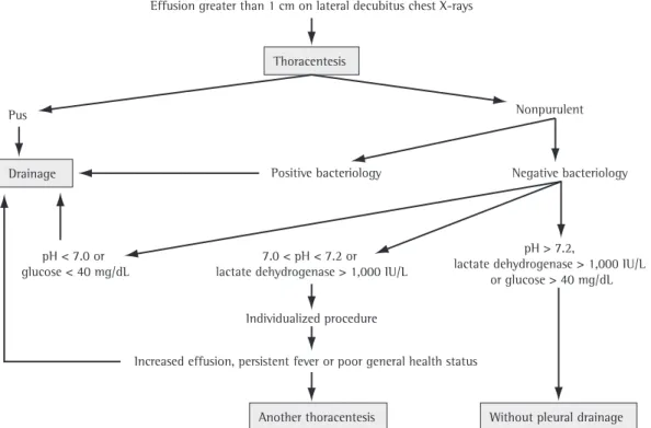
This was a retrospective study of 99 chil-dren (aged between 3 months and 12 years) submitted to thoracoscopy for the treatment of complicated parapneumonic pleural effusion at the fibrinopurulent stage between November of 1995 and July of 2005. The mean age was 2.6 years, and 60 (60%) were male. Thoracoscopy was performed at the Hospital de Clínicas de Porto Alegre (HCPA, Porto Alegre Hospital de Clínicas) and at the Hospital Moinhos de Vento (HMV, Moinhos de Vento Hospital) in the city of Porto Alegre, Brazil, as well as at the Hospital Geral de Caxias do Sul (HGCS, Caxias do Sul General Hospital) in the city of Caxias do Sul, Brazil. The management of these patients was performed following the same treatment algorithm (Figures 1 and 2).(4) At each hospital studied, the
following data were collected from the medical charts: age; gender; use of thoracentesis; use of antibiotics prior to surgery; macroscopic aspect, biochemical analysis, microscopy and culture of pleural fluid; time elapsed from the onset of the clinical symptoms to thoracoscopy; duration of chest tube drainage; length of hospital stay; efficacy of thoracoscopy; and need for a second surgical procedure.
The presence of pleural septations, charac-terizing the fibrinopurulent stage of complicated parapneumonic pleural effusion, was confirmed if one or more of the following criteria were met: lack of mobility of the pleural fluid on lateral decubitus chest X-rays (horizontal beam); air-fluid level identified prior to instrumentation of
Introduction
Parapneumonic pleural effusion is that occurring in association with pneumonia.(1)
Approximately 40% of children with pneumonia have pleural effusion,(2) and approximately 10%
of those children require surgical drainage.(2)
According to its aspect and content, parapneu-monic pleural effusion is classified as complicated or uncomplicated. Complicated parapneumonic pleural effusion is that in which pus or germs are seen in pleural fluid samples, either under microscopy or in culture, or in which biochemical analysis of the pleural fluid reveals a pH < 7.0, glucose < 40 mg/dL and lactate dehydroge-nase > 1,000 IU/L.(3,4) When there is pus in the
pleural space, the condition is designated pleural empyema.(1) Recent studies have confirmed that
parapneumonic pleural effusion, even without purulent content, behaves like empyema, with a strong tendency toward loculation and, conse-quently, a significant increase in morbidity and mortality.(5)
The evolution of complicated parapneu-monic pleural effusion has three well-defined stages, which, in fact, are progressive if the effusion is not treated appropriately.(6) The first
stage, known as the acute or exudative stage, is characterized by the presence of sterile inviscid pleural fluid, which is easily removed by drainage, with rapid lung expansion. In the second stage, known as the fibrinopurulent stage, the pleural fluid presents polymorphonuclear leukocytes, bacteria and cell remnants. In addition, there is fibrin formation and deposition in the pleura, as well as a tendency toward septum formation and loculation of the effusion. At this stage, lung expansion occurs only after the septations are disrupted and the infectious intrapleural content is completely removed. The last stage, known as the organized stage, is characterized by the pres-ence of fibroblasts on the pleurae, giving rise to a thick, inelastic membrane that covers the lung and reduces its expansion. At this stage, even after the pleural fluid is completely removed, complete lung expansion is not achieved.(7)
The treatment for complicated parapneu-monic pleural effusion is surgery,(7) and the
type of drainage depends on the stage of the effusion(4,8): at the acute stage, closed chest tube
costal space, in the posterior axillary line, in the lowest part of the fluid cavity. The last patients in our sample received insufflation with carbon dioxide through one of the trocars, with reduced intrathoracic pressure (3-5 mmHg), which allowed lung collapse and better viewing during thoracoscopy. After the thoracic cavity had been opened, the fluid content was completely aspi-rated. Subsequently, the mediastinoscope or the video equipment was introduced. The intratho-racic septations were identified, the septa were disrupted, and the pleural cavity was washed with saline solution. After the procedure, a chest tube was inserted through the incision that had previously been used to insert the equip-ment—the lowest incision in the hemithorax. All patients underwent simple pleural drainage.
Quantitative data are presented as mean and standard deviation. Symmetric contin-uous data were compared using the Student’s t-test for independent samples, and asym-metric continuous data were compared using the Wilcoxon-Mann-Whitney U test. Categorical variables were compared using Fisher’s exact test. The analyses were performed using the program Statistical Package for the Social Sciences, the pleural space; and septations seen on
ultra-sound or CT scans.(14,15)
In the children who presented complicated parapneumonic pleural effusion with septa-tions, thoracoscopy was performed as an initial procedure. Some of the children had previ-ously undergone closed chest tube drainage, and, due to persistent fever and radiological findings of pleural septations, subsequently underwent thoracoscopy.(5,16,17) The procedure
was performed under general anesthesia, with the child placed in the lateral decubitus posi-tion. Initially, selective intubation of the healthy lung was performed. However, since 2005, we have performed nonselective tracheal intubation only.
The surgical procedure was performed using a small mediastinoscope or trocars for video-assisted surgery. The mediastinoscope or the first trocar was introduced into the thoracic cavity through a small incision in the fourth intercostal space, in the anterior axillary line, preferably immediately below the nipple. The other instruments needed in order to manipulate and clean the pleural cavity using video-assisted surgery were introduced into the sixth
inter-Effusion greater than 1 cm on lateral decubitus chest X-rays
Thoracentesis
Another thoracentesis Without pleural drainage Drainage
Nonpurulent
Negative bacteriology Positive bacteriology
Pus
Individualized procedure
pH > 7.2,
lactate dehydrogenase > 1,000 IU/L or glucose > 40 mg/dL
Increased effusion, persistent fever or poor general health status 7.0 < pH < 7.2 or
lactate dehydrogenase > 1,000 IU/L pH < 7.0 or
glucose < 40 mg/dL
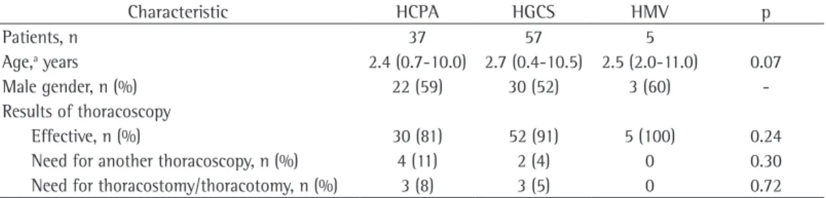
nopurulent stage, thoracoscopy was successful in 87 (88%) and a second surgical procedure was required in 12 (12%). The procedure was performed at the HCPA in 37 cases, at the HGCS in 57 and at the HMV in 5 (Table 2). There were no differences among the various hospitals in terms of age or gender of the children under-going the procedure; nor were there any such differences in terms of the efficacy of thora-coscopy or the need to repeat the procedure (Table 2). Although each of the three hospitals had its own surgical team, the same treatment algorithm was used in all cases, and no differ-ences were found among the hospitals in terms of the treatment results obtained for the chil-dren with complicated parapneumonic pleural effusion at the fibrinopurulent stage (Table 2).
Preoperative drainage was performed in 31 of the complicated parapneumonic pleural effusion patients in our sample. Of those, only 5 (16.1%) showed no improvement after thoracoscopy and subsequently required thoracostomy/thora-cotomy. In the remaining 26 patients (83.9%), the post-thoracoscopy evolution was satisfactory.
Thoracentesis was performed in 61 children (61%). Pleural fluid analysis (Table 1) revealed turbid or purulent fluid in 28 patients (28%). Germs were identified in the pleural secretions in 23 patients (23%). At the time of pleural fluid version 12.0 (SPSS Inc., Chicago, IL, USA). The
study was approved by the human research ethics committees of the hospitals involved.
Results
Table 1 shows that, among the 99 children submitted to thoracoscopy due to complicated parapneumonic pleural effusion at the
fibri-Complicated parapneumonic pleural effusion
Free effusion Septated or loculated pleural effusion
Chest tube drainage
Pleural lavage + closed drainage
Thoracoscopy
Thoracostomy Complete lung
expansion
Lung entrapment
Figure 2 - Algorithm of the types of drainage used in the study for the treatment of patients with complicated parapneumonic pleural effusion.(4)
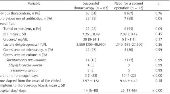
Table 1 - Complicated parapneumonic pleural effusion at the fibrinopurulent stage: comparison between the patients who improved after thoracoscopy and those requiring a second surgical procedure.
Variable Successful thoracoscopy (n = 87)
Need for a second operation (n = 12)
p
Previous thoracentesis, n (%) 53 (61) 8 (67) 0.76 No previous use of antibiotics, n (%) 25 (29) 7 (58) 0.05 Pleural fluid
Turbid or purulent, n (%) 22 (28) 6 (55) 0.09 pH, mean ± SD 7.25 ± 0.49 7.08 ± 0.42 0.45 Glucose,a mg/dL 30 (0-241) 5 (1-111) 0.17
Lactate dehydrogenase,a IU/L 2.559 (309-40.990) 1.340 (679-23.600) 0.36
Germs seen on microscopy, n (%) 22 (27) 2 (20) 0.99 Germs seen on culture, n (%)
Streptococcus pneumoniae 14 (16) 2 (17) 0.99
Staphylococcus aureus 4 (5) 0 0.99
Pseudomonas spp. 3 (3) 0 0.99 Duration of drainage,a days 3 (1-23) 10 (4-22) < 0.001
Time elapsed from the onset of the clinical symptoms to thoracoscopy (days), mean ± SD
7.97 ± 3.51 9.48 ± 4.45 0.18
Hospital stay,a days 14 (6-40) 26 (17-55) < 0.001
intervention. Indications for additional surgical intervention were fever and residual fluid in the pleural cavity, with or without septations.
The comparison between the children submitted to a single thoracoscopy and those who required a second surgical procedure can be seen in Table 1. There were no differences between the two groups in terms of the previous use of antibiotics, the presence of germs in pleural fluid samples (microscopy or culture), the finding of turbid or purulent fluid during thoracentesis or biochemical abnormalities (in pH, glucose and lactate dehydrogenase). The median duration of chest tube drainage and the median hospital stay were significantly higher in the children who required additional surgical intervention (Table 1). In all cases, the long-term evolution was favorable: the pleural infection resolved, and there was pleural thickening (to varying degrees) in the radiological follow-up evaluations on postoperative days 30 and 60.
Discussion
The effectiveness of thoracoscopy in our patients (88%) is within the range reported in the literature, according to which complicated parapneumonic pleural effusion improves in 71-100% of children undergoing surgery.(5,17-27)
In children for whom thoracoscopy was insuf-ficient to produce clinical improvement, and in whom a second surgical procedure was required, another thoracoscopy was always the first choice. This was not possible in some children, since complicated parapneumonic pleural effu-sion had evolved to the organized stage, with lung entrapment, and thoracotomy or thora-costomy was therefore required. Although both procedures can be performed at this stage of collection for study, 67 children (67%) were
receiving antibiotics (Table 1).
Regarding the radiological tests performed in order to diagnose and confirm the fibrinopu-rulent stage of complicated parapneumonic pleural effusion, 99 children (100%) underwent chest X-ray, 41 (42%) underwent ultrasound, and 5 (6%) underwent CT.
In the postoperative period, chest tube air leak occurred in 30 children (30%), with a mean duration of 5 days (range, 1-15 days). In addi-tion, chest tube bleeding, albeit not requiring blood replacement, occurred in 12 patients (12%), subcutaneous emphysema associated with trocar insertion occurred in 2 (2%), and surgical wound infection occurred in another 2 (2%). None of those complications required surgical intervention. An immediate second operation, unrelated to thoracoscopy, was required in 1 child with pneumonia and compli-cated parapneumonic pleural effusion caused by Staphylococcus aureus. That child also had puru-lent pericarditis and cardiac tamponade. Chest tube air leak in the postoperative period did not represent an unfavorable prognostic factor for the cure of complicated parapneumonic pleural effusion (p = 0.241).
Of the 12 children who required additional surgical intervention, 5 had undergone the initial procedure at the HGCS: 2 underwent another thoracoscopy; and 3 underwent thoracostomy/ thoracotomy. The remaining 7 children who required additional surgical intervention had undergone the initial procedure at the HCPA: 4 underwent another thoracoscopy; and 3 under-went thoracostomy/thoracotomy. None of the children who had undergone the initial proce-dure at the HMV required additional surgical
Table 2 - Characteristics of the patients with complicated parapneumonic pleural effusion and results of thoracoscopy, by hospital.
Characteristic HCPA HGCS HMV p
Patients, n 37 57 5
Age,a years 2.4 (0.7-10.0) 2.7 (0.4-10.5) 2.5 (2.0-11.0) 0.07
Male gender, n (%) 22 (59) 30 (52) 3 (60) -Results of thoracoscopy
Effective, n (%) 30 (81) 52 (91) 5 (100) 0.24 Need for another thoracoscopy, n (%) 4 (11) 2 (4) 0 0.30 Need for thoracostomy/thoracotomy, n (%) 3 (8) 3 (5) 0 0.72
HCPA: Hospital de Clínicas de Porto Alegre (Porto Alegre Hospital de Clínicas); HGCS: Hospital Geral de Caxias do Sul (Caxias do Sul General Hospital); and HMV: Hospital Moinhos de Vento (Moinhos de Vento Hospital). aData presented as
with complicated parapneumonic pleural effu-sion should be made with great caution, since the test usually requires sedation or general anesthesia and exposes the child to high doses of radiation.(28)
In the present study, isolation of germs in pleural fluid occurred in a small number of patients—only 23 children (Table 1). This is in agreement with findings reported in the literature,(4) In fact, this can result from the
use of antibiotics prior to pleural fluid culture, since most of the children in our sample were receiving antibiotics at the time of pleural fluid collection.
For children with complicated parapneu-monic pleural effusion in whom the radiological findings include loculated or septated fluid collection (fibrinopurulent stage), thoracos-copy is recommended.(4) For children without
radiological evidence of septated or blocked parapneumonic pleural fluid collection, thora-centesis is indicated in order to determine whether the parapneumonic pleural effusion is complicated; when there is turbid/purulent fluid, bacteria or biochemical alterations (pH < 7 or glucose < 40 mg/dL), surgical drainage of the effusion is indicated.(2) For such children (those
without evidence of septated fluid collection), thoracentesis is essential and should be prefer-ably performed before the initiation of antibiotic treatment, especially if there is a lateral decu-bitus chest X-ray finding of parapneumonic pleural effusion > 1 cm between the lung and the chest wall.(4,13)
Some of the patients in our sample were submitted to closed chest tube drainage prior to thoracoscopy. Since there was no improve-ment after chest tube drainage, with persistence of the infection and the occurrence of septated pleural fluid collection, they were subsequently submitted to thoracoscopy. These children were referred from other hospitals or were at the acute stage of complicated parapneumonic pleural effusion when initially submitted to chest tube drainage at our facility. In an initial evalu-ation, preoperative chest tube drainage could be presumed to be an unfavorable factor for the resolution of complicated parapneumonic pleural effusion by means of thoracoscopy due to the longer period of evolution of the infec-tious process. However, that was not observed in the present study, since only a great proportion complicated parapneumonic pleural effusion,
we chose to perform thoracostomy in children who had poor general health status or who were debilitated, in whom thoracotomy would signif-icantly increase surgical risk.(4)
There is no consensus regarding the ideal treatment for complicated parapneumonic pleural effusion, and various approaches have been described: antibiotic therapy alone or antibiotic therapy accompanied by thora-centesis; closed chest tube drainage, with or without instillation of fibrinolytic agents; thoracoscopy; thoracotomy; and open pleural drainage.(4,6,7) With the advances in and the
mini-aturization of the equipment for video-assisted surgery, thoracoscopy has become the recom-mended approach in children with complicated parapneumonic pleural effusion at the fibri-nopurulent stage.(9-11) However, there have been
no well-designed, large-scale studies to confirm this recommendation. The number of patients 12 years of age or younger was greater in the present study than in any other study in the literature.
In this retrospective study, the diagnosis of complicated parapneumonic pleural effusion was based on lateral decubitus chest X-rays revealing localized collection of fluid that did not move, even when the patient changed posi-tion. Ultrasound was used in 41 of the children evaluated, more frequently near the end of the study period, and proved to be the best test for identifying pleural fluid. In addition to detecting the presence of fluid, ultrasound was also essen-tial for detecting the presence of fibrin and septation, which define the fibrinopurulent stage of the effusion.(17) Despite being a diagnostic
method that depends on the examiner, ultra-sound performed by experienced professionals is the best way in which to determine the stage of the effusion, and, based on this finding, the type of surgical drainage to be performed can be chosen.(4,8) Chest CT was used in only 5
chil-dren and should be especially recommended to evaluate complications of parapneumonic pleural effusion, such as the extent of pneu-monia, pulmonary necrosis, pneumatoceles, lung abscess and bronchopleural fistula, as well as to rule out other diseases, such as subdiaphrag-matic abscess and effusions caused by tumors in the lung, chest wall, liver or mediastinum.(17)
The second most common complication was chest tube bleeding, which was, in a few cases, followed by subcutaneous emphysema and infection at the trocar insertion site. Despite the complications, none of those children required blood transfusion or even a second opera-tion. The presence of air leak demonstrates the severity of pneumonia in our patients, possibly with significant damage to the lung paren-chyma. In addition, the presence of air leak also demonstrates that thoracoscopy was probably performed in an advanced phase of the fibri-nopurulent stage, almost at the organized stage, at which point it becomes more difficult to free and debride the visceral pleura and this process is therefore more likely to cause damage to the lung parenchyma. In order to prevent this type of complication, it is important to perform thoracoscopy as early as possible in children with complicated parapneumonic pleural effusion at the fibrinopurulent stage. The occurrence of chest tube air leak was not a significant factor in the failure of thoracoscopy.
Regardless of the minor thoracoscopy-related complications observed in our study, thoracoscopy is recommended for children with complicated parapneumonic pleural effusion at the fibrinopurulent stage, since simple drainage, without cleaning of the pleural cavity, is not effective. In addition, thoracoscopy has many advantages over thoracotomy, especially in children(4,29): less postoperative pain; early return
to activities; reduced parental anxiety regarding postoperative care and length of hospital stay; less likelihood of pulmonary resection; less need for blood transfusion; excellent aesthetic results; and prevention of thoracotomy-related sequelae, encouraging pediatricians and pulmonologists to refer children with complicated parapneumonic pleural effusion for surgical evaluation in a more timely manner.
The comparison between the children who improved after thoracoscopy and those who required a second operation revealed statistically significant differences only in terms of the dura-tion of drainage and the length of hospital stay. These findings were expected since the perform-ance of more than one surgical procedure increases the duration of chest tube use and, consequently, the length of the hospital stay. However, it is noteworthy that the comparison between these children was impaired and that (83.9%) of the children submitted to
preopera-tive chest tube drainage presented improvement in the clinical symptoms after the first thoracos-copy and did not required further surgery.
Thoracoscopy can be performed using a mediastinoscope or using equipment for video-assisted surgery. In studies comparing thoracoscopy using a mediastinoscope with thoracoscopy using tools for video-assisted surgery in children with complicated parap-neumonic pleural effusion,(19) no statistically
significant differences have been found in terms of effectiveness, duration of the procedure, duration of postoperative chest tube drainage or hospital stay. Although video-assisted surgery is the procedure of choice, since it allows greater visualization of the thoracic cavity, as well as allowing intrathoracic insufflation with carbon dioxide, the use of a mediastinoscope is also possible, especially in hospitals in which the equipment for video-assisted surgery is unavail-able.(4)
In the past, we did not perform insuffla-tion with carbon dioxide through trocars for video-assisted thoracoscopy.(19) However, from
2005 onward, we began to perform this surgical maneuver, which allows the lung to be collapsed and improves visualization of the entire thoracic cavity. This artificial pneumothorax created by intrathoracic insufflation with carbon dioxide compresses the lung in such a way that selec-tive intubation, often difficult in small children, is unnecessary.(29,30) It is important remain on
the alert for decreased ventilation or hemody-namic instability during insufflation with carbon dioxide; in such cases, it is advisable to reduce carbon dioxide pressure or even discontinue its use.(20)
All of the video-thoracoscopic procedures in this study were performed using only two trocars. In children, especially when artificial pneumot-horax is induced by insufflation with carbon dioxide, there is usually no need to perform thoracoscopy using more than two trocars. In adolescent or adults, due to the greater size of the thoracic cavity, the insertion of more trocars might be necessary in order to increase surgical exposure.(20)
As has recently been reported in another study,(20) the most common
6. American Thoracic Society. Management of nontuberculous complicated pleural effusion - statement of the subcommittee in surgery. Am Rev Resp Dis. 1962;85:935-6.
7. Light RW. Parapneumonic effusions and empyema. Proc Am Thorac Soc. 2006;3(1):75-80.
8. Cirino LM, Francisco Neto MJ, Tolosa EM. Classificação ultra-sonográfica do derrame pleural e do empiema parapneumônico. Radiol Bras. 2002;35(2): 81-3. 9. Pinto Filho, D. Derrame pleural parapneumônico
complicado. In: Silva LC, editor. Condutas em Pneumologia. Rio de Janeiro: Revinter; 2001. p. 665-77.
10. Barret NR. The treatment of acute empyema. Ann R Coll Surg Engl. 1954;15(1):25-33.
11. Kalfa N, Allal H, Lopez M, Saguintaah M, Guibal MP, Sabatier-Laval E, et al. Thoracoscopy in pediatric pleural empyema: a prospective study of prognostic factors. J Pediatr Surg. 2006;41(10):1732-7.
12. Hoff SJ, Neblett WW, Edwards KM, Heller RM, Pietsch JB, Holcomb GW Jr, et al. Parapneumonic empyema in children: decortication hastens recovery in patients with severe pleural infections. Pediatr Infect Dis J. 1991;10(3):194-9.
13. Kosloske AM, Cartwright KC. The controversial role of decortication in the management of pediatric empyema. J Thorac Cardiovasc Surg. 1988;96(1):166-70. 14. Ben-Ami TE, O’Donovan JC, Yousefzadeh DK.
Sonography of the chest in children. Radiol Clin North Am. 1993;31(3):517-31.
15. Stark DD, Federle MP, Goodman PC, Podrasky AE, Webb WR. Differentiating lung abscess and empyema: radiography and computed tomography. AJR Am J Roentgenol. 1983;141(1):163-7.
16. Meier AH, Smith B, Raghavan A, Moss RL, Harrison M, Skarsgard E. Rational treatment of empyema in children. Arch Surg. 2000;135(8):907-12.
17. Kern JA, Rodgers BM. Thoracoscopy in the management of empyema in children. J Pediatr Surg. 1993;28(9):1128-32.
18. Davidoff AM, Hebra A, Kerr J, Stafford PW. Thoracoscopic management of empyema in children. J Laparoendosc Surg. 1996;6 Suppl 1:S51-4.
19. Fraga JC, Nunes G, Hinke T, Schopf L, Antunes CR. Toracoscopia em crianças com derrame parapneumônico complicado. Revista HCPA. 2000;20(1):13-20.
20. Kang DW, Campos JR, Andrade Filho Lde O, Engel FC, Xavier AM, Macedo M, et al. Thoracoscopy in the treatment of pleural empyema in pediatric patients. J Bras Pneumol. 2008;34(4):205-11.
21. de Campos JR, Andrade Filho LO, Werebe EC, Minamoto H, Quim AO, Filomeno LT, et al. Thoracoscopy in children and adolescents. Chest. 1997;111(2):494-7.
22. Klena JW, Cameron BH, Langer JC, Winthrop AL, Perez CR. Timing of video-assisted thoracoscopic debridement for pediatric empyema. J Am Coll Surg. 1998;187(4):404-8.
23. Grewal H, Jackson RJ, Wagner CW, Smith SD. Early video-assisted thoracic surgery in the management of empyema. Pediatrics. 1999;103(5):e63.
24. Merry CM, Bufo AJ, Shah RS, Schropp KP, Lobe TE. Early definitive intervention by thoracoscopy in pediatric empyema. J Pediatr Surg. 1999;34(1):178-80; discussion 180-1.
the characteristics studied might not have shown statistical differences due to the fact that the number of patients, especially those requiring a second surgical procedure, was small.
Possible limitations of the present study include its retrospective design, the long study period and the fact that surgical procedures were performed at three different hospitals. The difficulty in performing prospective studies of children with complicated parapneumonic pleural effusion in a short period of time is well known, since the number of patients treated at each hospital individually is not large. This multi-institutional study was only possible because the different surgery centers dealt with complicated parapneumonic pleural effu-sion in the same manner, as confirmed by the uniformity of the sample of children studied at the three hospitals—there were no differences among those children in terms of age, gender, efficacy of thoracoscopy or need for a second surgical procedure due to failure of the thora-coscopy. Further prospective randomized clinical trials with children with complicated parapneu-monic pleural effusion are needed in order to determine the precise role of thoracoscopy in such patients and, especially, the stage at which it should be performed.
This multi-institutional study showed that the effectiveness of thoracoscopy in children with parapneumonic pleural effusion at the fibrinopurulent stage was 88%. The procedure was safe, with a low rate of severe complica-tions. Thoracoscopy should be the treatment of choice for children with this type of effusion.
References
1. Eastham KM, Freeman R, Kearns AM, Eltringham G, Clark J, Leeming J, et al. Clinical features, aetiology and outcome of empyema in children in the north east of England. Thorax. 2004;59(6):522-5.
2. Light RW, Macgregor MI, Luchsinger PC, Ball WC Jr. Pleural effusions: the diagnostic separation of transudates and exudates. Ann Intern Med. 1972;77(4):507-13. 3. Moreira GO, Ribeiro JD, Tresoldi AT. Utility of a
scoring system and indicative variables for assessing the need for pleural drainage in pediatric patients with parapneumonic pleural effusion. J Bras Pneumol. 2005;31(3):205-11.
4. Fraga JC, Kim P. Abordagem cirúrgica da efusão pleural parapneumônica e suas complicações. J Pediatr. 2002; 78(Suppl 2):S161-S70.
28. Brenner DJ, Hall EJ. Computed tomography--an increasing source of radiation exposure. N Engl J Med. 2007;357(22):2277-84.
29. Subramaniam R, Joseph VT, Tan GM, Goh A, Chay OM. Experience with video-assisted thoracoscopic surgery in the management of complicated pneumonia in children. J Pediatr Surg. 2001;36(2):316-9.
30. Steinbrecher HA, Najmaldin AS. Thoracoscopy for empyema in children. J Pediatr Surg. 1998;33(5):708-10.
25. Doski JJ, Lou D, Hicks BA, Megison SM, Sanchez P, Contidor M, et al. Management of parapneumonic collections in infants and children. J Pediatr Surg. 2000;35(2):265-8; discussion 269-70.
26. Rescorla FJ, West KW, Gingalewski CA, Engum SA, Scherer LR 3rd, Grosfeld JL. Efficacy of primary and secondary video-assisted thoracic surgery in children. J Pediatr Surg. 2000;35(1):134-8.
27. Chen LE, Langer JC, Dillon PA, Foglia RP, Huddleston CB, Mendeloff EN, et al. Management of late-stage parapneumonic empyema. J Pediatr Surg. 2002;37(3):371-4.
About the authors
Sérgio Freitas
Professor of Pediatric Surgery. University of Caxias do Sul Foundation, Caxias do Sul, Brazil.
José Carlos Fraga
Associate Professor of Pediatric Surgery. Porto Alegre Hospital de Clínicas, Federal University of Rio Grande do Sul School of Medicine, Porto Alegre, Brazil.
Fernanda Canani