Application of Self-retaining Bidirectional Barbed Absorbable
Suture in Retroperito- neoscopic Partial Nephrectomy
_______________________________________________
Wang Ke, Zhang Yu-Lian, Lin Chun-Hua, Liu Dong-Fu, Men Chang-Ping, Wang Jian-Ming, Gao Zhen-Li
Department of Urology (WK, LCH, LDF, MCP, WJM, GZL) and Department of Gynecology (ZYL), Yantai Yuhuangding Hospital , Yantai 264000, China
ABSTRACT
ARTICLE
INFO
______________________________________________________________ ______________________
Objective: To investigate the safety and feasibility of self-retaining bidirectional barbed absorbable suture application in retroperitoneoscopic partial nephrectomy.
Materials and Methods: From Sep 2011 and Aug 2012, 76 cases of retroperitoneoscopic partial nephrectomy were performed at our hospital. The patients were divided into two groups: self-retaining barbed suture (SRBS) group (n = 36) and non-SRBS group (n = 40). There was no significant difference in age, sex, tumor size and location between the two groups. Clinical data and outcomes were analyzed retrospectively.
Results: All 76 cases of retroperitoneoscopic partial nephrectomy were successfully performed, without conversion to open surgery or serious intraoperative complica-tions. In the SRBS group, the suture time, warm ischemia time and operation blood loss were significantly shorter than that of non-SRBS group (p < 0.01), and operation time and hospital stay were shorter than that of non-SRBS group (p < 0.05).
Conclusions: The application of self-retaining bidirectional barbed absorbable suture in retroperitoneoscopic partial nephrectomy could shorten suture time and warm ische-mia time, with good safety and feasibility, worthy of being used in clinic.
Key words:
Nephrectomy; Carcinoma, Renal Cell
Int Braz J Urol. 2014; 40: 220-4
_____________________
Submitted for publication: February 01, 2013
_____________________
Accepted after revision: July 25, 2013
INTRODUCTION
With the fast development of laparosco-pic technique, laparoscolaparosco-pic partial nephrectomy (LPN) became a new way to treat T1 renal cell carcinoma (RCC) (1). Compared with open partial nephrectomy (OPN), LPN has many advances such as less postoperative pain therapy, shorter hospital stay time and quicker recovery (2-4). But it has an increased complication rate and longer warm ischemia time (5,6). Quill SRS bidirectional bar-bed suture (Quill Self-Retaining System; Angiote-ch Pharmaceuticals, Vancouver, British Columbia, Canada) consists of a delayed-absorbable material (polydioxanone) cut with barbs that prevents sli-ppage through tissue and avoids to knot, increases
efficiency, and shortens suture time. Quill SRS has been described for use in LPN and can decrease suture time and warm ischemia time (WIT). From September 2011 to August 2012, 76 cases of retro-peritoneoscopic partial nephrectomy (RPN) were performed at our hospital, and Quill SRS was used in 36 cases of them. Clinical data and outcomes were analyzed retrospectively.
MATERIALS AND METHODS
36) and non-SRBS (n = 40) group. There were no significant differences in age, sex, tumor size and location between the two groups (Table-1). All ca-ses were in stage T1N0M0 according to AJCC. Prior to the study, the protocol was approved by our local institutional ethics committee, and in accor-dance to the ethical guidelines of the 1975 Hel-sinki Declaration. Written, informed consent was obtained from all of the subjects.
Retroperitoneoscopic Partial Nephrec-tomy Procedure (left)
The patient was placed in the right lateral position. Port A (posterior axillar line under the 12th rib) was created using a home-made ballo-on and 500-800mL of CO2 was inflated. Port B (anterior axillar line under the 11st rib) was cre-ated and digitally guided. Port C (median axillar line, 1-2cm above the iliac crest) was created and a 10mm trocar was inserted. A 12mm trocar was inserted in port A. Initially the lumbar fascia was
sutured and next the skin and muscle were sutu-red. After the access of the peritoneal cavity the extraperitoneal and perirrenal fascias were sepa-rated using an ultrasonic scissor from up to down and from anterior to posterior location, and the peritoneal reflection and the Gerota fascia were clearly identified. Gerota fascia was dissected clo-se to the peritoneal reflection, beyond the renal superior pole and 3-4cm below the inferior kidney pole. At this site, the dissection must be careful in order to identify the ureter. The renal pedicle was dissected and a bulldog clamp was used to clamp the renal artery. The mass was excised using a la-paroscopic scissor maintaining a 0.5 - 1.0cm mar-gin. For SRBS group, a single barbed bidirectional suture 1-PDO 14x14 cm 1/2 was used to suture the kidney (Figure-1). One needle entered first throu-gh kidney surface and stopped at the middle of the whole suture. Continuous suture was used to close renal pelvis or calices; then the needle went out through contralateral surface of the kidney,
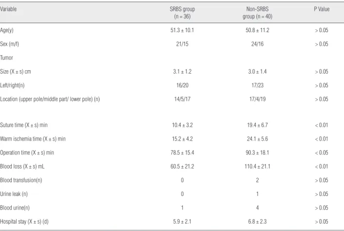
Table 1 - Comparisons between characteristics of operation and postoperative outcomes.
Variable SRBS group
(n = 36)
Non-SRBS group (n = 40)
P Value
Age(y) 51.3 ± 10.1 50.8 ± 11.2 > 0.05
Sex (m/f) 21/15 24/16 > 0.05
Tumor
Size (X ± s) cm 3.1 ± 1.2 3.0 ± 1.4 > 0.05
Left/right(n) 16/20 17/23 > 0.05
Location (upper pole/middle part/ lower pole) (n) 14/5/17 17/4/19 > 0.05
Suture time (X ± s) min 10.4 ± 3.2 19.4 ± 6.7 < 0.01
Warm ischemia time (X ± s) min 15.2 ± 4.2 24.1 ± 5.6 < 0.01
Operation time (X ± s) min 78.5 ± 15.4 90.3 ± 18.1 < 0.05
Blood loss (X ± s) mL 60.5 ± 21.2 110.4 ± 21.1 < 0.01
Blood transfusion(n) 0 2 > 0.05
Urine leak (n) 0 1 > 0.05
Blood urine(n) 1 4 > 0.05
and continuous suture was used again to close the kidney. Another needle was used to close the left part. After the suture, the left quill line was used to tie a knot, or a Hem-o-lock was used to close at the end of suture (Figure-2).
For the non-SRBS group, the tumor bed and the collecting system were sutured with a continuous 3-0 polyglactin suture then superfi-cial renorrhaphy was performed with running 3-0 polyglactin line intermittently, with Hem-o-lock clip for every suture. The bulldog clamp was remo-ved and there was no bleeding in the surgical field and the tumor was removed through the Port A.
Statistical analysis
Statistical analyses were performed with SPSS software for Windows (Statistical Product and Service Solutions, version 10.0, SSPS Inc, Chicago, IL, USA). Categorical variables were compared with
the chi-square test; continuous variables were com-pared with the Mann-Whitney U test. A value for P < 0.05 was considered statistically significant.
RESULTS
All 76 cases of retroperitoneoscopic par-tial nephrectomies were successfully performed, without conversion to open surgery or important intraoperative complications. All patients were followed for 1~11 months, without local recurren-ce and distant metastasis. In the SRBS group, the suture time, warm ischemia time and operation blood loss were shorter than that of non-SRBS group significantly; operation time and hospi-tal stay were also shorter than that of non-SRBS group (Table-1).
DISCUSSION
With the widespread application of B ul-trasound, CT and MRI exams, incidental renal cell carcinoma increased generally recently, which has characteristics of small size, low stage, slow gro-wth and low potential for metastasis, with better prognosis than symptomatic renal cell carcinoma; the operation is the gold standard treatment for most T1 RCC currently (7).
Partial nephrectomy (PN) has been a new treatment for T1a renal cell carcinoma(RCC). Some studies show that chronic kidney disease (CKD) has relations with cardiovascular diseases (8), and when GFR < 60mL/min, the risks of death and in hospital treatment increase (9). RN is considered as a risk factor for the genesis and the development of CKD; PN treatment keeps more kidney units left and decreases those affectted (10,11). RN is a risk factor for the genesis and worsening of CKD; the studies showed that RN could increase the death rate and renal failure of RCC patients (12,13), PN can get the same outcomes with RN in histology, and it can maintain the kidney and cardiovascular function better in a long term follow-up (11,14).
PN includes open partial nephrectomy (OPN), laparoscopic partial nephrectomy (LPN), and robot-assisted partial nephrectomy (RAPN). LPN has gained increased acceptance with equi-valent results at oncological and renal function
Figure 1 - a) Bi-directional barbed suture (Quill SRS); b) Barbs that change direction at the mid-point of the double-armed suture.
outcomes as OPN, with many advances such as less postoperative pain therapy, shorter hospital stay, and quick recovery (2-4).
LPN includes transperitoneal approach and retroperitoneoscopic approaches. Gill des-cribed the first retroperitoneoscopic partial ne-phrectomy in 1994 (15), Winfield finished the first retroperitoneoscopic partial nephrectomy in 1993 (16). The retroperitoneoscopic approach has advantages of easier controlling of kidney vessel, less disturbance of internal organs, and disad-vantages of smaller operation field, less anatomic landmarks. Anatomic, programmed and standard operation could make up the disadvantages of the retroperitoneoscopic approach (1).
Even though with more advantages, LPN keeps some challenge for many urologists, resul-ting in more intraoperative complications (blood and urine leak etc.) and longer WIT. The WIT is closely related with kidney function, while the WIT > 30 minutes, the kidney function was affec-ted more than 3-5 times (17,18). Suturing was the best way to keep kidney and to avoid urine leak, but it had great challenges (19,20). The good sutu-ring techniques could decrease the rate of compli-cations and shorten suturing time (3). Hem-o-lok substitution for knots was valid and safe (20,21), and could shorten suturing time and reduce WIT, but renal closures was still not tighter enough.
Bidirectional barbed sutures are manufac-tured from monofilament fibers via a microma-chining technique that cuts barbs into the suture around the circumference in a helical pattern. The barbs are separated from one another by a dis-tance of 0.88 to 0.98 mm and are divided into 2 groups that face each other in opposing directions from the suture midpoint. The use of knotless, bar-bed suture can securely suture tissues with less time, to close multiple layers tissues at the same time, and to decrease operation blood loss (22). Our study showed that Quill SRS barbed suture could improve efficiency in LPN, simplifying the suturing procedure, shortening suture time and WIT, decreasing blood loss, with a tighten renal closure, and decrease of the incidence of urine leaks, hemorrhage, or other complications. Quill SRS consists of a delayed-absorbable material (polydioxanone) cut with barbs, which could
pre-vent slippage through tissue and strengthen the suture, decreasing the chance of blood loss.
The application of Quill SRS bidirectional barbed absorbable suture in retroperitoneoscopic partial nephrectomy could shorten suturing time and warm ischemia time, with good safety and fe-asibility, worthy of being used generally in clinic.
CONFLICT OF INTEREST
None declared.
REFERENCES
1. Xie M, Wang K, Men CP: Anatomic and programmed retroperitoneal laparoscopic partial nephrectomy( with 125 cases of reports). Chinese Journal of Endourology. Electronic Version. 2012: 6: 11-4.
2. Gill IS, Matin SF, Desai MM, Kaouk JH, Steinberg A, Mascha E, et al.: Comparative analysis of laparoscopic versus open partial nephrectomy for renal tumors in 200 patients. J Urol. 2003; 170: 64-8.
3. Gill IS, Kavoussi LR, Lane BR, Blute ML, Babineau D, Colombo JR Jr, et al.: Comparison of 1,800 laparoscopic and open partial nephrectomies for single renal tumors. J Urol. 2007; 178: 41-6.
4. Gong EM, Orvieto MA, Zorn KC, Lucioni A, Steinberg GD, Shalhav AL: Comparison of laparoscopic and open partial nephrectomy in clinical T1a renal tumors. J Endourol. 2008; 22: 953-7.
5. Kane CJ, Mallin K, Ritchey J, Cooperberg MR, Carroll PR: Renal cell cancer stage migration: analysis of he National Cancer Data Base. Cancer. 2008; 113: 78-83.
6. Collins S, McKiernan J, Landman J: Update on the epidemiology and biology of renal cortical neoplasms. J Endourol. 2006; 20: 975-85.
7. Ljungberg B, Cowan NC, Hanbury DC, Hora M, Kuczyk MA, Merseburger AS, et al.: EAU guidelines on renal cell carcinoma: the 2010 update. Eur Urol. 2010; 58: 398-406. 8. Go AS, Chertow GM, Fan D, McCulloch CE, Hsu CY: Chronic
kidney disease and the risks of death, cardiovascular events, and hospitalization. N Engl J Med. 2004; 351: 1296-305. Erratum in: N Engl J Med. 2008; 18: 4.
10. Medina-Polo J, Romero-Otero J, Rodríguez-Antolín A, Domínguez-Esteban M, Passas-Martínez J, Villacampa-Aubá F, et al.: Can partial nephrectomy preserve renal function and modify survival in comparison with radical nephrectomy? Scand J Urol Nephrol. 2011; 45: 143-50.
11. Russo P, Huang W: The medical and oncological rationale for partial nephrectomy for the treatment of T1 renal cortical tumors. Urol Clin North Am. 2008; 35: 635-43.
12. Thompson RH, Boorjian SA, Lohse CM, Leibovich BC, Kwon ED, Cheville JC, et al.: Radical nephrectomy for pT1a renal masses may be associated with decreased overall survival compared with partialnephrectomy. J Urol. 2008; 179: 468-71; discussion 472-3.
13. Huang WC, Levey AS, Serio AM, Snyder M, Vickers AJ, Raj GV, et al.: Chronic kidney disease after nephrectomy in patients with renal cortical tumours: a retrospective cohort study. Lancet Oncol. 2006; 7: 735-40.
14. Crépel M, Jeldres C, Perrotte P, Capitanio U, Isbarn H, Shariat SF, et al.: Nephron-sparing surgery is equally effective to radical nephrectomy for T1BN0M0 renal cell carcinoma: a population-basedassessment. Urology. 2010; 75: 271-5. 15. Gill IS, Delworth MG, Munch LC: Laparoscopic retroperitoneal
partial nephrectomy. J Urol. 1994; 152: 1539-42.
16. Winfield HN, Donovan JF, Godet AS, Clayman RV: Laparoscopic partial nephrectomy: initial case report for benign disease. J Endourol. 1993; 7: 521-6.
17. Becker F, Van Poppel H, Hakenberg OW, Stief C, Gill I, Guazzoni G, Montorsi F, et al.: Assessing the impact of ischaemia time during partial nephrectomy. Eur Urol. 2009; 56: 625-34.
18. Yossepowitch O, Eggener SE, Serio A, Huang WC, Snyder ME, Vickers AJ, et al.: Temporary renal ischemia during nephron sparing surgery is associated with short-term but not long-term impairment in renalfunction. J Urol. 2006; 176: 1339-43; discussion 1343.
19. Sammon J, Petros F, Sukumar S, Bhandari A, Kaul S, Menon M, et al.: Barbed suture for renorrhaphy during robot-assisted partial nephrectomy. J Endourol. 2011; 25: 529-33. 20. Orvieto MA, Chien GW, Laven B, Rapp DE, Sokoloff MH,
Shalhav AL: Eliminating knot tying during warm ischemia time for laparoscopic partial nephrectomy. J Urol. 2004; 172: 2292-5.
21. Benway BM, Cabello JM, Figenshau RS, Bhayani SB: Sliding-clip renorrhaphy provides superior closing tension during robot-assisted partial nephrectomy. J Endourol. 2010; 24: 605-8.
22. Gözen AS, Arslan M, Schulze M, Rassweiler J: Comparison of laparoscopic closure of the bladder with barbed polyglyconate versus polyglactin suture material in the pigbladder model: an experimental in vitro study. J Endourol. 2012; 26: 732-6.
_______________________ Correspondence address: Zhang Yu-lian, MD Department of Gynecology Yantai Yuhuangding Hospital,