Beneficial effect of pentoxifylline into the testis of rats
in an experimental model of unilateral hindlimb ischemia/
reperfusion injury
_______________________________________________
Mohammad Ashrafzadeh Takhtfooladi
1, Fariborz Moayer
2, Hamed Ashrafzadeh Takhtfooladi
21 Young Researchers and Elites Club, Science and Research Branch, Islamic Azad University, Tehran, Iran; 2 Department of Pathobiology, College of Veterinary Medicine, Karaj Branch, Islamic Azad University,
Alborz, Iran
ABSTRACT
ARTICLE
INFO
______________________________________________________________ ______________________
Objective: The objective of the present study was to investigate the role of pentoxifylline (PTX) on remote testicular injury caused by unilateral hind limb ischemia/reperfusion of rats.
Materials and Methods: Twenty healthy male Wistar rats were allocated randomly into two groups: ischemia/reperfusion (IR group) and ischemia/reperfusion + pentoxifylline (IR+PTX group). Ischemia was induced by placement of a rubber tourniquet at the greater trochanter for 2h. Rats in IR+PTX group received PTX (40 mg/kg IP) before the reper-fusion period. At 24h after reperreper-fusion, testes were removed and levels of superoxide dismutase (SOD), malondialdehyde (MDA), catalase (CAT) and myeloperoxidase (MPO) activity were determined in testicular tissues. Three rats of each group were used for wet/ dry weight ratio measurement. Testicular tissues were also examined histopathologically under light microscopy.
Results: Activities of SOD and CAT in testicular tissues were decreased by ischemia/ reperfusion (P<0.05). Significantly increased MDA levels in testicular tissues were de-creased by PTX treatment (P<0.05). MPO activity in testicular tissues in the IR group was significantly higher than in the IR+PTX group (P<0.05). The wet/dry weight ratio of testicular tissues in the IR group was significantly higher than in the IR+PTX group (P<0.05). Histopathologically, there was a statistically significant difference between two groups (P<0.05).
Conclusions: According to histological and biochemical findings, we conclude that PTX has preventive effects in the testicular injury induced by hind limb ischemia/reperfusion.
Key words:
Pentoxifylline; Hindlimb; Ischemia; Reperfusion; Testicular Diseases; Oxidative Stress
Int Braz J Urol. 2015; 41: 576-83
_____________________
Submitted for publication: May 30, 2014
_____________________
Accepted after revision: October 10, 2014
INTRODUCTION
Hind limb ischemia/reperfusion injury may occur clinically after a release of tourniquets du-ring orthopedic surgery, or extrication of a trauma victim who is compressed with a heavy weight for a prolonged period (crush syndrome). Several re-ports have indicated that the pathophysiology of
is associated with a systemic inflammatory res-ponse and determines the effect on remote or-gans (liver, lung, kidney, myocardium and testis) structure and function (4-9). The development of remote organ dysfunction was observed only following reperfusion, which implies that humoral and/or cellular mediators produced locally in the limb were responsible for mediating remote organ injury (10-12).
Pentoxifylline (PTX) is a methylxanthine derivative with multiple hemorheologic properties. PTX through effects of increasing intracellular cyclic AMP on red blood cells improve the oxy-gen delivery to ischemic tissues and also increases the cyclic AMP on polymorph nuclear leukocytes and decreases oxygen free radical production (10, 13-16). In addition, PTX limit the inflammatory response with reduction in cellular activation, phagocytosis and endothelium adhesion. There are evidences that PTX also reduces the nitric oxi-de oxi-destruction (15). Recent studies have indicated that PTX improves ischemia/reperfusion injury in many organs (17-19). However, the effects of PTX on remote testicular injury caused by skeletal muscle ischemia/reperfusion are not clear. The ob-jective of the present study was to investigate the role of PTX on remote testicular injury caused by unilateral hind limb ischemia/reperfusion of rats. For this purpose, the biochemical and pathologi-cal effects of skeletal muscle ischemia/reperfusion and PTX in testicular tissues of rats have been in-vestigated. The results of our investigation would help to clarify the potential importance of the use of PTX in situations of oxidative damage. These findings may encourage the use of antioxidants to reduce remote organ injury after skeletal muscle ischemia/reperfusion.
MATERIALS AND METHODS
Animals
The study was conducted on 20 healthy male Wistar rats, 12-16 weeks old and weighing between 270-300g. All rats of this study were kept according to the norms of the Islamic Azad University College of Veterinary Medicine Tehran Iran laboratory of animal experimentations; this investigation was approved by the Committee of
Ethics in Research with animals of Islamic Azad University. The study was designed so as to mini-mize the number of animals required for the expe-riments. The rats were housed in individual cages under temperature controlled standard conditions, 12h/12h light/dark cycle with free access to stan-dard rodent food and filtrated tap water.
Experimental Design
overdose of intraperitoneal pentobarbital by in-jection (300mg/kg).
Preparation of Testicular Tissue Homoge-nates
The left testicular tissues were washed three times in cold normal saline solution (0.9%). Then, the tissues were homogenized in ice-cold Tris-HCl buffer solution, within a homogenizer for 2min at 11200×g. The homogenate was centrifu-ged at 3500×g for 60min and a supernatant was obtained. The levels of MPO were determined in the supernatant, and MDA levels were studied in the homogenate. For a further extraction proce-dure, the supernatant was extracted in ethanol/ chloroform mixture (5/3 v/v). After a second cen-trifugation at 3500×g for 20min, the clear upper layer was taken and used for SOD activity deter-mination (21).
BIOCHEMICAL ASSAYS
Measurement of SOD Activity
The principle of the SOD activity determi-nation method was based on the inhibition of ni-troblue tetrazolium reduction described by Sun et al. (22) and modified by Durak et al. (23). One unit of SOD was defined as the enzyme activity cau-sing 50% inhibition in the nitroblue tetrazolium reduction rate. The SOD activity was expressed as units per mg tissue protein.
Measurement of MDA levels
The MDA levels in testicular tissues were analyzed by a method based on the reaction with thiobarbituric acid at 95ºC (24). In the thiobarbi-turic acid test reaction, MDA or MDA-like subs-tances and thiobarbituric acid react together to produce a pink pigment with an absorption ma-ximum of 532nm. The results were expressed as nanomol per gram wet tissue (nmoL/g tissue).
Measurement of CAT activity
CAT activity was determined according to Aebi’s method (25). The principle of the assay is based on determination of the rate constant k (s -1) of H
2O2 decomposition at 240nm. Results were
expressed as k (rate constant) per gram of protein.
Measurement of MPO activity
Testicular injury was quantified by me-asuring testicular MPO activity, the activity of infiltrated polymorphonuclear leukocytes, using a protocol modified from a previous report (26). MPO activity was determined after adding O--dianisidine dihydrochloride and hydrogen pe-roxide. The MPO activity was expressed as units per gram tissue.
Wet/dry weight assay
The wet/dry weight ratio, the tissue ede-ma index, was measured to evaluate testicular injury. Briefly, freshly harvested testes were ghed, placed in an oven for 24h at 60˚C and wei-ghed again when dry (26). The wet/dry weight ratio was then calculated.
HISTOPATHOLOGICAL EVALUATION
The right extracted testes were immedia-tely placed into 10% neutral formaldehyde solu-tion. The tissue specimens were placed in paraffin blocks, sectioned at 5µm, and stained with he-matoxylin and eosin (H&E) for light microscopic
analysis. An experienced pathologist, who was blinded to the experiment and data, examined the samples histopathologically. The histological parameters were scored according to Cosentino et al. (27) classification as follows: Grade-I: Showed normal testicular architecture with an orderly, ar-rangement of germinal cells; Grade-II: Injury sho-wed less orderly, non-cohesive germinal cells and closely packed seminiferous tubules;
Grade-III: Injury exhibited disordered slou-ghed germinal cells with shrunken pyknotic nuclei and less distinct seminiferous tubule borders;
Grade-IV: Injury defined seminiferous tu-bules that were closely packed with coagulative necrosis of the germinal cells.
Statistical analysis
significant, Bonferroni adjusted Mann-Whitney U test was used in the paired comparison. Data are shown as the mean±standard deviation with sig-nificance considered at 0.05.
RESULTS
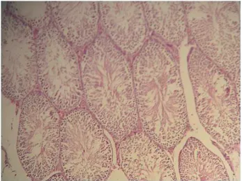
The experimental procedure was well tole-rated and no animal died during the experiment. One of the most interesting observations at the his-topathological examination of the testicular tissues is the presence of sloughed germinal cells within the seminiferous tubules and disorganization in rat testes after the unilateral hind limb ischemia/reper-fusion. The testicular injury score increased signifi-cantly in the IR group compared with the IR+PTX group (P<0.05) (Table-1). In the IR group, coagulati-ve necrosis with loss of seminiferous tubule epithe-lium, edema and sloughed germinal cells were pre-dominant features in sections (Figure-1). However, the rats in the IR+PTX group had essentially normal seminiferous tubule morphology (Figure-2).
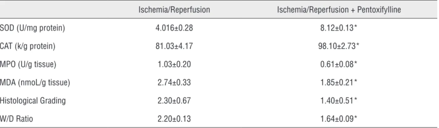
Activities of SOD and CAT in testicular tis-sues were decreased by ischemia/reperfusion, but ad-ministration of PTX increased these levels (P<0.05) (Table-1). The tissue levels of MDA increased in the IR group in comparison to the IR+PTX group (Ta-ble-1). MPO activity in testicular tissues in the IR group was significantly higher than in the IR+PTX group (P<0.05) (Table-1). The wet/dry weight ratio of testicular tissues in the IR group was significantly higher than in the IR+PTX group (P<0.05) (Table-1).
DISCUSSION
The systemic inflammatory response syn-drome is a consequence of many conditions, such as surgery, trauma, burn, shock, or bacterial in-fection (28), that results in the development of potentially fatal complication known as multiple organ dysfunction syndrome. Hind limb ischemia/ reperfusion has been extensively used in our labo-ratory as a model of systemic inflammatory res-ponse syndrome, which closely resembles the acu-te traumatic and ischemic insult seen in sysacu-temic inflammatory response syndrome patients (29). It has been demonstrated that hind limb ischemia/ reperfusion causes cellular injury in remote organs (kidney, lung, and myocardium) and contributes to the development of multiple organ dysfunction syndrome (5, 29). As far as we know, there are only a few reports demonstrating testicular remote in-jury following muscle ischemia/reperfusion inin-jury (8). The results of Takhtfooladi et al. (8) indicated that skeletal muscle ischemia/reperfusion induces severe testicular damage and N-acetylcysteine has protective effects on testicular injury after hind limb ischemia/reperfusion. Their data supported this view that temporary occlusion of the femo-ral artery induced testicular injury in rats (8). Pre-vious studies indicate that inflammatory response and injury to remote organs can be caused by the systemic release of the pro-inflammatory media-tors and free oxygen radicals upon reperfusion of ischemic limbs (5, 30). Despite decades of research
Table 1 - The tissue CAT and MPO activities and levels of SOD and MDA in testicular tissues and score of testicular histological changes and muscle wet/dried weight (W/D) ratio.
Ischemia/Reperfusion Ischemia/Reperfusion + Pentoxifylline
SOD (U/mg protein) 4.016±0.28 8.12±0.13*
CAT (k/g protein) 81.03±4.17 98.10±2.73*
MPO (U/g tissue) 1.03±0.20 0.61±0.08*
MDA (nmoL/g tissue) 2.74±0.33 1.85±0.21*
Histological Grading 2.30±0.67 1.40±0.51*
W/D Ratio 2.20±0.13 1.64±0.09*
in this area, ischemia/reperfusion injury remains a clinically challenging problem.
PTX is one of the phosphodiesterase inhi-bitors that have been reported to increase intra-cellular cyclic AMP and reduce superoxide anion production by both monocytes and polymorpho-nuclear cells dose dependently in vitro (31). En-dres et al. (32) indicated that PTX led to a marked increase in cyclic AMP levels, whereas cyclic GMP levels were only marginally elevated in lipopoly-saccharide stimulated human monocytes. PTX has received considerable attention with respect to its action on leukocytes in many organs (33-35). Reignier et al. (19) reported that after reperfusion, myeloperoxidase activity and blood neutrophil count were lower with PTX than with saline, and changes in the filtration coefficient were correla-ted to the percent changes in blood neutrophils during reperfusion. They suggested that this effect may be mainly caused by a decrease in seques-tration of neutrophils in the lung during reperfu-sion. Their group also reported that PTX preven-ted endothelial injury during ischemia/reperfusion by decreasing neutrophil sequestration in isolated perfused rat and rabbit lungs and in pigs after left lung allotransplantation (33).
Previous studies have also confirmed the potential antioxidant effects of PTX (36-38). Re-cently administered PTX was shown to protect against ischemia/reperfusion injuries in local and
remote organs and it was suggested that this pro-tective effect may be due to its ability to inhibit of free radical generation (12, 20). There is growing evidence regarding its beneficial effects in amelio-rating testicular ischemia/reperfusion injury (39). In an experimental study Savas et al. (39) sugges-ted that the administration of PTX at a dose of 50 mg/kg 15 min. before spermatic cord torsion may have a protective effect in rat experimental testicular torsion/detorsion models. Their results suggest that pentoxifylline treatment attenuates reperfusion damage on both sides, possibly with its effects on blood flow and neutrophils (39). This observation was supported by Pozor et al. (40), who demonstrated that PTX may be a potential protective agent for preventing the negative chan-ges related to oxidative stress in testicular injury caused by spermatic cord torsion in miniature horse stallions. However, the protective effect of PTX on testes from skeletal muscle ischemia/re-perfusion injury has not been studied to date. In the current study, we tested the hypothesis that PTX could protect the testes from remote organ injury after skeletal muscle ischemia/reperfusion.
Oxidative stress is associated with an in-creased rate of cellular damage induced by oxygen and oxygen-derived oxidants, commonly known as reactive oxygen species (41, 42). The major targets of reactive oxygen species are membrane lipids, in a process known as lipid peroxidation.
Figure 1 - Light microscopic view of testis tissues from IR group showing coagulative necrosis with loss of seminiferous tubule epithelium, edema and sloughed germinal cells. H&E staining; magnification of 10 × 10.
It is also acknowledged that testicular tissues and spermatozoa are very sensitive to reactive oxygen species attack and lipid peroxidation. The suscep-tibility of testicular tissues to oxidation is attribu-ted to the high polyunsaturaattribu-ted fatty acid content of sperm membranes (42, 43). Many tissues con-tain powerful endogenous scavengers that provide protection against free radical damage, including SOD, CAT, glutathione peroxidase, ascorbic acid and α-tocopherol (44). Sikka et al. (45) have re-ported that adequate levels of antioxidants such as SOD, CAT and possibly glutathione peroxidase and reductase, maintain the scavenging potential in gonads and seminal fluids, which is referred to as oxidative stress status. Tissue CAT and MPO ac-tivities, and SOD and MDA levels are considered to indicate oxidative stress. In the present study, SOD level and CAT activity were decreased significan-tly in IR group compared with IR+PTX group. The tissue MDA level and MPO activity in testes were increased significantly in IR group compared with IR+PTX group. According to the observations of this study, the histopathological injury score was significantly decreased in IR+PTX group com-pared with that of IR group. In the PTX treated group, histopathological features such as edema, congestion, hemorrhage, and necrosis of the ger-minal cells were markedly less than in IR group. The findings in this study indicate that reperfusion of the ischemic limb leads, within 24h of reperfu-sion, to a systemic response as demonstrated by the biochemical and histological impairment of the testis. Also, data on CAT and MPO activity, and SOD and MDA levels suggested a protective effect of PTX against testicular remote injuries af-ter unilaaf-teral hind limb ischemia/reperfusion. The-se data together with previous findings (39) con-firm the potent anti-oxidation capacity of PTX. These data also support the concept that PTX may be an effective therapeutic adjunct to reperfusion injury. The clinical relevance of this manuscript refers to the previous use of PTX in situations re-quiring procedures ischemia with reperfusion to reduce or prevent distant organs damage. There-fore, the long-term effect of PTX warrants further investigation.
CONCLUSIONS
The results of this study showed that uni-lateral hind limb ischemia/reperfusion induced testicular injury in rats. Nevertheless, PTX ad-ministration significantly decreased testes injury induced by skeletal muscle ischemia/reperfusion according to our histological and biochemical fin-dings. PTX is already in clinical use for the treat-ment of vascular diseases and these results suggest the possibility of clinical application of PTX in testicular remote organ injury following skeletal muscle ischemia/reperfusion. Further studies are needed to clarify clinical usefulness of this agent.
CONFLICT OF INTEREST
None declared.
REFERENCES
1. Oredsson S, Arlock P, Plate G, Qvarfordt P. Metabolic and electrophysiological changes in rabbit skeletal muscle during ischaemia and reperfusion. Eur J Surg.1993;159:3-8. 2. Roberts JP, Perry MO, Hariri RJ, Shires GT. Incomplete
recovery of muscle cell function following partial but not complete ischemia. Circ Shock. 1985;17:253-8.
3. Kishi M, Tanaka H, Seiyama A, Takaoka M, Matsuoka T, Yoshioka T, et al. Pentoxifylline attenuates reperfusion injury in skeletal muscle after partial ischemia. Am J Physiol. 1998;274:H1435-42.
4. Coe DA, Freischlag JA, Johnson D, Mudaliar JH, Kosciesza SA, Traul DK, et al. Pentoxifylline prevents endothelial damage due to ischemia and reperfusion injury. J Surg Res. 1997;67:21-5.
5. Yassin MM, Harkin DW, Barros D’Sa AA, Halliday MI, Rowlands BJ. Lower limb ischemia-reperfusion injury triggers a systemic inflammatory response and multiple organ dysfunction. World J Surg. 2002;26:115-21.
6. Takhtfooladi MA, Jahanshahi A, Sotoudeh A, Jahanshahi G, Takhtfooladi HA, Aslani K. Effect of tramadol on lung injury induced by skeletal muscle ischemia-reperfusion: an experimental study. J Bras Pneumol. 2013;39:434-9. 7. Takhtfooladi MA, Jahanshahi A, Jahanshahi G, Sotoudeh
8. Sotoudeh A, Takhtfooladi MA, Jahanshahi A, Asl AH, Takhtfooladi HA, Khansari M. Effect of N-acetylcysteine on lung injury induced by skeletal muscle ischemia-reperfusion. Histopathlogical study in rat model. Acta Cir Bras. 2012;27:168-71.
9. Takhtfooladi MA, Jahanshahi A, Sotoudeh A, Daneshi MH, Khansari M, Takhtfooladi HA. The antioxidant role of N-acetylcysteine on the testicular remote injury after skeletal muscle ischemia and reperfusion in rats. Pol J Pathol. 2013;64:204-9.
10. Emrecan B, Tulukoglu E, Bozok S, Kestelli M, Onem G, Küpelioglu A, et al. Effects of Iloprost and pentoxifylline on renal ischemia-reperfusion in rabbit model. Eur J Med Res. 2006;11:295-9.
11. Gradl G, Gaida S, Finke B, Lindenblatt N, Gierer P, Menger MD, et al. Supernatant of traumatized muscle induces inflammation and pain, but not microcirculatory perfusion failure and apoptotic cell death. Shock. 2005;24:219-25. 12. Teruya R, Fagundes DJ, Oshima CT, Brasileiro JL, Marks
G, Ynouye CM, et al. The effects of pentoxifylline into the kidneys of rats in a model of unilateral hindlimb ischemia/ reperfusion injury. Acta Cir Bras. 2008;23:29-35.
13. Stafford-Smith M. Evidence-based renal protection in cardiac surgery. Semin Cardiothorac Vasc Anesth. 2005;9:65-76. 14. Vadiei K, Brunner LJ, Luke DR. Effects of pentoxifylline in
experimental acute renal failure. Kidney Int. 1989;36:466-70. 15. Dávila-Esqueda ME, Martínez-Morales F. Pentoxifylline
diminishes the oxidative damage to renal tissue induced by streptozotocin in the rat. Exp Diabesity Res. 2004;5:245-51. 16. Gunduz Z, Canoz O, Per H, Dusunsel R, Poyrazoglu MH,
Tez C, et al. The effects of pentoxifylline on diabetic renal changes in streptozotocin-induced diabetes mellitus. Ren Fail. 2004;26:597-605.
17. Adams JG Jr, Dhar A, Shukla SD, Silver D. Effect of pentoxifylline on tissue injury and platelet-activating factor production during ischemia-reperfusion injury. J Vasc Surg. 1995;21:742-8.
18. Horton JW, White DJ. Free radical scavengers prevent intestinal ischemia-reperfusion-mediated cardiac dysfunction. J Surg Res. 1993;55:282-9.
19. Reignier J, Mazmanian M, Detruit H, Chapelier A, Weiss M, Libert JM, et al. Reduction of ischemia-reperfusion injury by pentoxifylline in the isolated rat lung. Paris-Sud University Lung Transplantation Group. Am J Respir Crit Care Med.1994;150:342-7.
20. Takhtfooladi H, Moayer F, Abarkar M: Effects of pentoxifylline on skeletal muscle inj ury induced by acute hindlimb ischaemia–reperfusion: a histopathological study in a rat model. Comp Clin Pathol. 2014; 24(3) In Press.
21. Parlaktas BS, Atilgan D, Gencten Y, Akbas A, Markoc F, Erdemir F, et al. The effects of carvedilol on ischemia-reperfusion injury in the rat testis. Int Braz J Urol. 2014;40:109-17.
22. Sun Y, Oberley LW, Li Y. A simple method for clinical assay of superoxide dismutase. Clin Chem. 1988;34:497-500. 23. Durak I, Yurtarslanl Z, Canbolat O, Akyol O. A methodological
approach to superoxide dismutase (SOD) activity assay based on inhibition of nitroblue tetrazolium (NBT) reduction. Clin Chim Acta. 1993;214:103-4.
24. Wasowicz W, Nève J, Peretz A. Optimized steps in fluorometric determination of thiobarbituric acid-reactive substances in serum: importance of extraction pH and influence of sample preservation and storage. Clin Chem. 1993;39:2522-6.
25. Aebi H. Catalase. In: Bergmeyer HU (ed.), Methods of Enzymatic Analysis. Academic Press, New York, 1974; pp. 673-7. 26. Yang CH, Tsai PS, Wang TY, Huang CJ.
Dexmedetomidine-ketamine combination mitigates acute lung injury in haemorrhagic shock rats. Resuscitation. 2009;80:1204-10. 27. Cosentino MJ, Nishida M, Rabinowitz R, Cockett AT.
Histological changes occurring in the contralateral testes of prepubertal rats subjected to various durations of unilateral spermatic cord torsion. J Urol. 1985;133:906-11.
28. Carden DL, Granger DN. Pathophysiology of ischaemia-reperfusion injury. J Pathol. 2000;190:255-66.
29. Wunder C, Brock RW, McCarter SD, Bihari A, Harris K, Eichelbrönner O, et al. Inhibition of haem oxygenase activity increases leukocyte accumulation in the liver following limb ischaemia-reperfusion in mice. J Physiol. 2002;540:1013-21. 30. Lawlor DK, Brock RW, Harris KA, Potter RF. Cytokines
contribute to early hepatic parenchymal injury and microvascular dysfunction after bilateral hindlimb ischemia. J Vasc Surg. 1999;30:533-41.
31. Bessler H, Gilgal R, Djaldetti M, Zahavi I. Effect of pentoxifylline on the phagocytic activity, cAMP levels, and superoxide anion production by monocytes and polymorphonuclear cells. J Leukoc Biol. 1986;40:747-54.
32. Endres S, Fülle HJ, Sinha B, Stoll D, Dinarello CA, Gerzer R, et al. Cyclic nucleotides differentially regulate the synthesis of tumour necrosis factor-alpha and interleukin-1 beta by human mononuclear cells. Immunology. 1991;72:56-60. 33. Chapelier A, Reignier J, Mazmanian M, Detruit H, Dartevelle P,
Parquin F,et al. Pentoxifylline and lung ischemia-reperfusion injury: application to lung transplantation. Université Paris-Sud Lung Transplant Group. J Cardiovasc Pharmacol. 1995;25,Suppl 2:S130-3.
34. Ciuffetti G, Mercuri M, Ott C, Lombardini R, Paltriccia R, Lupattelli G, et al. Use of pentoxifylline as an inhibitor of free radical generation in peripheral vascular disease. Results of a double-blind placebo-controlled study. Eur J Clin Pharmacol. 1991;41:511-5.
36. Bhat VB, Madyastha KM. Antioxidant and radical scavenging properties of 8-oxo derivatives of xanthine drugs pentoxifylline and lisofylline. Biochem Biophys Res Commun. 2001;288:1212-7.
37. Horvath B, Marton Z, Halmosi R, Alexy T, Szapary L, Vekasi J, et al. In vitro antioxidant properties of pentoxifylline, piracetam, and vinpocetine. Clin Neuropharmacol. 2002;25:37-42. 38. Lin SL, Chen YM, Chien CT, Chiang WC, Tsai CC, Tsai TJ.
Pentoxifylline attenuated the renal disease progression in rats with remnant kidney. J Am Soc Nephrol. 2002;13:2916-29. 39. Savas C, Dindar H, Bilgehan A, Ataoglu O, Yucesan S.
Pentoxifylline attenuates reperfusion injury in testicular torsion. Scand J Urol Nephrol. 2002;36:65-70.
40. Pozor MA, Muehlhaus J, King A, Macpherson ML, Troedsson MH, Bailey CS. Effect of pentoxifylline treatment on testicular perfusion and semen quality in Miniature horse stallions. Theriogenology. 2011;76:1027-35.
41. Tinkel J, Hassanain H, Khouri SJ. Cardiovascular antioxidant therapy: a review of supplements, pharmacotherapies, and mechanisms. Cardiol Rev. 2012;20:77-83.
42. Erdemir F, Atilgan D, Firat F, Markoc F, Parlaktas BS, Sogut E. The effect of sertraline, paroxetine, fluoxetine and escitalopram on testicular tissue and oxidative stress parameters in rats. Int Braz J Urol. 2014;40:100-8.
43. Jungwirth A, Giwercman A, Tournaye H, Diemer T, Kopa Z, Dohle G, et al. European Association of Urology guidelines on Male Infertility: the 2012 update. Eur Urol. 2012;62:324-32. 44. Demopoulos HB, Flamm ES, Pietronigro DD, Seligman ML.
The free radical pathology and the microcirculation in the major central nervous system disorders. Acta Physiol Scand Suppl. 1980;492:91-119.
45. Sikka SC, Rajasekaran M, Hellstrom WJ. Role of oxidative stress and antioxidants in male infertility. J Androl. 1995;16:464-8.