The effect of Sertraline, Paroxetine, Fluoxetine and
Escitalopram on testicular tissue and oxidative stress
parameters in rats
_______________________________________________
Fikret Erdemir, Dogan Atilgan, Fatih Firat, Fatma Markoc, Bekir Suha Parlaktas, Erkan Sogut
Department of Urology (FE, DA, FF, BSP), Department of Pathology (FM) and Department of Biochemis-try (ES) Gaziosmanpasa University, Turkey
ABSTRACT
ARTICLE
INFO
______________________________________________________________ ______________________
Introduction: The aim of this study was to evaluate the effect of selective serotonin reup-take inhibitors (SSRIs) on testicular tissue and serum malondialdehyde (MDA) levels in rats. Materials and methods: A total of 40 male Wistar albino rats, 5.5-6 months old, were equally divided at random into five groups: group 1 was the control group, group 2 re-ceived sertraline 10mg/kg (p.o), group 3 was administered fluoxetine 10mg/kg (p.o), group 4 received escitalopram 10mg/kg (p.o), and group 5 (n = 8) was administered paroxetine 20mg/kg. Each dose was administered orally for two months. Johnsen’s criteria were used to categorize spermatogenesis. Johnsen’s method assigns a score of 1 to 10 to each tubule cross-section examined. In this system, a Johnsen score of 9 and 10 indicates normal histo-logy. Serum luteinizing hormone (LH), follicle-stimulating hormone (FSH), and testosterone levels were evaluated. Serum MDA levels were also measured.
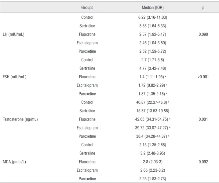
Results: The mean Johnsen scores were 9.36 ± 0.33, 9.29 ± 0.32, 8.86 ± 0.48, 9.10 ± 0.56, and 8.33 ± 0.90 in control group, sertraline group, fluoxetine group, escitalopram group, and paroxetine group, respectively. The Johnsen score was significantly lower for paroxe-tine group compared with the control group (p < 0.05). The mean FSH level increased only in the sertraline group. With the exception of the fluoxetine group, the testosterone levels were lower in all groups compared with the control group. The total testosterone level was significantly lower in the sertraline group compared with the control group [40.87 (22.37-46.8) vs. 15.87 (13.53-19.88), p < 0.01]. There were no significant differences between the groups with respect to the MDA and LH levels (p = 0.090 and p = 0.092).
Conclusion: These data suggest that SSRIs have a negative effect on testicular tissues. This negative impact is markedly greater in the paroxetine group. To determine the exact me-chanism of action of these drugs on testicular tissue, well-designed randomized controlled clinical studies are needed on a larger population.
Key words:
Male; Infertility; etiology [Subheading]; Spermatozoa
Int Braz J Urol. 2014; 40: 100-8
_____________________ Submitted for publication: March 08, 2013
_____________________ Accepted after revision: November 05, 2013
INTRODUCTION
The incidence of infertility ranges from 10% to 15% among couples (1,2). Examining the etiology of infertility among couples reveals isola-ted female factors to be the cause in 40% to 50%
by germ cells in the seminiferous tubules. The ba-sic process includes stimulation of Sertoli cells by follicle-stimulating hormone (FSH), which is se-creted from the pituitary gland, and the stimula-tion of testosterone synthesis following Leydig cell stimulation by luteinizing hormone (LH), which is also secreted from the pituitary. For this reason, FSH, LH, and testosterone are used as markers of spermatogenesis and testicular activity in males. Abnormalities or disruptions in sperm production are indicated by deterioration of sperm number and movement and may result in infertility (3). Many factors affect the male reproductive system and lead to infertility. Among the etiological fac-tors of male infertility, varicocele, sexual facfac-tors, congenital anomalies, urogenital infections, endo-crine disorders, and immunological factors are all important, and idiopathic causes of semen alte-ration account for up to 75% of cases (4). Addi-tionally, obesity, radiation, climate, environment, occupation, and drug usage may also affect male fertility (5-8). Antidepressants and antipsycho-tic agents are extensively used, both in the short and long term, depending on various indications. Among the agents used in psychiatric practice are monoamine oxidase inhibitors, tricyclic an-tidepressants, selective serotonin reuptake inhi-bitors (SSRIs), serotonin and noradrenaline reup-take inhibitors, selective noradrenaline reupreup-take inhibitors, noradrenaline and dopamine reuptake inhibitors, noradrenergic and specific serotonergic antidepressants, and benzodiazepines, with SSRI medications accounting for the majority of drugs prescribed (9). SSRI drugs include fluoxetine, flu-voxamine, sertraline, paroxetine, citalopram, and escitalopram. SSRIs are frequently prescribed, and their use is directly proportional to the frequency of mental disorders, observed in 6-18% of the po-pulation (10,11). Approximately 80% of psychia-tric disorders diagnosed are depression and an-xiety, for which SSRIs are the first-line drug in treating these conditions (10,11). Both disorders are observed more frequently at reproductive ages, i.e., between the ages of 15 and 50 years. Indeed, SSRIs are frequently used, and they constituted 65% of all new drugs prescribed for 20.5 million psychiatric patients in the year 2000. Approxima-tely 6 million males have been reported to use
SS-RIs (11). Among these, sertraline, paroxetine, and fluoxetine are very well known drugs used to treat premature ejaculation, particularly in urological practice (12).
Oxidative stress is a disturbance in the ba-lance between the production of reactive oxygen species (ROS) and antioxidant defenses, which can damage DNA, proteins, and lipids, ultimately lea-ding to apoptosis or necrosis in living cells. Several factors cause oxidative stress, including drugs. In the last decade, the SSRI class of drugs has been reported to be associated with sexual dysfunction, gastrointestinal disorders, insomnia, headaches, anorexia, weight loss, nausea, diarrhea, and palpita-tions as well as both male and female infertility (13). The relationship between the serotonergic system and male reproductive system has been evaluated only in a limited number of studies. In this study, the effects of SSRIs on testicular tissue and serum malondialdehyde (MDA) levels were investigated.
MATERIALS AND METHODS
This study was approved by the local ethics committee (ethical approval number 2011-HA-DYEK-047). A total of 40 male Wistar albino rats, 5.5-6 months old, were used in the study. The ex-perimental animals were housed at 18-22ºC throu-ghout the study period of 8 weeks and had free access to rat food and tap water ad libitum. All sur-gical procedures were performed under xylazine/ ketamine anesthesia in sterile conditions. The rats were randomly divided into five groups of eight as follows: group 1 was the accepted control group, group 2 received 10mg/kg sertraline, group 3 was administered 10mg/kg fluoxetine, group 4 received 10mg/kg escitalopram, and group 5 was red 20mg/kg paroxetine. Each dose was administe-red orally for two months, as previously reported in the literature (14-16). All groups were compared to the control group. In addition, the sertraline group, fluoxetine group, escitalopram group, and paroxe-tine group were compared to identify their differen-ces in Johnsen scores.
embe-dded in paraffin blocks. The sections were cut by a rotary microtome and stained with hematoxylin and eosin. The stained sections were studied un-der a light microscope to evaluate spermatogenesis. Johnsen’s criteria were used to categorize sperma-togenesis. This system describes the preservation of spermatogenesis, on a scale from 1 to 10, according to the absence or presence of the main cell types ar-ranged in order of maturity. A Johnsen score of 9 or 10 indicates normal histology, a score of 8 signifies hypospermatogenesis, a score of 3-7 implies ma-turation arrest, a score of 2 indicates germinal cell aplasia (Sertoli cells only), and a score of 1 repre-sents tubular fibrosis (Table-1). The germinal epi-thelium of at least 50 tubules was assessed for each testis, and the mean Johnsen’ score was calculated for each rat. Blood samples from the inferior vena cava were stored in heparin-free tubes for bioche-mical analyses. After centrifugation (2000 x g for 15 min at +4ºC), the serum samples were stored and frozen at -70ºC.
Biochemical Analysis
Blood samples were drawn into Vacutainer serum separator tubes and allowed to clot for 20 minutes at room temperature before the serum was separated by centrifugation (1500 x g for 10 min at
4ºC). The serum samples were then separated from the clot within one hour of blood collection, trans-ferred to a clean test tube, and stored at -70ºC until examination. Rat LH, FSH (Cusabio Biotech, na), and testosterone (Uscn Life Science Inc., Chi-na) were measured using ELISA kits according to the manufacturers’ instructions. Serum MDA levels were also measured using a method based on reac-tion with thiobarbituric acid (TBA) at 90-100ºC (17). Serum MDA levels were considered to indicate lipid peroxidation and oxidative stress.
Statistical analysis
The Kruskal-Wallis test was used to com-pare continuous data between groups. For multiple comparisons, the Bonferroni-adjusted Mann-Whit-ney U test was employed. Continuous data are gi-ven as the median and interquartile range (quarter 1 to quarter 3). A p-value of < 0.05 was considered significant. Analyses were performed using SPSS 19 (IBM SPSS Statistics 19, SPSS Inc., IBM Co., So-mers, NY).
RESULTS
The mean Johnsen scores were 9.36 ± 0.33, 9.29 ± 0.32, 8.86 ± 0.48, 9.10 ± 0.56, and
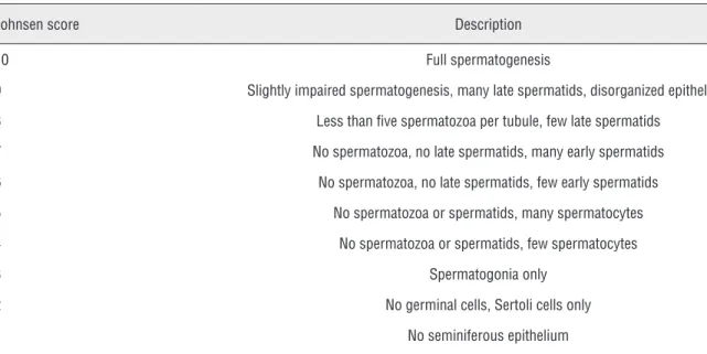
Table 1 - Modified Johnsen score system.
Johnsen score Description
10 Full spermatogenesis
9 Slightly impaired spermatogenesis, many late spermatids, disorganized epithelium
8 Less than five spermatozoa per tubule, few late spermatids
7 No spermatozoa, no late spermatids, many early spermatids
6 No spermatozoa, no late spermatids, few early spermatids
5 No spermatozoa or spermatids, many spermatocytes
4 No spermatozoa or spermatids, few spermatocytes
3 Spermatogonia only
2 No germinal cells, Sertoli cells only
8.33 ± 0.90 for the control group, sertraline group, fluoxetine group, escitalopram group, and paroxeti-ne group, respectively (Table-2). The Johnsen score was significantly lower for paroxetine group when compared with control group (Figures 1 and 2 and Table-2). There were no statistically significant di-fferences in Johnsen score between the other groups (p > 0.05). FSH levels were lower in the fluoxetine group, escitalopram group, and paroxetine group compared with the control group (p < 0.001). The mean FSH level increased only in the sertraline
group. In contrast, the FSH levels were significantly decreased in groups 3 and 5 compared with group 2 (p < 0.001) (Table-3). With the exception of the fluoxetine group, the testosterone levels were lo-wer in all groups compared with the control group. The total testosterone level was significantly lower in the sertraline group compared with the control group [40.87 (22.37-46.8) vs. 15.87 (13.53-19.88), p < 0.01] (Tab3). The serum LH and serum MDA le-vels did not significantly differ between the groups (p = 0.090 and p > 0.092, respectively).
Table 2 - The spermatogenesis results according to Johnsen Score System in the testicular tissues of rats.
Groups n Mean ± Std. Deviation p
Control 8 9.36 ± 0.33
0.021
Sertraline 8 9.29 ± 0.32
Fluoxetine 8 8.86 ± 0.48
Escitalopram 8 9.10 ± 0.56
Paroxetine 8 8.33 ± 0.90a,b,d
Total 40 8.99 ± 0.65
a: Different from Group 1, b: Different from Group 2, d: Different from Group 4.
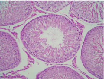
Figure 1a - Spermatogenesis with a Johnsen score of 9.8 in the control group. A seminiferous tubule with normal sper-matogenesis is seen.
Figure 2a - Spermatogenesis with a Johnsen score of 7.8 in paroxetine group (H-E, x100).
Figure 2b - Spermatogonia, spermatocytes and many early spermatids are seen. No late spermatids and spermatozoa are present (H-E, x400).
DISCUSSION
Infertility is defined as the inability of cou-ples to achieve pregnancy within 12 months des-pite regular and unprotected intercourse (1). Drugs have been reported to play a possible role in the etiology of male infertility. Recently, it has been reported that SSRIs may affect semen parameters (13). SSRIs increase the amount of serotonin in the synaptic clefts by inhibiting serotonin reup-take pumps. To elucidate the relationship between infertility and the use of SSRIs, we need to inves-tigate the relationship between serotonin and the urinary system. In the urinary system, serotonin receptors have been reported to be located in the vas deferens and are responsible for contraction (18). Serotonin receptors have also been identified in the testes, where they have been shown to play a role in the regulation of testicular blood flow (19). Moreover, serotonin receptors are also present in the epididymis on epithelial, neuroendocrine, and mast cells (20). In the epididymis, the serotonin receptors 5-HT2A and 5-HT3 play a role in sperm maturation. Tryptophan hydroxylase, which con-verts tryptophan to serotonin, has been detected in epithelial and neuroendocrine cells in the epididy-mis and is also likely to support the local synthesis of serotonin in this region. Studies suggest that serotonin receptors are located in Sertoli cells and
are likely to play a role in spermatogenesis (21). Testosterone is known to be synthesized by LH released from Leydig cells in the interstitial area and to play a role in spermatogenesis. Serotonin receptors, particularly 5-HT2, have been co-loca-lized with LH, and studies have shown that sero-tonin receptors can bind LH to Leydig cells or play a role in the synthesis of testosterone in Leydig cells. Therefore, serotonin may affect sperm func-tion. The relationship between serotonin receptors and spermatozoa has also been investigated. It has been speculated that 5-HT2A and 5-HT3, detected in the tail of the sperm, play a role in the activity of spermatozoa (22). According to several studies, a lack or excess of serotonin may also lead to a deterioration of sperm parameters (23,24).
The first study on the relationship between male infertility and the use of antidepressants was published more than four decades ago. In 1966, Simpson incidentally identified a spermatogenesis disorder in a patient using trimipramin, a tricyclic antidepressant, with the diagnosis of schizophre-nia (25). Several years later, subsequent studies confirmed this observation (26). Most of the en-suing studies were conducted in the mid-1980s using tricyclic antidepressants, as it was before the introduction of SSRIs into clinical practice.
dys-Table 3 - The Serum hormones and MDA levels of all groups and statistical comparisons.
Groups Median (IQR) p
LH (mIU/mL)
Control 6.22 (3.16-11.03)
0.090
Sertraline 3.55 (1.64-6.33)
Fluoxetine 2.57 (1.92-5.17)
Escitalopram 2.45 (1.04-3.89)
Paroxetine 2.52 (1.59-5.72)
FSH (mIU/mL)
Control 2.7 (1.71-3.6)
<0.001
Sertraline 4.77 (3.42-7.48)
Fluoxetine 1.4 (1.11-1.95)a
Escitalopram 1.72 (0.82-2.29)a
Paroxetine 1.87 (1.35-2.16)a
Testosterone (ng/mL)
Control 40.87 (22.37-46.8)a
0.001
Sertraline 15.87 (13.53-19.88)
Fluoxetine 42.05 (34.31-54.75)a
Escitalopram 39.72 (33.07-47.27)a
Paroxetine 38.4 (34.28-44.37)a
MDA (µmol/L)
Control 2.15 (1.35-2.88)
0.092
Sertraline 3.2 (2.48-3.95)
Fluoxetine 2.8 (2.03-3)
Escitalopram 2.65 (2.23-3.2)
Paroxetine 2.25 (1.83-2.73)
IQR = Interquartile range (quarter 1 to quarter 3).a:There was statistical significant differences from group 2.
LH = Luteinizing hormone; FSH =Follicle-stimulating hormone; MDA = Malondialdehyde
function. The negative effects of increased serum and urinary serotonin levels on semen parameters have been reported in many studies (23,27). In this context, in their study investigating 70 infer-tile patients aged 20 to 40 years old, Gonzales et al. reported that increased serum serotonin levels are associated with deterioration in sperm num-ber and function (23). In a clinical study, Tanrikut and Schlegel reported the detailed examination of two patients with primary infertility and a history of SSRI use. Semen analysis conducted after the
showed DNA fragmentation, which has been sho-wn to be significantly associated with the use of SSRIs (28). In another study conducted by Tanri-kut et al., 35 healthy male subjects with a mean age of 33.9 ± 11.1 years were administered pa-roxetine for 5 weeks (30). This study showed no significant change in semen parameters following drug intake, whereas the DNA fragmentation rate increased from 13.8% to 30.3% after drug intake. Consequently, the researchers reported that SSRIs result in misreading of the DNA code by inhibiting DNA binding by AP-2 (30).
In one study, 74 male patients (group 1) who were receiving treatment for depression (ci-talopram, esci(ci-talopram, fluoxetine, paroxetine, or sertraline) and were known to be previously fer-tile were compared with 44 healthy ferfer-tile adults (group 2) who were not undergoing depression therapy. In that work, the sperm counts were found to be 61.2 ± 11.4 million and 186.2 ± 31.4 million in groups 1 and 2, respectively, while the rates of motile sperm were determined to be 48.2% ± 4.6% and 66.2% ± 4.4%, respectively. These di-fferences were found to be statistically significant (31). In the same study, the group receiving tre-atment was shown to have significantly poorer sperm morphology. It was also observed that the deterioration of semen parameters was directly proportional to the duration of drug use. In a large study that evaluated 530 infertile male patients, SSRI usage alone was shown to be associated with motility disorders among several factors affecting infertility such as age, smoking, and body mass index (32). The effect of SSRIs on semen parame-ters has been evaluated in experimental studies. In a study by Kumar et al., fluoxetine, sertraline, flu-voxamine, and citalopram were demonstrated to negatively affect semen parameters and showed a spermicidal effect (33). They proposed that the SS-RIs bound to sulfhydryl groups in the sperm mem-brane and impaired ATP synthesis in the sperm by interacting with phospholipids. Similarly, in our study, the Johnsen scores were decreased in all groups compared with the control group.
As reported previously, a deficiency that occurs in Sertoli or Leydig cells will be indicated by an FSH, testosterone, or LH surge (3). SSRIs can affect reproductive hormones. In one study, rats
were administered fluoxetine orally for 60 days, which led to decreased testicular, epididymal, and prostate volumes and reduced sperm counts and motility. Significantly decreased serum testostero-ne and FSH levels were also detected at the end of the investigation (34). The same study also sho-wed reduced pregnancy rates in rats administered fluoxetine compared with the control group. Our study showed that serum FSH levels were decre-ased in the fluoxetine group, escitalopram group, and paroxetine group compared with the control group, whereas they were increased in the sertra-line group. Several mechanisms have been pro-posed to explain the reduction in hormone levels, including serotonin inhibition of LH binding to Leydig cells (35). The previously mentioned stu-dy by Tanrikut et al., which included 35 healthy male volunteers who received paroxetine for 5 weeks, also demonstrated reduced levels of serum testosterone and estradiol (30). Notably, however, the FSH, LH, and prolactin levels were all normal. Other studies have also reported that SSRIs do not affect serum hormone levels (36).
Oxidative stress is associated with an in-creased rate of cellular damage induced by oxygen and oxygen-derived oxidants, commonly known as reactive oxygen species (ROS) (37). The major targets of ROS are membrane lipids, in a process known as lipid peroxidation. Oxidative stress is a pathophysiologic process that is common in a number of disease states (37). It is also acknow-ledged that testicular tissues and spermatozoa are very sensitive to ROS attack and lipid peroxida-tion. The susceptibility of testicular tissues to oxi-dation is attributed to the high polyunsaturated fatty acid content of sperm membranes (1). The effect of drugs on semen parameters has been re-ported in rat models and clinical studies (38). Se-rum MDA levels are considered to indicate lipid peroxidation and oxidative stress. In this study, however, there was no statistical relationship be-tween SSRIs and oxidative stress.
Our study demonstrated that spermatogenesis was affected in all groups, but the most prominent effect was observed in the paroxetine group. To elucidate the exact mechanism by which SSRIs and serotonin affect sperm, more experimental studies and larger randomized trials are needed.
CONFLICT OF INTEREST
None declared.
REFERENCES
1. Jungwirth A, Diemer T, Dohle GR, Giwercman A, Kopa Z, Tournaye H. EAU guidelines on male infertility. Eur Urol. 2013:7.
2. Mascarenhas MN, Flaxman SR, Boerma T, Vanderpoel S, Stevens GA: National, regional, and global trends in infertility prevalence since 1990: a systematic analysis of 277 health surveys. PLoS Med. 2012; 9:e1001356.
3. Sofikitis N, Giotitsas N, Tsounapi P, Baltogiannis D, Giannakis D, Pardalidis N. Hormonal regulation of spermatogenesis and spermiogenesis. J Steroid Biochem Mol Biol. 2008; 109 :323-30. 4. Dohle GR, Colpi GM, Hargreave TB, Papp GK, Jungwirth
A, Weidner W; et al.: EAU guidelines on male infertility. Eur Urol. 2005; 48: 703-11.
5. Cabler S, Agarwal A, Flint M, du Plessis SS: Obesity: modern man’s fertility nemesis. Asian J Androl. 2010; 12: 480-9. 6. Hougaard KS, Hannerz H, Feveile H, Bonde JP: Increased
incidence of infertility treatment among women working in the plastics industry. Reprod Toxicol. 2009; 27: 186-9. 7. Rendtorff R, Hohmann C, Reinmuth S, Müller A, Dittrich R,
Beyer M, et al.: Hormone and Sperm Analyses after Chemo- and Radiotherapy in Childhood and Adolescence. Klin Padiatr. 2010 ; 222: 145-9.
8. Dohle GR: Male infertility in cancer patients: Review of the literature. Int J Urol. 2010; 17: 327-31.
9. Sheehan DV, Mao CG: Paroxetine treatment of generalized anxiety disorder. Psychopharmacol Bull. 2003; 37 (Suppl) 1:64-75. 10. Carrasco JL, Sandner C: Clinical effects of pharmacological
variations in selective serotonin reuptake inhibitors: an overview. Int J Clin Pract. 2005; 59: 1428-34.
11. Pirraglia PA, Stafford RS, Singer DE: Trends in Prescribing of Selective Serotonin Reuptake Inhibitors and Other Newer Antidepressant Agents in Adult PrimaryCare. Prim Care Companion J Clin Psychiatry. 2003; 5: 153-7.
12. Hellstrom WJ: Update on treatments for premature ejaculation. Int J Clin Pract. 2011; 65: 16-26.
13. Brambilla P, Cipriani A, Hotopf M, Barbui C: Side-effect profile of fluoxetine in comparison with other SSRIs, tricyclic and newer antidepressants: a meta-analysis ofclinical trial data. Pharmacopsychiatry. 2005; 38: 69-77.
14. Bilge S, Bozkurt A, Bas DB, Aksoz E, Savli E, Ilkaya F, Kesim Y: Chronic treatment with fluoxetine and sertraline prevents forced swimming test-induced hypercontractility of rat detrusormuscle. Pharmacol Rep. 2008; 60: 872-9.
15. Snoeren EM, Refsgaard LK, Waldinger MD, Olivier B, Oosting RS: Chronic paroxetine treatment does not affect sexual behavior in hormonally sub-primed female rats despite 5-HT₁(A) receptor desensitization. J Sex Med. 2011; 8: 976-88.
16. Hui J, Zhang Z, Liu S, Xi G, Zhang X, Teng G, et al.: Adolescent escitalopram administration modifies neurochemical alterations in the hippocampus of maternally separated rats. Eur Neuropsychopharmacol. 2010; 20: 875-83.
17. Esterbauer H, Cheeseman KH: Determination of aldehydic lipid peroxidation products: malonaldehyde and 4-hydroxynonenal. Methods Enzymol. 1990; 186: 407-21. 18. Hay DW, Wadsworth RM: The contractile effects of
5-hydroxytryptamine on the rat isolated vas deferens.Br J Pharmacol. 1982; 77: 605-13.
19. Collin O, Damber JE, Bergh A: 5-Hydroxytryptamine--a local regulator of testicular blood flow and vasomotion in rats. J Reprod Fertil. 1996 ; 106: 17-22.
20. Jiménez-Trejo F, Tapia-Rodríguez M, Queiroz DB, Padilla P, Avellar MC, Manzano PR, et al.: Serotonin concentration, synthesis, cell origin, and targets in the rat caput epididymis during sexual maturation andvariations associated with adult mating status: morphological and biochemical studies. J Androl. 2007; 28: 136-49.
21. Syed V, Gomez E, Hecht NB: Messenger ribonucleic acids encoding a serotonin receptor and a novel gene are induced in Sertoli cells by a secretedfactor(s) from male rat meiotic germ cells. Endocrinology. 1999; 140: 5754-60.
22. Fujinoki M: Serotonin-enhanced hyperactivation of hamster sperm. Reproduction. 2011; 142: 255-66.
23. Gonzales GF, Garcia-Hjarles M, Velasquez G: Hyperprolactinaemia and hyperserotoninaemia: their relationship to seminal quality. Andrologia. 1992; 24: 95-100.
24. Parisi E, De Prisco P, Capasso A, del Prete M: Serotonin and sperm motility. Cell Biol Int Rep. 1984; 8: 95.
25. Simpson GM, Blair JH, Iqbal J, Iqbal F: A preliminary study of trimiprimine in chronic schizophrenia. Curr Ther Res Clin Exp. 1966; 8: 225-31.
26. Kurland AA, Pinto A, Destounis N, Babikow PW: Effects of trimeprimine (surmontil) on spermatogenesis and mood in normal volunteers. Curr Ther Res Clin Exp. 1970; 12: 186-91.
28. Tanrikut C, Schlegel PN: Antidepressant-associated changes in semen parameters. Urology. 2007; 69: 185.e5-7.
29. Rowland D, McMahon CG, Abdo C, Chen J, Jannini E, Waldinger MD, et al.: Disorders of orgasm and ejaculation in men. J Sex Med. 2010; 7: 1668-86.
30. Tanrikut C, Feldman AS, Altemus M, Paduch DA, Schlegel PN: Adverse effect of paroxetine on sperm. Fertil Steril. 20100; 94: 1021-6.
31. Safarinejad MR: Sperm DNA damage and semen quality impairment after treatment with selective serotonin reuptake inhibitors detectedusing semen analysis and sperm chromatin structure assay. J Urol. 2008; 180: 2124-8.
32. Relwani R, Berger D, Santoro N, Hickmon C, Nihsen M, Zapantis A, et al.: Semen parameters are unrelated to BMI but vary with SSRI use and prior urological surgery. Reprod Sci. 2011; 18: 391-7.
33. Kumar VS, Sharma VL, Tiwari P, Singh D, Maikhuri JP, Gupta G, et al.: The spermicidal and antitrichomonas activities of SSRI antidepressants. Bioorg Med Chem Lett. 2006; 16: 2509-12.
34. Bataineh HN, Daradka T: Effects of long-term use of fluoxetine on fertility parameters in adult male rats. Neuro Endocrinol Lett. 2007; 28: 321-5.
35. Das TK, Mazumder R, Biswas NM: Effect of intraventricular injection of 5,6-dihydroxytryptamine on spermatogenesis and plasma testosterone levels in the rat. J Endocrinol. 1985; 106: 395-400.
36. Bell S, Shipman M, Bystritsky A, Haifley T: Fluoxetine treatment and testosterone levels. Ann Clin Psychiatry. 2006; 18: 19-22.
37. Tinkel J, Hassanain H, Khouri SJ: Cardiovascular antioxidant therapy: a review of supplements, pharmacotherapies, and mechanisms. Cardiol Rev. 2012; 20: 77-83.
38. Morakinyo AO, Iranloye BO, Daramola AO, Adegoke OA: Antifertility effect of calcium channel blockers on male rats: association with oxidative stress. Adv Med Sci. 2011; 56: 95-105.