Introduction
Cystic disease of the popliteal artery (CDPA) and pop-liteal artery entrapment syndrome (PAES) are the leading causes of lower limb claudication in young patients. CDPA has been little studied due to its rarity. Its etiology is not clear, and its existence must always be remembered by the vascular surgeon. he Brazilian literature presents some case reports1-5, references in meetings6-8,and a personal
communication on this theme2.
Case description
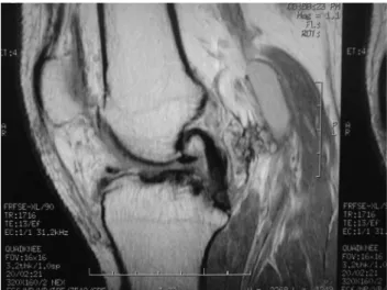
RLS, 39-year-old Caucasian male, soccer teacher, complained of calf pain for 1 year and its worsening for 8 years, when he started presenting paleness and 100-m intermittent claudication of right lower limb, followed by right foot paresthesia. The patient’s history indicated right ruptured cruciate ligament 18 years earlier, con-firmed by magnetic resonance imaging, which showed anterior ruptured cruciate ligament, injured meniscus,
severe arthrosis on the right knee and mild arthrosis on the left knee (Figure 1). The patient did not present risk factors for atherosclerotic disease. Physical examination revealed important muscle hypertrophy on both calves. Distal arterial pulses were present, but disappeared dur-ing passive dorsiflexion maneuvers of the feet and exten-sion of the knees.
Doppler ultrasonography imaging suggested right popliteal artery aneurysm with disappearance of arterial low ater prolonged calf contraction (Figures 2 and 3). Arteriography of the right inferior limb showed absence of medial popliteal artery deviation (Figure 4).
Magnetic resonance imaging conirmed the aneu-rysmal formation in the right popliteal artery, ruling out anomalous muscle insertions (Figure 5).
Retrospectively, subtle signs of lateral entrapment of the popliteal artery in juxta-articular position were identi-ied by angiographic images.
he treatment was open surgical intervention via pos-terior approach of the right popliteal fossa. A large multi-locular adventitial cyst was observed in the popliteal artery,
Abstract
Although its rarity, the adventitial cystic disease of the popliteal artery (ACDPA) must be remembered in the diferential diagnosis of intermittent claudication in young patient’s lower limbs. Brazilian literature presents a few cases of this disease. his study is a case report of ACDPA in a male patient, with a literature review, comparing to popliteal artery entrapment syndrome.
Keywords: Intermittent claudication, popliteal artery, cysts.
Resumo
Apesar de sua raridade, a doença cística da artéria poplítea (DCAP) deve ser lembrada no diagnóstico diferencial de claudicação intermitente de membros inferiores em pacientes jovens. A literatura brasileira apresenta poucos relatos dessa doença. Este trabalho reportou o caso de um paciente masculino portador de DCAP e revisou a literatura, traçando um paralelo com a síndrome de aprisionamento da artéria poplítea.
Palavras-chave: Claudicação intermitente, artéria poplítea, cistos.
Study carried out at Clínica Albernaz and at Hospital Regina, Novo Hamburgo (RS), Brazil.
1 Vascular Surgeon specialized at the Brazilian Society of Angiology and Vascular Surgery (SBACV), Novo Hamburgo (RS), Brazil. 2 Physician, Novo Hamburgo (RS), Brazil.
3 Vascular Surgeon specialized at the SBACV; Professor of Vascular Surgery at Universidade Luterana do Brasil (ULBRA), Canoas (RS), Brazil.
No conlict of interest was declared concerning the publication of this article. Received on: Feb 12, 2010. Accepted on: Apr 28, 2010
between its distal and medium thirds. he rest of the vessel had no anatomical alterations (Figure 6).
he exploration of the popliteal fossa did not identify Baker cyst or muscle compression.
Dissection of the popliteal artery demonstrated an im-portant involvement of the vessel by the cystic formation, without precise limits between them (Figure 7A), thus caus-ing lumen compression due to the irm consistency of the cysts. he slight adherence to the adjacent tissues allowed
Figure 1 – Arthrosis and intra-articular degenerative cysts
Figure 2 – Color-assisted Doppler echography of the right lower limb
revealing hypoechoic image compatible with cyst
Figure 3 – Echographic evidence of arterial low disappearance after vigorous contraction of the calves
Figure 5 - Magnetic resonance imaging showing cyst and
angioreso-nance with slight lateral fulillment failure
Figure 6 – Absence of communication of the cyst with adjacent
struc-tures
release of the cystic mass without rupture or macroscopic extravasation of the content. Resection of the impaired ar-terial segment and interposition of the inverted ipsilateral internal saphenous vein segment were the chosen surgical techniques. Macroscopic analysis of the surgical specimen demonstrated a viscous substance in the cystic formation (Figure 7B). Histopathological analysis conirmed the ad-ventitial mucinous degeneration with a material containing protein and histiocytes. he patient has had a 49-month follow-up and has reported absence of symptoms and of physical restriction.
Discussion
Lower limb intermittent claudication in young patients without evident etiological factors for arteriosclerotic dis-ease is rare. he most common etiologies are PAES and CDPA, which is considered to be the cause of 1 in 1,200 cases of intermittent claudication2. Extrinsic compression and embolism of the popliteal artery, thromboangiitis ob-literans and ibromuscular dysplasia3,4 are also diagnostic possibilities.
Some particularities of the diferential diagnosis be-tween PAES and CDPA are henceforth discussed taking into account the age, sex, clinical features, alterations found in semiologic maneuvers and imaging examinations.
Age
Symptoms of PAES are common in young individuals until the third decade of life, while those of CDPA occur preferentially between the fourth and the sixth decades. It is worth emphasizing that entrapment may go unnoticed for years, hence this hypothesis should not be rejected in individuals of all age groups1.
Genre
Both diseases are more common in males: PAES pres-ents a frequency of 8:1 and CDPA of 4.7:12.
Clinical presentation
here are cases of PAES resulting only from calf mus-cular hypertrophy. However, the vast majority of cases are caused by congenital anomaly of the anatomical relation between the popliteal vessels and the remaining adjacent structures. In the described case, the presence of muscular hypertrophy made the diagnosis more diicult.
In both CDPA and PAES, there may be any degree of ischemic manifestations, depending on the extension of in-volvement and on the time of disease development. Both CDPA and PAES may also present asymptomatic periods. As a justiication of this inding, some authors suggest that, in some cases, the synovial capsule and the cyst may com-municate, thus modifying the intermittent pressure in its interior9. Spontaneous remission of the disease in a ten-year follow-up has been reported10.
In PAES, there is bilaterality in almost 25% of the cases11, while in CDPA there is only one case reported in literature12.
Semiologic maneuvers
In PAES, disappearance of distal pulses with knee extension and passive dorsiflexion of the feet are
fre-quently found, as reported by McDonald13. The
maneu-ver may be performed with the aid of computed tomog-raphy scan or ultrasonogtomog-raphy11. However, one should be aware of the possibility of false negative results11 in the presence of hypertrophy of the calves, as confirmed
Figure 7 – Segment of the popliteal artery presenting adventitial cysts (A) and evacuated mucoid content (B)
the image of a cyst may be mistaken for aneurysm or pseu-doaneurysm, and the internal viscous substance may be mistaken for hypoechogenic mural thrombi. On the other hand, post-stenosis aneurysmal injuries may be found in 6.7% of the entrapments18. In CDPA, the arteriography can reveal compression of the artery afected by the cyst – the scimitar sign15.
A detailed study can identify small alterations in im-ages. In PAES, there may be medial deviation of the artery, presence of collateral circulation and decrease or absence of blood low with active lexion of the feet. here are reports of entrapment without arterial deviation and with post-ste-nosis aneurysmal dilatation19.
Etiopathogeny of the cystic disease of the popliteal artery
CDPA is an uncommon morphological alteration. he disease has a higher incidence in the popliteal artery. here are reports of cases with involvement of the external, femoral, radial, ulnar and brachial iliac arteries20, and even
veins21. Some authors report the presence of mucoid cysts
originated in other sites of the vessel wall, besides the ad-ventitia, as in the intima-media layers20,22. he etiology is unknown and controversial.
In 1998, South African authors presented a theory called “uniier hypothesis” considering that the cysts were traces of ganglion cells in non-axial vessel junctions23.
Other authors indicate that repeated trauma is a pos-sible etiologic factor2. he present case support this hypoth-esis, here represented by the degenerative alterations and ligamentous lesion of the right knee resulting from past trauma, as shown by magnetic resonance imaging.
Some authors9,22,24 report cystic communication with the knee. his fact supports the hypothesis that adventitial cysts are actually synovial cysts that iniltrated the adventi-tia. Conversely, there is no explanation for the diference in the amount of hyaluronic acid found in the synovial luid in comparison to the content of the cysts25. he presence of macroscopic communications of the cyst with other adja-cent structures was not identiied.
one must observe the structure of the reminiscent artery and pay attention to the possibility of unsuccessful inter-vention and recurrence, which occurs in 10% of cases in some reports10. Due to the viscosity of the luid, simple as-piration may not be easy9. In this case, adventitia resection is an alternative27. Angioplasty must only be considered in exceptional cases28,29. When there is arterial occlusion, i-brosis or partial destruction of its wall by the cyst, excision and replacement by a venous grat are recommended, an alternative considered to be reliable and safe, with lower re-currence rate3,4,29.
Conclusion
his paper reported the case of a patient presenting a clinical picture compatible with CDPA (which etiology re-mains unknown), who underwent successful conventional surgical treatment.
References
1. Mafei FH, Barbosa AG, Rollo HA, Neser A, Lastoria S. Adventitial cystic disease of the popliteal artery in Brazil: additional data on the geographical distribuition of the disease. Angiology. 1982;33:339-42.
2. Castiglia V. Doença cística das artérias. In: Mafei FHA, Lastória S, Yoshida WB, Rollo HA. Doenças vasculares periféricas. Rio de Janeiro: Medsi; 2002. p. 1291-304.
3. Cardoso EJ, Teixeira RJ, Galego G, Boabaid RS. Doença cística da adventícia da artéria poplítea. Arquivos Catarinenses de Medicina. 1991;20:181-3.
4. Miranda Junior F, Francisco Junior J, Burihan E. Cisto de artéria poplítea. Rev Bras Cir. 1982;72:221-2.
5. Rollo HA, Gama JC, Lastoria S, Yoshida WB, Mafei FH. Cisto de ad-ventícia em artéria poplítea. Relato de dois casos. AMB Rev Assoc Med Bras. 1982;28:79-81.
6. Alioti R, Prado RF, Boemer VM e col. Doença cística da ar-téria poplítea: relato de dois casos. J Vasc Bras. 2005;4: S119.
tery. J R Soc Med. 2004;97:77-8.
11. Jasinski RW, Masselink BA, Partridge RW, Deckinga BG, Bradford PF. Adventitial cystic disease of the popliteal artery. Radiology. 1987;163:153-5.
12. Ortiz M WR, Lopera JE, Gimenéz CR, Restrepo S, Moncada R, Castañeda-Zúñiga WR. Bilateral adventitial cystic disease of the popliteal artery: a case report. Cardiovasc Intervent Radiol. 2006;29:306-10.
13. McDonald PT, Easterbrook JA, Rich NM, et al. Popliteal artery en-trapment syndrome. Clinical, noninvasive and angiographic diag-nosis. Am J Surg. 1980;139:318-25.
14. Ishikawa K, Mishima Y, Kobayashi S. Cystic adventitial disease of the popliteal artery. Angiology. 1961;12:357-66.
15. Fox CJ, Rasmussen TE, O’Donnell SD. Cystic adventitial disease of the popliteal artery. J Vasc Surg. 2004;39:1351.
16. Brodmann M , Stark G, Pabst E, et al. Cystic adventitial degenera-tion of the popliteal artery - the diagnostic value of duplex. Eur J Radiol. 2001;38:209-12.
17. Papavassiliou VG, Nassim A, Awad EM, Bell PR. Adventitial cystic disease of the popliteal artery: diagnosis and treatment. A case re-port. J Cardiovasc Surg (Torino). 2002;43:399-401.
18. Castiglia V. Síndrome do aprisionamento da artéria poplítea. In: Mafei FHA, Lastória S, Yoshida WB, Rollo HA. Doenças vasculares periféricas. Rio de Janeiro: Medsi; 2002. p. 1305-16.
19. Elias DA, White LM, Rubenstein JD, Christakis M, Merchant N. Clinical evaluation and mr imaging features of popliteal artery entrapment and cystic advential disease. AJR Am J Roentgenol. 2003;180:627-32.
20. Wali MA, Dewan M, Renno WM, Ezzeddin M. Mucoid degenera-tion of the brachial artery: case report and a review of literature. J R Coll Surg Edinb. 1999;44:126-9.
21. Dix FP, McDonald M, Obomighie J, et al. Cystic adventitial disease of the femoral vein presenting as deep vein thrombosis: a case re-port and review of the literature. J Vasc Surg. 2006;44:871-4.
24. Galle C, Cavenaile JC, Hoang AD, et al. Adventitial cystic disease of the popliteal artery communicating with the knee joint. A case report. J Vasc Surg. 1998;28:738-41.
25. Rispoli P, Moniaci D, Zan S, et al. Cystic adventitial disease of the popliteal artery. Report of 1 case and review of the literature. J Cardiovasc Surg (Torino). 2003;44(2):255-8.
26. Asciutto G, Mumme A, Marpe B, Hummel T, Geier B. Diferent approaches in the treatment of cystic adventitial disease of the popliteal artery. Chir Ital. 2007;59:467-73.
27. Stierli P, Mauch J, Koella C, Huber A, Eugster T, Gürke L. Circumferential removal of the adventitia for cystic degeneration of the popliteal artery. Br J Surg. 2005;92:56-7.
28. Setacci F, Sirignano P, de Donato G, Chisci E, Palasciano G, Setacci C. Adventitial cystic disease of the popliteal artery: experience of a single vascular and endovascular center. J Cardiovasc Surg (Torino). 2008;49:235-9.
29. Khoury M. Failed angioplasty of a popliteal artery stenosis second-ary to cystic adventitial disease – a case report. Vasc Endovascular Surg. 2004;38:277-80.
Correspondence:
Daiane Taís Schlindwein Albernaz Rua Marcílio Dias, 1.431, sala 53 Novo Hamburgo (RS), Brasil Fone (51) 3594-1837 E-mail: [email protected]
Authors’ contributions
Study conception and design: DTSA