ABSTRACT
Purpose: To compare the outcomes and costs of stress urinary incontinence (SUI) surgery using a hand-made sling (Mar-lex®) versus a commerciallyavailable suburethral polypropylene sling (Advantage®).
Materials and Methods: Thirty-nine women with SUI due to bladder neck hypermobility and/or sphincter incompetence diagnosed by clinical examination and urodynamic studies were divided into two groups: group 1 (n = 19) consisted of patients from an academic center (Department of Urology, University Hospital of Federal University of Maranhao, and group 2 (n = 20) patients from private practice. The hand-made polypropylene suburethral sling was used in group 1 and the commercial sling in group 2. The patients were evaluated 30, 60 and 90 days after surgery.
Results: The mean duration of surgery was 43 min. in group 1 and 51 min. in group 2. No postoperative voiding dificul -ties were observed in group 1 (100%), as well as, in 94.7% of patients of group 2. A bladder catheter was not required in any of the patients of the two groups at the end of the study. The level of satisfaction was 100% in group 1, whereas, one patient of group 2 considered the surgery to be unsuccessful. Urodynamic studies showed low amplitude uninhibited contraction in 11.1% of patients of group 1 and 10.5% of group 2. No complications were observed in either group. Conclusion: The hand-made polypropylene mesh (Marlex®) can be used for sling procedures, saving costs and yielding results similar to that obtained with commercial sling systems.
Key words: surgical tape; urinary incontinence; suburethral sling; outcome; polypropylenes Int Braz J Urol. 2011; 37: 519-527
INTRODUCTION
Urinary incontinence is the involuntary loss of urine through the urethra causing physical and emotional distress in patients(1). In contrast, stress urinary incontinence (SUI) is the involun-tary loss of urine through the urethra resulting from increased abdominal pressure and urethral occlusion mechanism dysfunction in the absence of detrusor muscle contraction (1).
Epidemiological studies suggest pregnan-cy and vaginal delivery as possible primary etio-logical factors of SUI. Alterations in pelvic sup-port, perineal body and anal sphincter caused by vaginal delivery may contribute to the occurrence
of SUI (2). In most women, the pelvic musculature returns to normal within 2 months after delivery. However, in a small portion of women sequelae might remain that progress to prolapse and urinary incontinence (3). Although epidemiological data indicate a higher incidence of SUI among mul-tiparous women (4,5), this disorder is observed in 16 to 31% of nulliparous women (6,7). Hormonal alterations resulting from aging, obesity, smok-ing, chronic cough, and constipation are associated with SUI (8).
SUI can be classiied into three types ac -cording to leak-point pressure: type I is deined as urine loss at an abdominal pressure higher than 90 cm H2O; in type II urine loss occurs at a pressure
Comparison of the outcomes of the sling technique using a
commercial and hand-made polypropylene sling
Luciane Maria Oliveira Brito, Antonio de Pádua Silva Sousa, José Albuquerque de
Figueire-do Neto, Thaiana Bezerra Duarte, George Figueire-do Lago Pinheiro, Maria Bethânia da Costa Chein
520
of 90 to 60 cm H2O as a result of urethralhyper-mobility, and in type II urine loss occurs at a pres-sure lower than 60 cm H2O (9). The diagnosis of SUI is made clinically and anamnesis is the most important tool. The patient’s history, including sur-gical, gynecological and obstetric history, should be obtained (10). The minimum parameters for the investigation of urinary incontinence recommend-ed by the American Association of Urology consist of a detailed clinical history including micturition data and/or questionnaires about micturition hab-its, physical examination in the presence of a full bladder, micturition diaries, pad tests, and urody-namic study (11).
Behavioral alterations are recommended for the treatment of SUI. The cessation of smoking is advised since this habit causes respiratory dis-eases such as asthma, chronic obstructive pulmo-nary disease and chronic cough that have perineal repercussions (12). Weight control is necessary since obesity is a risk factor for the development of SUI, with overweight increasing the intra-ab-dominal pressure that inluences the perineal mus -culature (13). Pharmacological treatment of SUI includes estrogens, alpha-adrenergic agonists and tricyclic antidepressants. Other, less frequently used drugs are alpha-adrenergic receptor antago-nists and alpha2-adrenergic agoantago-nists (14).
Anatomical changes in the pelvic loor responsible for urinary incontinence in women should be corrected by surgical procedures that are aimed at the stabilization of the urethra. In-trinsic disorders of urethral sphincter mechanisms should be treated by interventions that promote urethral coaptation (1). Numerous surgical tech-niques have been developed for the correction of SUI, including conventional open surgeries and minimally invasive procedures such as periure-thral injection therapy and procedures that use organic and synthetic materials to support the ure-thra, known as slings. The last procedure is cur-rently the treatment of choice for the correction of SUI of any etiology.
Due to the eficiency of sling procedures, the present study proposes the use of a low-cost, hand-made polypropylene sling that could be used in the public health system offering similar results
to those obtained with commercial synthetic sling systems that are much more expensive in Brazil (15).
MATERIALS AND METHODS
From December 2007 and December 2009, a non-randomized study was conducted in Mara-nhão, Brazil; recruiting patients from the academic University Hospital (HU) of Federal University of Maranhão (UFMA), and from a local private urol-ogy practice.
A total of 39 women with urinary inconti-nence were included in the study. They were diag-nosed according to the McGuire classiication (9) (type I: urine loss at an abdominal pressure higher than 90 cm H2O; type II: urine loss at a pressure of 90 to 60 cm H2O; type III: urine loss at pressure less than 60 cm H
2O). Exclusion criteria included medical history of diabetes mellitus, major pelvic surgery accompanied by bladder denervation, pre-vious radiotherapy, and malignant neoplasms of the bladder. Patients with active urinary infection and bladder stones diagnosed by urethrocystosco-py were treated and then included in the study.
Two groups were considered: group 1 consisted of 20 patients from the Urology Service of HU-UFMA, and group 2 included 19 patients from the private clinic. Group 1 was treated with a hand-made sling consisting of a Marlex® mesh measuring 1.5 cm in width and 30 cm in length. In group 2, the commercial Advantage™ Transvagi-nal Mid-Urethral Sling System (Boston Scientiic Corporation, Maple Grove, MN) was used. This system was chosen because the ixation principle is similar to that of the Marlex® sling. In both cas-es, ixation of the prosthesis occurs by incorpora -tion of the mesh into surrounding tissues through proliferation of ibrotic tissue that gives support to the suburethral sling. Group 2 had patients were recruited from a private practice due to the acqui-sition of the commercially available slings used in this study.
Before the surgical procedure, a complete history and physical examination, preoperative laboratory testing, urodynamic study, and urethro-cystoscopy was illed out. Urodynamic analysis was performed according to the guidelines of the
International Continence Society using 0.9% sa-line at a temperature of 37ºC, two Nelaton® 6F urethral catheters (one for luid infusion and one for the measurement of bladder pressure) and a Nelaton 8F rectal catheter. The pressure produc-ing stress urinary loss (PUL) was obtained after removal of the infusion catheter when the patient reported the irst micturition desire. On this occa -sion, the patient was asked to increase abdominal pressure using a Valsalva maneuver. PUL was de-ined as the lowest abdominal pressure detected in the absence of detrusor contraction which was able to produce urinary loss (ICS, 1991). All pa-tients were submitted to urethrocystoscopy and none of them presented bladder lithiasis or neo-plastic alterations.
After hospital discharge, the patients were evaluated 30, 60 and 90 ± 2 days after the surgi-cal procedure. Another form was illed out during these assessments and urodynamic analysis was performed on the last assessment (90 days after surgery). In this analysis, urinary residues less than 100 mL were deined as a normal result, indi -cating absence of signiicant urethral compression. Higher values were considered to indicate over-treatment and required reassessment.
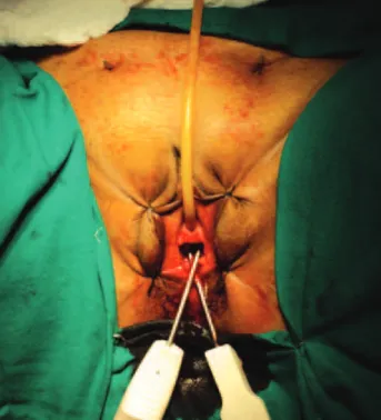
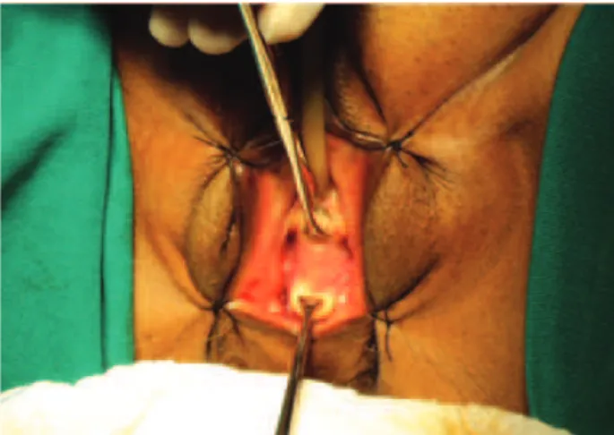
All surgeries were performed by the same surgeon according to the technique described by Petros (16). Regional block was used for anesthe-sia and antibiotic prophylaxis (1 g cefazolin, in-travenously) was administered 30 min before and up to 48 h after the end of surgery. The polypro-pylene sling used for urethra support consisted of a Marlex® mesh measuring 1.5 cm in width and 30 cm in length (Figure1). Using a special needle (Figure2), one end of the sling was passed through a vaginal incision in the direction of the abdominal wall, with the sling following a retropubic trajec-tory and exteriorizing through a 1 cm incision in the skin (Figure3). The procedure was repeated on the opposite side, forming a loop to support the middle third of the urethra. No additional stitch-es were used for ixation of the ends of the sling preventing hypermobility of the urethra. A Kelly clamp was placed between the urethra and sling to prevent unnecessary compression of the former (Figure 4). Urethrocystoscopy was performed
dur-ing each passage of the needle to rule out possible bladder injury which, if detected, was immediately corrected. The vaginal mucosa was closed with continuous 3.0 chromic catgut suture. The indwell-ing bladder catheter was maintained until the sec-ond postoperative day. The same surgical steps for the commercial sling system (Advantage®).
Figure 1 - Making the hand-made polypropylene sling for surgery.
Figure 2 - Stamey needles introduction.
Bra-522
zil according to electronic price quotation (http:// www2.ciashop.com.br/cpassos). This mesh was divided into 20 segments of 1.5 x 30 cm, with one segment being used per surgery, corresponding to a cost per patient of approximately R$ 13.05 (R$ 261.00/20 segments). In contrast, the commercial sling system (Advantage®) costs R$ 1.800.00 ac-cording to the Financial Sector of Covenants Box Health Care Emnployess of the Bank of Brazil (CASSI.).The results are presented in tables. The Epi-Info program, version 3.3.2, was used for sta-tistical analysis, adopting a level of signiicance of p < 0.05. The chi-square test was used for the calculation of signiicance in the univariate com -parison of proportions.
The study was approved by the Ethics Com-mittee of HU-UFMA (process 33104-0636/2005).
RESULTS
During the study period, 39 patients were submitted to surgical treatment of SUI, 20 in group 1 who received the hand-made sling and 19 in group 2 who received the commercial sling system.
With respect to age, patients aged 30 to 39 years (n = 5, 25%) and 50 to 59 years (n = 5, 25%) predominated in group 1. In contrast, in group 2 there was a predominance of patients aged 50 to 59 years (n = 7, 36.8%).
SUI was the only cause of urinary inconti-nence in 100% (n = 20) of cases of group 1 and in
Outcomes of the hand made sling
Figure 3 - Incision of mid urethra for introduction of sling.
Figure 4 – Passage of hand-made polypropylene sling. 84.2% (n = 16) of group 2. In group 2, only three (15.8%) patients presented with urgency in addi-tion to SUI.
The mean duration of surgery was 43 min. in group 1 and 51 min. in group 2. The mean dura-tion of hospitalizadura-tion was 52.8 hr in group 1 and 49.14 hr in group 2.
Postoperative voiding dificulties were re -ported in three (15%) patients of group 1 during the irst 30 days after the procedure, and one patient (5.3%) in group 2. At the end of follow-up, all pa-tients of group 1 were able to void normally, and only one (5.3%) in group 2 continued to experience voiding dificulties, with no signiicant differenc -es between groups.
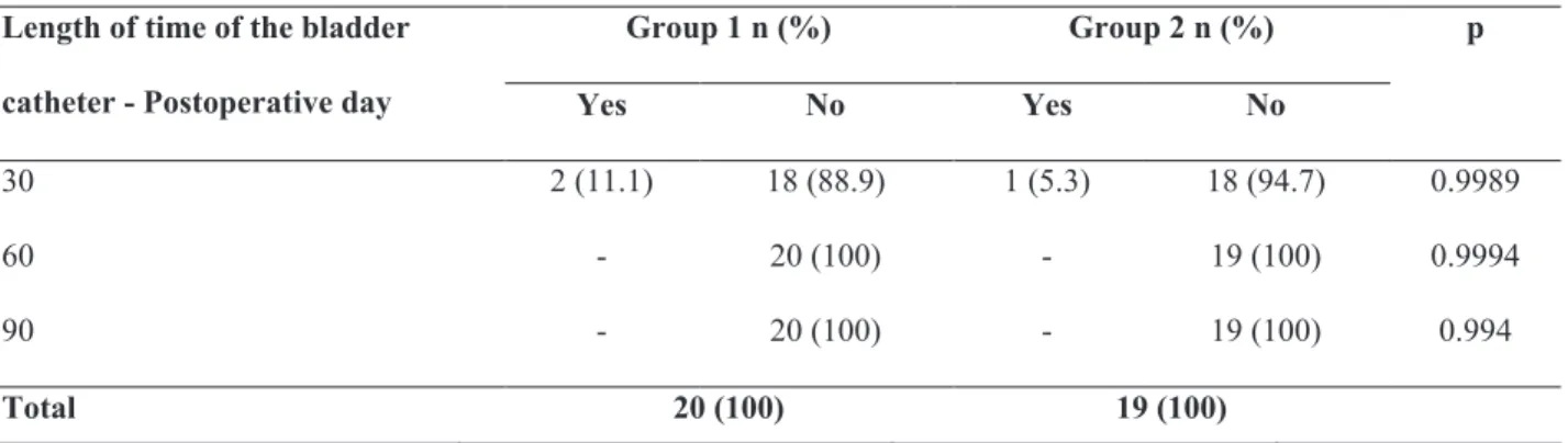
A postoperative catheter was necessary in two (11.1%) patients in group 1 and in one (5.3%) patient in group 2 up to 30 days after surgery. None of the patients of either group required a catheter at the end of follow-up. The differences in the results ob-served between groups were not signiicant (Table-1). Only two (11.1%) patients in group 1 did not have a normal urinary stream 30 days after surgery,
whereas a normal urinary stream was observed in all patients of the two groups on other assessments. The differences in the results observed between groups were not signiicant (Table-2).
523
involuntary urine loss and only one (5.3%) in group2. The differences in the results between groups were not signiicant (Table-3).
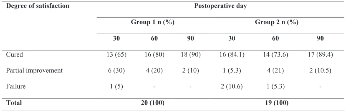
With respect to the degree of satisfaction with surgery, surgical failure was not observed in any of the patients of either group at the end of follow-up. At the end of the study, 18 (90%) patients of group 1 and 17 (89.4%) of group 2 were satisied and con -sidered themselves cured. Partial improvement (1 to 2 episodes of urine loss per day) at the end of fol-low-up was reported by two patients each in group 1 and group 2, with no signiicant differences between groups (Table-4).
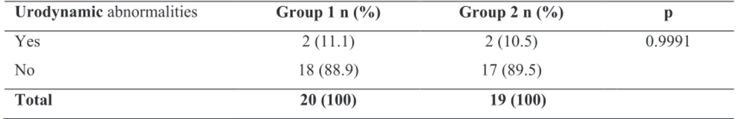
Urodynamic study was performed on postop-erative day 90 and revealed low amplitude uninhib-ited contractions in two (11.1%) patients in group 1 and two (10.5%) in group 2, with no signiicant dif -ferences between groups (Table-5). In both groups, patients with uninhibited bladder contractions of low
intensity did not reported loss of urine or change in urinary stream.
DISCUSSION
Sling surgery has proven to be eficient for treatment of SUI (15), the present study proposes a low-cost hand-made polypropylene sling that could be used routinely in the public health system with similar results obtained with commercial synthetic sling systems that are much more expensive.
In the present study, no signiicant differ -ence in the variables analyzed were observed be-tween the two groups treated with the hand-made (group 1) and commercial slings (group 2) over the 90-day follow-up. A higher number of patients in their sixth decade of life were observed in both groups, as described in other studies (17,18). This inding conirms the predominance of the disease in Table 1 - Length of time of indwelling bladder catheter after sling procedure for treatment of SUI.
Table 2 - Postoperative spontaneous void after sling surgery for the treatment of stress urinary incontinence. Group 1: hand-made sling; Group 2: commercial sling system.
Group 1: hand-made sling; Group 2: commercial sling system.
–
Normal urinary stream -
Postoperative day
Group 1 n (%) Group 2 n (%) p
Yes No Yes No
30 18 (88.9) 2 (11.1) 19 (100) - 0.9985
60 20 (100) - 19 (100) - 0.9994
90 20 (100) - 19 (100) - 0.9994
Total 20 (100) 19 (100)
–
n (%) n (%)
20 (100) 19 (100)
Length of time of the bladder
catheter - Postoperative day
Group 1 n (%) Group 2 n (%) p
Yes No Yes No
30 2 (11.1) 18 (88.9) 1 (5.3) 18 (94.7) 0.9989
60 - 20 (100) - 19 (100) 0.9994
90 - 20 (100) - 19 (100) 0.994
Total 20 (100) 19 (100)
2
n (%) n (%)
20 (100) 19 (100)
524
21 patients submitted to sling surgery, the frequency of postoperative temporary voiding dificulties was 28.6% (21). In the present study, temporary voiding dificulties during the irst 30 days after the proce -dure were observed in 15% of patients in group 1, but in only 5.3% of patients in group 2. At the end of follow-up, all patients of group 1 were able to void, whereas one patient of group 2 still experi-enced voiding dificulties. This inding suggests that voiding dificulty maybe a natural occurrence due to the surgical technique and is not related to the material used for the suburethral sling.
With respect to involuntary urine loss, none of the patients in group 1 experienced urine loss at the end of the observation period and only one (5.3%) patient in group 2 reported this symp-tom. In another study, 87% were completely dry this age group. In the present study, similar to the
hospitalization period, it was decided to leave the bladder catheter in place for 48h, as observed in 95% of the patients of both groups and in agree-ment with another Brazilian study (19).
In a recent study, eight of 128 patients sub-mitted to sling surgery presented urinary retention and underwent clean intermittent catheterization until postoperative day 25, when spontaneous void returned, except for two patients who required ure-throlysis (20). In the present study, only two (11.1%) patients in group 1 did not show a normal urinary stream during the irst 30 days after surgery. Howev -er, all patients of the two groups presented a normal urinary stream on the subsequent assessments (60 and 90 days) and reported satisfactory micturition at the end of the study. In another series involving
Outcomes of the hand made sling
Table 3 - Postoperative urinary incontinence after sling surgery for the treatment of stress urinary incontinence.
Table 4 - Postoperative satisfaction after sling surgery for the treatment of stress urinary incontinence.
Group 1: hand-made sling; Group 2: commercial sling system. Group 1: hand-made sling; Group 2: commercial sling system.
2
–
n (%) n (%)
20 (100) 19 (100)
2
–
n (%) n (%)
20 (100) 19 (100)
3
Urine loss - Postoperativeday
Group 1 n (%) Group 2 n (%) p
Yes No Yes No
30 2 (11.1) 18 (88.9) 1 (5.3) 18 (94.7) 0.9989
60 1 (5.0) 19 (95.0) 1 (5.3) 18 (94.7) 0.9993
90 - 20 (100.0) 1 (5.3) 18 (94.7) 0.9995
Total 20 (100) 19 (100)
Group 1: hand-made sling; Group 2: commercial sling system.
4
Degree of satisfaction Postoperative day
Group 1 n (%) Group 2 n (%)
30 60 90 30 60 90
Cured 13 (65) 16 (80) 18 (90) 16 (84.1) 14 (73.6) 17 (89.4)
Partial improvement 6 (30) 4 (20) 2 (10) 1 (5.3) 4 (21) 2 (10.5)
Failure 1 (5) - - 2 (10.6) 1 (5.3) -
and no longer experienced urine loss and 6.8% presented signiicant improvement, whereas surgi -cal failure and persistent urine loss were observed in 5% (22). In another study including 45 patients that underwent sling surgery for the treatment of SUI, 74% of the patients experienced no urine loss, 11.2% presented one to two episodes a day, and 14.8% had three or more episodes a day (23).
With respect to the degree of satisfaction with the surgery, 18 (90%) patients of group 1 were satisied and considered themselves cured and two (20%) reported improvement (1 to 2 episodes of urine loss per day). In group 2, 17 (89.4%) pa-tients considered themselves cured and were satis-ied and two (10.5%) reported improvement, with a reduction in the episodes of urine loss to 1-2 per day. In another Brazilian study, 29 of 30 patients submitted to surgery for the treatment of SUI us-ing a hand-made polypropylene slus-ing reported sat-isfaction with the surgery over a follow-up period of 15 months (24). Raz et al. (25), evaluating 26 patients submitted to vaginal sling surgery, ob-served excellent outcomes in 20 (77%) patients, very good outcomes in two (8%), improvement in one (4%), and failure in three (12%). These re-sults demonstrate the eficacy of the technique, ir -respective of the type of material used for fabrica-tion of the sling.
In a recent study involving 80 patients sur-gically treated for SUI using a tension-free sling, only one patient developed a hyperactive blad-der, accompanied by the loss of large volumes of urine (19). This patient required a bladder catheter for 9 days and the symptoms only improved after the introduction of anticholinergic medication. In
the present study, the urodynamic alterations ob-served on postoperative day 90 consisted of low amplitude uninhibited detrusor contractions in two (11.1%) patients in group 1 and two (10.5%) in group 2. This abnormality was not clinically signiicant and was tolerated by the patients, who did not require a bladder catheter. The symptoms disappeared after the introduction of anticholiner-gic medication. No complications were observed in either group and, therefore, no blood products transfusion was necessary.
Perforation of the bladder, which occurred in one patient in group 1 during passage of the needle, was identiied by cystoscopy and procedure was continued. The patient had a good recovery and required a bladder catheter for 6 days. Bladder per-foration is one of the most common complications of sling surgery. In a series of 20 patients submitted to sling surgery using a hand-made sling, bladder perforation was observed in two patients (18). Dur-ing the follow-up there was no evidence of erosion of the urethra or vaginal mucosa in patients who had the hand-made polypropylene sling for surgery.
CONCLUSIONS
Despite the short postoperative follow-up (90 days) in the present study, investigations us-ing the same material and a longer follow-up of 13 months (18), 15 months (24) and 23 months (17) reported similar cure rates of SUI or improvement of clinical symptoms (95%, 96% and 89%, respec-tively). These results demonstrate the long-term ef-icacy of a cost-effective hand-made polypropylene mesh sling for the treatment of SUI.
Urodynamic abnormalities Group 1 n (%) Group 2 n (%) p
Yes 2 (11.1) 2 (10.5) 0.9991
No 18 (88.9) 17 (89.5)
Total 20 (100) 19 (100)
Table 5 - Presence of urodynamic abnormalities on postoperative day 90 after sling surgery for the treatment of stress urinary incontinence.
526
Outcomes of the hand made sling
CONFLICT OF INTEREST
None declared.
REFERENCES
1. Haylen BT, de Ridder D, Freeman RM, Swift SE, Berghmans B, Lee J, et al.: An International Urogy-necological Association (IUGA)/International Conti-nence Society (ICS) joint report on the terminology for female pelvic loor dysfunction. Neurourol Uro -dyn. 2010; 29: 4-20.
2. Meyer S, Schreyer A, De Grandi P, Hohlfeld P: The effects of birth on urinary continence mechanisms and other pelvic-loor characteristics. Obstet Gyne -col. 1998; 92: 613-8.
3. Allen RE, Hosker GL, Smith AR, Warrell DW: Pel-vic loor damage and childbirth: a neurophysiological study. Br J Obstet Gynaecol. 1990; 97: 770-9.
4. Thomas TM, Plymat KR, Blannin J, Meade TW: Prevalence of urinary incontinence. Br Med J. 1980; 281: 1243-5.
5. Sommer P, Bauer T, Nielsen KK, Kristensen ES, Hermann GG, Steven K, et al.: Voiding patterns and prevalence of incontinence in women. A question-naire survey. Br J Urol. 1990; 66: 12-5.
6. Bo K, Maehlum S, Oseid S, Larsen S: Prevalence of stress urinary incontinence among physically ac-tive and sedentary female students. Scan J. Sport Sci. 1989; 11: 113-6.
7. Wolin LH: Stress incontinence in young, healthy nul-liparous female subjects. J Urol. 1969; 101: 545-9. 8. Hunskaar S, Burgio K, Clark A, Lapitan MC, Nelson
R, Sillén U, et al.: Epidemiology of Urinary (UI) and Faecal (FI) Incontinence and Pelvic Organ Prolapse (POP). In: Abrams P, Cardozo L, Khoury S, Wein A (ed.), Incontinence 3rd International Consultation on Incontinence. United Kingdom, Health Publication, 2005; pp; 255-312.
9. McGuire EJ, Cespedes RD, O’Connell HE: Leak-point pressures. Urol Clin North Am. 1996; 23: 253-62. 10. Harvey MA, Versi E: Predictive value of clinical
eval-uation of stress urinary incontinence: a summary of the published literature. Int Urogynecol J Pelvic Floor Dysfunct. 2001; 12: 31-7.
11. Blaivas JG, Appell RA, Fantl JA, Leach G, McGuire EJ, Resnick NM, et al.: Standards of eficacy for
evaluation of treatment outcomes in urinary inconti-nence: recommendations of the Urodynamic Society. Neurourol Urodyn. 1997; 16: 145-7.
12. Tampakoudis P, Tantanassis T, Grimbizis G, Papal-etsos M, Mantalenakis S: Cigarette smoking and uri-nary incontinence in women--a new calculative meth-od of estimating the exposure to smoke. Eur J Obstet Gynecol Reprod Biol. 1995; 63: 27-30.
13. Wingate L, Wingate MB, Hassanein R: The relation between overweigh and urinary incontinence in post-menopausal women: A case control study. J North Am Menopause Soc. 1994; 1: 199-203.
14. DuBeau CE: Therapeutic/pharmacologic approaches to urinary incontinence in older adults. Clin Pharma-col Ther. 2009; 85: 98-102.
15. D’ancona CAL, Castro N, Sabaneff J, Querne FAO. Urinary Incontinence: Routine Brazilian Society of Urology. 2006. [cited Nov 28 2009] Available at: http://www.projetodiretrizes.org.br/6_volume/30-In-contiUrinProp.pdf.
16. Petros PP: The intravaginal slingplasty operation, a minimally invasive technique for cure of urinary in-continence in the female. Aust N Z J Obstet Gynae-col. 1996; 36: 453-61.
17. Rodrigues FR, Maroccolo Filho R, Maroccolo RR, Paiva LC, Diaz FA, Ribeiro EC: Pubovaginal sling with a low-cost polypropylene mesh. Int Braz J Urol. 2007; 33: 690-4.
18. Sola V, Pardo J, Ricci P, Guiloff E: Tension free mono-ilament macropore polypropylene mesh (Gynemesh PS) in female genital prolapse repair. Int Braz J Urol. 2006; 32: 410-4; discussion 415.
19. Sartori JP, Martins JA, Castro Rde A, Sartori MG, Girão MJ: Pubovaginal sling and tension-free vaginal tape for surgical treatment of stress urinary inconti-nence in women. Rev Bras Ginecol Obstet. 2008; 30: 127-34.
20. Gauruder-Burmester A, Popken G: The MiniArc sling system in the treatment of female stress urinary in-continence. Int Braz J Urol. 2009; 35: 334-41; author reply 341-3.
21. Martins JA, Castro RA, Grão MJBC, Sartori MGF, Baracat EC, Lima GR. Stress Urinary Incontinence Correction with Sling: First Results. Rev Bras Gine-col Obstet 2000, 22: 301-305.
23. Meloni WA, Tome AL, Karam-Jr AM, Bestane MC, Bestane WJ: Surgical treatment of female stress uri-nary incontinence with pubovaginal sling: better qualites of life. Rev Médica Ana Costa 2004 [cited Nov 28 2009] Available at: http://www.revistamedi-caanacosta.com.br/9(1)/artigo_1.htm.
______________________ Correspondence address: Dr. Luciane Maria Oliveira Brito Programa de Pós-Graduação em Saúde Materno-Infantil
Praça Gonçalves Dias nº 21 Prédio de Medicina, 2º andar Sao Luis, MA, 65020-240, Brazil Fax: + 55 98 3232-0286
E-mail: [email protected]
24. Rapp DE, Govier FE, Kobashi KC: Outcomes fol-lowing mid-urethral sling placement in patients with intrinsic sphincteric deiciency: comparison of Sparc and Monarc slings. Int Braz J Urol. 2009; 35: 68-75; discussion 75.
25. Raz S, Siegel AL, Short JL, Snyder JA: Vaginal wall sling. J Urol. 1989; 141: 43-6. Erratum in: J Urol 1989; 141: 1214. Synder JA [corrected to Snyder JA].
_____________________
Submitted for publication: June 16, 2010
___________________