Detection of leptospires from infected urine and tissue
samples
in vitro by modiied Fontana silver stain
Detecção de leptospiras na urina e nos tecidos infectados
in vitro
por
impregnação com prata Fontana modificada
Islay Rodríguez1; Iuley Rodríguez2; Carmen Fernández3; José E. Rodríguez4; Jorge Cantillo5
ABSTRACT
Introduction: The microbiological diagnosis of leptospirosis comprises bacteriological and serological methods. The former ones allow the direct detection of leptospires and are considered presumptive with the exception of culture. Therefore, they constitute invaluable tools for rapid
diagnosis, mainly in samples from deceased subjects. Objective: To evaluate a modiied Fontana silver staining method in experimentally
infected samples. Material and methods: Human and animal (hamster) urine samples were experimentally infected with different strains of
Leptospira interrogans sensu lato. Liquid culture medium, leptospira cultures, experimentally infected and non-infected human urine samples, clariied and non-clariied imprints, and clariied and non-clariied suspension smears from tissues of experimentally infected and non-infected hamsters were applied for the ass essment of silver staining. The analytical sensitivity of the assay was compared with dark ield microscopy and
culture. Other bacterial and fungi species were also used. Results: The modiied Fontana silver staining allowed the accurate observation of the
well-deined leptospire helical structure. On leptospire cultures from infected human samples, we could observe until (1-10) × 103 leptospires/ml,
higher sensitivity in comparison with direct dark ield microscopy and lower in comparison with culture. The best results in tissues were obtained on clariied imprints and non-clariied suspension smears. Morphological and stainable structures compatible with leptospires were not observed
in the samples without them. Conclusion: This procedure allowed differentiating the characteristic morphology of leptospires. As its application
suggests, it consists of a simple and easily conducted procedure with stable reagents.
Key words: leptospira; silver staining; leptospirosis; diagnosis.
First submission on 06/06/12; last submission on 28/09/12; accepted for publication on 04/10/12; published on 20/02/13
1. Doctor in Health Sciences; researcher and professor of Microbiology at the Tropical Medicine Institute Pedro Kourí (IPK). 2. Microbiologist; laboratory specialist at IPK.
3. Doctor in Health Sciences; researcher and professor of Microbiology at IPK. 4. Senior Microbiology technician at IPK.
5. Veterinary Medical doctor; head of the Department of Experimental Animals at IPK.
INTRODUCTION
Leptospirosis is a worldwide zoonosis, which bears considerable medical and economic relevance. It is caused by spirochetes
belonging to the Leptospira interrogans sensu lato (sl) (pathogenic
group)(1, 5). It has been acknowledged as a neglected and likewise
emerging infectious disease in the last 20 years, hence its recent major outbreaks in different countries(1, 9, 12).
Leptospires are usually present in the blood during the irst week of infection and are excreted in the urine from the second week onwards up to a month or even more. Major causes of death include renal failure,
cardiopulmonary failure, and widespread hemorrhage(1, 5, 19).
The direct demonstration of its causative agent is an invaluable tool for rapid diagnosis. However, current bacteriological methods
for the detection of leptospires in clinical specimens such as blood, cerebrospinal luid, urine and post mortem tissue have some disadvantages: leptospires are poorly stained with conventional methods; dark ield microscopy is relatively unreliable due to possible errors caused by leptospira-like artifacts; culturing is time-consuming to be of value for a rapid diagnosis(1, 5, 17, 19).
Leptospires can be stained in formalin ixed tissue sections by Dobell method for blocks, or Warthin-Starry, Faine, or Dieterle methods for cutting sections from parafin blocks. The use of parafin
blocks is a tedious and time-consuming procedure(5, 19).
MATERIAL AND METHODS
Strains
Leptospiral and other bacterial or fungal strains were used in this
study. Details are described in Table 1.
Pure cultures of reference leptospiral strains with 7-10 days of growth at 28-30ºC were applied. Leptospires were inoculated into Ellinghausen-McCullough-Johnson-Harris (EMJH) medium and enriched with 10% salts, albumin, vitamins, antimicrobial agents and Tween (SAVAT) supplement.
Specimens of leptospiral culture and experimentally infected urine
Ten different cultures of pathogenic leptospira were diluted with equal quantities of EMJH-SAVAT medium. In order to determine the concentration of leptospires in each sample, the
optical density (OD) to λ = 420 nm was measured. Subsequently,
decimal and serial dilutions were conducted (10-1 to 10-8) for each one.
Urine specimens from 10 healthy persons were taken from each subject. These samples were divided in two fractions, one of them was alkalinized (pH 7-8) with NaOH 4N and the other one was kept at its natural state (pH 5-6.5). All these fractions were mixed with 10 different cultures of pathogenic leptospire in equal quantity. The
concentration of leptospires was determined and decimal and serial dilutions were performed as described above.
Non-inoculated EMJH-SAVAT medium and urine (alkalinized and natural) diluted 1:2 with medium were used as controls.
Three aliquots from each dilution and control were observed by dark ield microscopy, inoculated in EMJH-SAVAT medium, incubated at 28-30ºC and periodically checked macro and microscopically for 40 days.
A loopful of each leptospira culture dilutions or leptospira urine mix dilutions was placed at one end of a clean slide and a ilm was drawn in one stroke. The smear was air dried and labeled carefully in order to avoid the risk of infection through direct contact of ingers with the smear surface.
The remaining volumes were centrifuged for 10 minutes at 6,500 rpm, the supernatants were centrifuged for 30 minutes at 12,000 rpm, and a smear from each inal sediment was made on the slide.
Experimental infection of animal tissues with leptospires
Ten golden male Syrian hamsters (Mesocricetus aureatus)
weighing 10-30 g were used based on the three Rs of Russell and
Burch (replacement, reduction and reinement)(2). Eight animals
were intraperitoneally inoculated with 1 ml of 108 cells/ml culture
of Copenhageni M20, Tropica 299, Canicola Hond Utrecht IV and
TABLE 1 – Classiication of leptospiral and other fungi and bacterial strains used in this study
Leptospiral strains Serovar Strain
L. interrogans sl Tropica 299
Copenhageni M20
Shermani 1342k
Canicola Hond Utrecht IV
Ballum Mus 127
Hardjo Hardjoprajitno
Pyrogenes Salinem
Bratislava Jez Bratislava
Tarassovi Perepelitsin
Bataviae Swart
L. biflexa sl Patoc Patoc I
Other bacteria and fungi Strain
Escherichia coli Field strain*
Klebsiella sp. Field strain*
Proteus sp. Field strain*
Staphylococcus aureus Field strain*
Candida albicans Field strain*
Aspergillus fumigatus Field strain*
Ballum Mus 127 strains of Leptospira interrogans sl (two animal/ serovar), and the other two were inoculated with non pathogenic leptospira and sterile medium, respectively (controls).
The animals were observed daily in order to detect the deceased ones until day 20. The surviving animals underwent cervical dislocation. Kidneys, liver, spleen, lungs and heart were taken aseptically from all animals.
Imprints of the surface of the suspected tissues were performed on clean slides and air dried. They were clariied by treatment with 10% acetic acid for 5-10 minutes, rinsed with water and air dried. The specimens were further clariied, when it was necessary, with 0.25% trypsin in buffer pH 7.8 for 3-5 minutes.
The different tissues were disrupted by use of syringe and collected in tubes with sterile phosphate buffered solution (PBS). The tubes were vortexed and later centrifuged for 10 minutes at 3,000 rpm. Leptospires were identiied by dark ield microscopy from the supernatant and smears for copies on slides were also performed. Only one copy of each tissue was clariied.
Three aliquots from each supernatant were inoculated into EMJH-SAVAT medium and incubated at 28-30ºC for 40 days. They were periodically checked by dark ield microscopy.
Other microorganisms
One drop of sterile distilled water and a loopful of each culture were put on clean slides, mixed and different smears were performed.
Staining procedure
We used the modiied procedure reported by Gangadhar and
Rajasekhar in 1998(6). Briely, the slides with the smears or imprints
were placed vertically in a 100 ml capacity beaker containing the ixing reagent (1 ml glacial acetic acid + 2 ml formalin [40% formaldehyde solution] + 100 ml distilled water) for 2 minutes, then they were removed by forceps, blotted onto tissue paper and dipped in a beaker containing absolute alcohol or methanol for 3 minutes. The under surface of the slides were cleaned with ilter paper and the smears were air dried. The slides were then dipped in a 100 ml beaker containing the mordant (1 g phenol + 5 g tannic acid + 100 ml distilled water) pre-heated to 75ºC in a hot water bath and allowed to react for 1 minute. Afterwards, they were rinsed in distilled water, the under surface wiped and air dried. The slides were dipped in a 100 ml beaker containing the ammoniated silver solution (see preparation) pre-heated to 75ºC in a hot water bath and allowed to react for 1 minute. The slides were then rinsed with distilled water, the under surface cleaned, air dried and examined under bright ield microscope using oil immersion lens.
Preparation of ammoniated silver solution: 100 ml of 0.6% silver nitrate solution prepared in distilled water was divided into 60 and 40 ml aliquots and transferred to separate glass lasks. To the 60 ml aliquot, a few drops of 10% ammonia solution was added and shaken to obtain a brown precipitate. This was followed by further addition of 0.5 to 1 ml or more of ammonia solution till all the precipitate
was dissolved. Small quantities of silver nitrate solution from the 40 ml aliquot were transferred to the above ammoniated silver nitrate solution till a stable precipitate reappeared. Remaining unused silver nitrate solution from the 40 ml aliquot was discarded.
Statistical analysis
Data were stored in an Excel spread sheet (Microsoft Ofice) for analysis. Absolute frequencies and percentages were calculated, and comparison test for proportions was conducted using EPIDAT 3.1 statistical software.
Ethical aspects
The experiment was performed in accordance with the Ethics
Committee of Instituto Pedro Kourí. The human urine samples were
collected with the volunteers’ previous consent and they were used exclusively in this investigation. Cervical dislocation was used as a physical method of euthanasia for the surviving animals according to the recommendations of Directorate-General of the Environment, Nuclear Safety, and Protection (DGXI) of the European Commission
for euthanasia of experimental animals(3). This procedure was
conducted by a highly experienced specialist in a rapid and effective way. All procedures were performed in a laminar low cabinet in a biosafety level II facility.
RESULTS
By use of modiied Fontana silver impregnation, the leptospires were stained brown and appeared as long curved organisms with one or both characteristically hooked extremities against a clear white or pale yellow background. The inal helical structure of the organism
was clearly visible under oil immersion (Figure).
The results of quantitative examination by dark-ield microscopy, culture and silver staining of leptospiral cultures and experimentally
infected natural or alkaline urine samples are presented inTables 2,
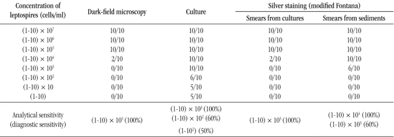
3 and 4. Leptospires could be identiied by silver staining at a density
of approximately 103 per ml in sediments from culture and urine
samples. Statistical differences (p < 0.05) were detected among the
proportions.
Hamsters infected with Copenhageni or Tropica serovars of
Leptospira began to show symptoms of illness between days 3 and 5, and all died before day 7. Abundant jaundice, increased organ size and hemorrhages were macroscopic and histopathological changes observed in these animals. In contrast, the animals infected with Ballum or Canicola serovars, similarly to control hamsters, did not show any symptom and they were sacrificed at day 20.
The results of the different methods and variants for the direct demonstration of leptospires in organs of hamsters with leptospirosis
are demonstrated in Table 5. The best results were obtained by silver staining from clariied imprints and non-clariied smears of the
studied tissues, although there were no statistical differences among the proportions (p > 0.05).
When studying the organs of animals infected with Ballum and Canicola serovars, we only detected leptospires by silver staining (modiied Fontana) in spleen and lung of one animal infected by Canicola serovar, and we isolated leptospires from kidney of one animal infected by Ballum serovar.
The results of silver staining using clariied imprints and non-clariied smears were compared and the correlation (positive and negative results) was 60% (12/20). These results were also compared with culture, and the correlation was 35% (7/20) for both.
The speciicity of silver staining (modiied Fontana) was determined using the negative control specimens (EMJH-SAVAT medium, natural and alkalinized urine and tissue from control animals without leptospires). In all cases leptospires or similar artifacts were not detected.
Silver staining was also applied with other microbial forms (bacteria, yeast and fungi), and all of them were stained distinguishing their different forms.
TABLE 2 – Proportion of positive results and sensitivity of different methods for detection of leptospires from cultures
Concentration of
leptospires (cells/ml) Dark-ield microscopy Culture
Silver staining (modiied Fontana) Smears from cultures Smears from sediments (1-10) × 107
(1-10) × 106 (1-10) × 105 (1-10) × 104 (1-10) × 103
(1-10) × 102 (1-10) × 10
(1-10)
10/10 10/10 10/10 2/10 0/10 0/10 0/10 0/10
10/10 10/10 10/10 10/10 10/10 6/10 5/10 5/10
10/10 10/10 10/10 2/10 0/10 0/10 0/10 0/10
10/10 10/10 10/10 10/10 6/10 0/10 0/10 0/10
Analytical sensitivity
(diagnostic sensitivity) (1-10) × 105 (100%)
(1-10) × 103 (100%)
(1-10) × 102 (60%)
(1-10) × 105 (100%) (1-10) × 104 (100%) (1-10) × 103 (60%)
(1-102) (50%)
TABLE 3 – Proportion of positive results and sensitivity of different methods for detection
of leptospires from experimentally infected natural urine samples (acid pH) Concentration of
leptospires (cells/ml)
Dark-ield microscopy Culture Silver staining (modiied Fontana)
Smears from urine samples
Smears from sediments
(1-10) × 107 10/10 5/10 10/10 10/10
(1-10) × 106 10/10 5/10 10/10 10/10
(1-10) × 105 9/10 3/10 10/10 10/10
(1-10) × 104 3/10 4/10 2/10 9/10
(1-10) × 103 0/10 2/10 0/10 4/10
(1-10) × 102 0/10 1/10 0/10 1/10
(1-10) × 10 0/10 2/10 0/10 1/10
(1-10) 0/10 2/10 0/10 1/10
Analytical sensitivity
(diagnostic sensitivity) (1-10) × 105 (90%) Non-determined (1-10) × 105 (100%)
TABLE 5 – Proportion of positive results in the detection of leptospires from tissues infected experimentally with Copenhageni or Tropica serovars
Tissue Silver staining (modiied Fontana) Dark ield microscopy Culture
Clarified imprints Clarified smears Non-clarified smears
Kidney 2/4 1/4 2/4 2/4 2/4
Liver 2/4 1/4 3/4 0/4 1/4
Spleen 1/4 1/4 2/4 0/4 2/4
Lung 2/4 4/4 2/4 2/4 1/4
Heart 3/4 0/4 1/4 0/4 3/4
Total 10/20 7/20 10/20 4/20 9/20
TABLE 4 – Proportion of positive results and sensitivity of different methods for detection
of leptospires from experimentally infected alkaline urine samples
Concentration of
leptospires (cells/ml) Dark-ield microscopy Culture
Silver staining (modiied Fontana) Smears from
urine samples
Smears from sediments
(1-10) × 107 10/10 5/10 10/10 10/10
(1-10) × 106 10/10 4/10 10/10 10/10
(1-10) × 105 9/10 4/10 10/10 10/10
(1-10) × 104 2/10 3/10 3/10 9/10
(1-10) × 103 0/10 2/10 1/10 4/10
(1-10) × 102 0/10 1/10 0/10 2/10
(1-10) × 10 0/10 1/10 0/10 2/10
(1-10) 0/10 2/10 0/10 1/10
Analytical sensitivity
(diagnostic sensitivity) (1-10) × 105 (90%) Non-determined (1-10) × 105 (100%)
(1-10) × 104 (90%) (1-10) × 103 (40%)
DISCUSSION
The standard stains for spirochetes have long been silver deposition methods, whose action depends on the reduction of silver
salts, usually stabilized silver nitrate, by reducing surface sugars(7).
The conventional protocol for Fontana technique is cumbersome and results in clouding of the smear due to excessive deposition of silver nitrate on other argyrophilic matter. Furthermore, development
of artifacts due to lack of uniformity in staining is also observed(6).
The key to success is to deal with the preparation as a photographic negative, washing out all reagents thoroughly before proceeding to the next step, and avoiding exposure to strong light or to extraneous reducing substances during the procedure. All traces of ethanol, methanol, or formalin used as a ixative must be thoroughly rinsed with water. The slide should be free of excess water at each step to avoid dilution of reagents. If these precautions are not observed, or if the leptospires are suspended and ixed in a medium that reduces the silver nitrate, the background will be dark instead of very pale yellow or colorless(2, 7).
The sensitivity of modiied Fontana silver staining was compared with the isolation by culture and the dark ground microscopy using pure cultures of leptospires and human urine samples infected with
leptospires. The isolation was the most sensitive assay for detecting leptospires, allowing the conirmation of results obtained by silver staining in those samples where there was a low concentration of leptospires. However, it was not possible to determine its sensitivity for urine specimens since leptospires are dificult to be cultured in artiicial media. Additionally, the conditions of urine, namely the toxicity due to acid pH or due to the alkalinization process with a sodium hydroxide solution may have contributed to this result. The usefulness of differential centrifugation for the detection of leptospires in urine was demonstrated.
method as the solely diagnostic procedure is not recommended in routine practice(8, 14, 18).
Gangadhar and Rajasekhar evaluated the modiied Fontana silver impregnation staining using several local isolates of leptospires and highlighted its effectiveness in terms of clear background of stained slides and demonstration of leptospire ine coils. Besides, all
results were reproducible(6).
We also evaluated this procedure for detecting leptospires from tissues experimentally infected with different leptospiral serovars. Golden Syrian hamsters were used because they are regarded as the most susceptible laboratory animals to the leptospiral illness and they reproduce with great idelity the pathological clinical process
of leptospirosis(10), although the production of symptoms and the
lethal activity are closely related with the virulence of the leptospiral
strain(13).
In the present work, the use of trypsin on smear samples, in which the quantity of cellular material is poor in relation to the imprints, was maybe the cause of the lower sensitivity of this method in clariied smears.
The dark ield microscopy for detecting leptospires from tissue suspension did not yield as good results as culture and silver staining, because the reading was dificult because of the presence of cell debris and blood remains in the samples.
When comparing the results from imprints and non-clariied smears for silver staining with those from culture, there was low correlation between the values. Compatible structures with leptospires were observed by silver staining, but isolation was not obtained by culture using the same sample. This could be due to the existence of some lithic elements present in the tissue suspension, as it is similarly
observed when blood is cultured(7), which affects the viability and
multiplication of leptospires. The contamination with other bacterial groups given the nutritional enrichment of culture medium may also have inluenced results. On the other hand, compatible structures with leptospires were not observed by silver impregnation but isolated by cultivation. This may have occurred due to the fact that the quantity of leptospires in samples was not enough to be visualized by silver impregnation, but it was enough to multiply and achieve its isolation. The heart was the organ where leptospires were found most frequently through stained and clariied imprints, therefore it should be considered in order to establish the diagnosis of leptospirosis, mainly when the patient had an early death. Other organs such as kidney,
spleen and liver are also commonly affected by leptospiras(2, 11). We also
observed the invasiveness of leptospires in lungs at experimental level, since in recent leptospirosis outbreaks this organ is reported as
one of the most affected, its damage being the main cause of death(4,
15, 16).
When we applied the modiied Fontana silver staining to non-inoculated culture medium, non-infected urine and tissue samples from control animals, morphological and stainable structures compatible with leptospires were not observed. However, it is worth mentioning that analysis of tissue samples requires some experience in observing leptospiras stained by this procedure, inasmuch as there are artifacts that may lead to misinterpretations. Therefore, it is very important to observe the characteristic morphology in order to distinguish leptospira from other elements.
The silver staining technique was not speciic for Leptospira and
also stained various other bacterial or fungical forms. Characteristic morphology of leptospires (size and nature of the helix) is particularly
helpful in distinguishing Leptospira from other microorganisms
and artifacts, which are easily mistaken for leptospira(7),though this
method does not identify altered microorganisms or their antigenic products.
The modiied Fontana silver staining procedure may have a potential as an aid to the diagnosis of leptospirosis in tissue samples (imprints) submitted to histopathology, and it does not require the preparation of parafin-embedded formalin ixed tissues. This is the irst evaluation of modiied Fontana silver staining in urine and animal tissue samples.
This technique has proved to be advantageous. Clouding of the smear, development of artifacts or cracks of staining can be avoided. The eficacy of the slide reading is higher. The characteristic morphology of the organism is clearly discernible against a clear white background. Even small quantities can be easily detected. The reagents are stable and reusable for long periods. The major advantage is its simplicity, inasmuch as it can be performed in any laboratory with no special equipment, but it must be developed by professionals skilled in observing leptospires.
CONCLUSIONS
The modiied Fontana silver staining could be an alternative tool for leptospirosis screening in poorly-resourced laboratories in developing countries, where this disease is a serious human and veterinary health problem. Furthermore, it has proved to be cost effective in comparison with molecular or immunohistochemical methods for the detection of leptospires, once it allows the direct use of imprints from tissues.
RESUMO
Introdução: O diagnóstico microbiológico da leptospirose inclui métodos bacteriológicos e sorológicos; os primeiros permitem a detecção
direta de leptospiras e, à exceção do cultivo, são considerados como presuntivos, mas constituem ferramentas valiosas para o diagnóstico rápido, principalmente nos falecidos. Objetivo: Avaliar a coloração de prata Fontana modificada em amostras experimentalmente infectadas. Material e métodos: Urinas humanas e hamsters foram infectados experimentalmente com diferentes cepas de Leptospira
REFERENCES
1. ADLER, B.; DE LA PEÑA-MOCTEZUMA, A. Leptospira and leptospirosis. Vet Microbiol,v. 140, n. 3-4, p. 287-96, 2010.
2. BALLS, M.; FESTING, M. F. W.; FLECKNELL, P. A. The three Rs: developments in laboratory animal science. London: Laboratory Animals Ltd, 2001. 3. CLOSE, B. et al. Recommendations for euthanasia of experimental animals. London: Laboratory Animals Ltd, 1996/1997.
4. DOLHNIKOFF, M. et al. Pathology and pathophysiology of pulmonary manifestations in leptospirosis. Braz J Infect Dis,v.11, n. 1, p. 142-8, 2007. 5. FAINE, S. et al. Leptospira and leptospirosis. Melbourne: Medi Sci, 1999. 6. GANGADHAR, N.; RAJASEKJAR, M. A modiied silver impregnation staining for leptospiras. Indian Vet J,v. 75, p. 349-51, 1998.
7. HARTSKEERL, R. A. et al. International course on laboratory methods for the diagnosis of leptospirosis. Havana: Palcograf, 2008.
8. HERNÁNDEZ-RODRÍGUEZ, P. et al. A comparison between polymerase chain reaction (PCR) and traditional techniques for the diagnosis of leptospirosis in bovines. J Microbiol Methods,v. 84, n. 1, p. 1-7, 2011. 9. HOTEZ, P. J. et al. The neglected tropical diseases of Latin America and the Caribbean: a review of disease burden and distribution and a roadmap for control and elimination. PLoS Negl Trop Dis,v. 2, n. 9, p. e300, 2008. 10. INFANTE, J. et al. Los biomodelos aplicados al desarrollo de vacunas y sueros en el Instituto Finlay. Rev Hisp Anim,v. 3, p. 30-40, 1998.
MAILING ADDRESS
Islay Rodríguez
Tropical Medicine Institute Pedro Kourí; Autopista Novia del Mediodía km 6 1/2, La Lisa; PO Box 601, Marianao 13; Havana – Cuba; e-mail: [email protected] 11. LEON, A. et al. Identiication of pathogenic Leptospira strains in tissues of a premature foal by use of polymerase chain reaction analysis. J Vet Diagn Invest,v. 18, n. 2, p. 218-21, 2006.
12. MCBRIDE, A. J. et al. Leptospirosis. Curr Opin Infect Dis,v. 18, n. 5, p. 376-86, 2005.
13. OLIVA, R. et al. Pathologic-clinical characterization of leptospirosis in a golden Syrian hamster model. Arch Med Res, v. 25, n. 2, p. 165-70, 1994. 14. RAO, P. S.; SHASHIBHUSHAN, P.; SHIVANANDA, P. G. Comparison of darkground microscopy with serological tests in the diagnosis of leptospirosis with hepatorenal involvement. A preliminary study Indian. J Pathol Microbiol, v. 41, n. 4, p. 427-9, 1998.
15. SEIJO, A. et al. Lethal leptospiral pulmonary hemorrhage. An emerging disease in Buenos Aires, Argentina. Emerg Infect Dis,v. 8, n. 9, p. 1003-4, 2002.
16. VIJAYACHARI, P. et al. Leptospira interrogans serovar Valbuzzi: a cause of severe pulmonary haemorrhages in the Andaman Islands. J Med Microbiol, v. 52, n. 10, p. 913-8, 2003.
17. VIJAYACHARI, P.; SUGUNAN, A. P.; SHRIDAM, A. N. Leptospirosis: an emerging global public health problem. J Biosci, v. 33, n. 4, p. 557-69, 2008. 18. VIJAYACHARI, P. et al. Evaluation of darkground microscopy as a rapid diagnostic procedure in leptospirosis. Indian J Med Res,v. 114, p.54-8, 2001. 19. WHO-ILS. Human leptospirosis: guidance for diagnosis, surveillance and control. Rio de Janeiro: Panamerican Centre of Aftosa Fever- VP/PAHO/WHO, 2008.
infectadas e não infectadas, impressões clarificadas e não clarificadas e esfregaços de suspensões clarificadas e não clarificadas a partir de tecidos de hamsters infectados e não infectados para o experimento. A sensibilidade analítica do ensaio foi comparada com a microscopia de campo escuro e de cultura. Outras espécies de bactérias e fungos também foram utilizadas. Resultados: A coloração de prata de Fontana modificada permitiu observar claramente e bem definida a estrutura helicoidal das leptospiras. Nas culturas destas e de urinas humanas infectadas, pôde-se observar até (1-10) × 103 leptospiras/ml, sensibilidade superior à da microscopia de campo
escuro e inferior à da cultura. Os melhores resultados nos tecidos foram obtidos em impressões clarificadas e a partir de esfregaços de suspensões não clarificadas. Estruturas morfológicas e tingidas compatíveis com leptospiras não foram observadas nas amostras livres destas. Conclusão: Esse procedimento permitiu diferenciar a morfologia característica das leptospiras. É um procedimento simples, fácil de realizar e com reagentes estáveis pelo o que a sua aplicação é sugerida.