Original Article
Artigo Original
Ana Paula Reimann1 Larissa Thaís Donalonso Siqueira1 Ana Vitória Rondon1 Alcione Ghedini Brasolotto1 Kelly Cristina Alves Silverio1
Descritores
Voz Disfonia Dor Medição da Dor Relaxamento
Keywords
Voice Dysphonia
Pain Pain Measurement
Relaxation
Correspondence address:
Kelly Cristina Alves Silvério
Departamento de Fonoaudiologia, Faculdade de Odontologia de Bauru, Universidade de São Paulo
Alameda Dr. Octávio Pinheiro Brisolla, 9-75, Bauru (SP), Brasil, CEP: 17012-901. E-mail: [email protected]
Received: 01/18/2015
Study carried out at the Department of Speech-Language Pathology and Audiology at the School of Odontology of Bauru, Universidade de São Paulo – USP – Bauru (SP), Brazil.
(1) Department of Speech-Language Pathology and Audiology at the School of Odontology of Bauru, Universidade de São Paulo – USP – Bauru (SP), Brazil.
Financial support: Fundação de Amparo à Pesquisa do Estado de São Paulo – Fapesp. (2012/02901-2)
Conlict of interests: nothing to declare.
Immediate effect of laryngeal manual
therapy in dysphonic individuals
Efeito imediato da terapia manual
laríngea em indivíduos disfônicos
ABSTRACT
Purposes: To investigate the immediate effect of Laryngeal Manual Therapy (LMT) in musculoskeletal pain, in voice and sensations referred to individuals with behavioral dysphonia and individuals without it. Methods: 30 individuals ranging from 18 to 45 years old were selected and sorted into two groups: the dysphonic group (DG) – 15 individuals with functional or organofunctional dysphonia, and the control group (CG) – 15 individuals without vocal complaints and with non-impaired voices. The individuals answered a pain questionnaire and their voices were subsequently registered. The initial evaluation was repeated after the LMT. The LMT was applied for 20 minutes. After the LMT, the individuals were self-evaluated in terms of sensations in their voices, larynxes, articulations and respiration. Results: After the application of LMT, the DG reported signiicant improvement of pain in the following areas: temporal, larynx, posterior neck, wrists/hands/ingers, upper and lower back, hip/thigh, which did not occur in CG. The perceptual analysis of the vowel /a/ revealed no signiicant difference in any parameter in both groups after the LMT. The analysis of the speech showed that there was an increase of the roughness parameter after the application of LMT just in the DG. The DG individuals reported better sensations in the larynx and articulations after the submission to LMT, which did not occur in CG. Conclusion: this study clariied that TML immediately reduces the intensity of corporal pain in dysphonic individuals, which did not occur in individuals without any vocal impairments. Although the perceptual analysis reveals an increase of the roughness in the quality of the voice, positive sensation in the larynx and articulation were reported by dysphonic individuals after the application of TML.
RESUMO
INTRODUCTION
Individuals with dysphonia related to vocal behavior, called functional or organofunctional(1), may have changes in
cervi-cal and perilaryngeal muscles(2,3), muscle pain at rest or
dur-ing function(2,4), hyperactivity of extrinsic laryngeal muscles(5),
limitation in the amplitude of the cervical movement(2,4), and
even postural changes(5,6).
Traditionally, the treatment of dysphonia related to mus-cle change uses body techniques, laryngeal massage and mas-sage on the shoulder girdle, postural changes in the neck and shoulder, besides techniques to soften the production and the stabilization of emission(7).
With the speciic goal of minimizing the symptoms related to the lack of balance in cervical and perilaryngeal muscles, techniques using manual therapy have been developed by pro-fessionals from several ields, such as speech-language patholo-gists(8-11), osteopathic physicians(12), and physical therapists(13).
Manual therapy has been used more often to treat dysphonia, when muscle and postural imbalances are present(14-17). Therefore,
literature also begins to provide scientiic evidence showing the eficacy of this type of intervention(10,11,17-19).
In 1990, the manual circumlaryngeal technique to reduce musculoskeletal tension associated with vocal hyperfunction was described(20). The main objective of manual
circumlaryn-geal technique is to relax the laryncircumlaryn-geal muscles, excessively tense, which ends up inhibiting the balance of phonatory function. The high position of the larynx in the neck may inluence the vocal function, changing length control and the rigidity of vocal folds, which contributes to the imbalance of the vocal quality(21,22).
Despite being different from the manual circumlaryngeal therapy, the laryngeal manual therapy(11) (LMT) also aims at
relaxing cervical and perilaryngeal muscles; however, it works with the sternocleidomastoid and the suprahyoid muscles and the thyrohyoid membrane region(11). Therefore, LMT works
irst with the sternocleidomastoid muscles, and, only then, the suprahyoid and the laryngeal muscles are manipulated(11).
Positive results have been reached by this technique, such as the reduced frequency and intensity of vocal discomfort and improved vocal quality(11). LMT proposal is recent(11); so, it
requires more scientiic studies to support clinical practice in order to know its effects better.
The objective of this study was to verify the immediate effect of LMT on musculoskeletal pain, on vocal quality, and on the sensations self-reported by individuals with behavioral functional/organofunctional dysphonia and by individuals without dysphonia.
METHODS
This is a clinical, prospective, and nonrandomized study. Thirty adult individuals, from both genders, aged between 18 and 45 years were analyzed and divided into two groups: dys-phonic group (DG) composed of 15 individuals (12 women with mean age of 27.6 years, and 3 men with mean age of 26.6 years), complaining of vocal changes and functional or
organofunctional dysphonia. They were submitted to otorhi-nolaryngological examination and showed cleft, thickening, polyps, vocal nodules, and cysts (Chart 1); and the control group (CG), with 15 individuals (12 women with mean age of 20.8 years, and 3 men with mean age of 25.3 years), without vocal complaints and healthy voices, observed by a speech–lan-guage pathology and audiology vocal assessment. The groups were paired according to gender and age.
To compose the DG and the CG, the exclusion criteria fol-lowed were: individuals aged more than 45 years, those with neurological dysphonia or who had presented any general neuro-logical alteration; people who underwent surgery in the larynx; individuals with report of thyroid changes (hypo or hyperthy-roidism); people reporting changes or those who underwent treatments in the cervical spine; those reporting any sort of heart conditions; and people who do weight lifting and smokers.
The CG also excluded individuals who presented history of dysphonia or those who declared feeling discomfort in the larynx after the intensive use of the voice.
The research was in accordance with the National Health Council (Resolution 196/96) and was initiated after being submitted to and approved by the Ethics Committee of the Dentistry College of Bauru, at Universidade de São Paulo (CEP – 099/2011). All individuals signed the informed consent form. Participants were investigated as to the location and intensity of musculoskeletal pain and were then submitted to the vocal recording, which allowed the auditory-perceptual and acous-tic analysis of voice. The procedures will be described next.
Investigation of musculoskeletal pain
A protocol called Musculoskeletal Pain Questionnaire(23) was
used to investigate the location of the pain, through the design of the body parts corresponding to the items to be checked. The investigated parts were: temporal region, masseter mus-cles, submandibular region, larynx, anterior and posterior neck regions, shoulders, upper back, hips/thighs, knees, and ankles/ feet. In this protocol, individuals indicated the part where pain was present in the last 12 months, and, for each body part, there was a 100-millimeter analog visual scale to measure the intensity of pain; in case it was present at the time, the proto-col was being applied. For each pain location, individuals used a vertical line on the scale to point to what would characterize the pain and the limit to the left referred to no pain and to the
Chart 1. Distribution of the dysphonic group according to the otorhinolaryngological diagnosis
Otorhinolaryngological diagnosis
Dysphonic group
n (%)
Bilateral thickening and median-posterior triangular chink 2 (13.3) Bilateral nodules and median-posterior triangular chink
or in dumbbell 5 (33.3)
Median-posterior triangular chink 4 (26.7)
Pseudocyst or cyst, contralateral nodule reaction, and
median-posterior chink or in dumbbell 3 (20)
right to the worst possible pain. Then, these markings were measured with a ruler, in millimeters, to perform the statisti-cal analysis. This procedure was repeated after LMT was con-ducted. The individuals had no access to the initial markings made before the procedure.
Vocal assessment
Individuals from both groups were submitted to the voice recording in an acoustically treated studio. For that procedure, the software of professional audio edition – Sound Forge 10.0 was used, in a sampling rate of 44,100 Hz, 16-bit mono chan-nel, and AKG microphone, model C 444 PP, attached to the computer. The vowel “a” was produced alone and was sus-tained, after a deep breath and spontaneous talk for 30 seconds, responding to the question: “What did you do yesterday?” and “Let me know about your work,” being all emissions in closer to those that are common for the individual in terms of fre-quency and intensity.
Auditory-perceptual analysis
For the auditory-perceptual analysis, vocal recordings were randomized and sent to three judges – speech-language pathologists specialized in voice, with experience in the ield, and double-blinded. The following parameters were analyzed: general level of voice quality (global impression of voice qual-ity), roughness (irregularity in the sound source), breathiness (audible air escape), tension (sensation of vocal effort), and instability (changes in intensity and frequency). In the analysis of spontaneous speech, the following were added: resonance (molding and projecting sound in space) and speech articulation (motor adjustments to produce speech sounds). For each assessed parameter, a protocol was used and the evaluator marked with an “x,” in a table corresponding to each subject, which vocal parameter was better: if emission “a” was better, if emission “b” or if “a” and “b” were the same. After the evaluations, the results of each evaluator were translated in order to identify the moments “before LMT” and “after LMT.” For the statisti-cal analysis of auditory-perceptual analyses, the response that was mostly agreed by the judges was chosen. When they did not agree, the option “no change” was selected.
Acoustic analysis
The computerized voice analysis was conducted with the software Multi Dimensional Voice Program (MDVP) – Model 5105, by KayPENTAX, and the sample chosen for analysis was the emission of the vowel “a,” ruling out the beginning and the end of the emission and observing: fundamental frequency (f0), disturbance measures: jitter in %, shimmer in %, and noise measurement: noise-to-harmonics ratio (NHR).
Application of LMT
After the irst evaluations, LMT was conducted for 20 minutes, while the individual was comfortably sitting on a
chair. The therapist stood behind the person and began to mas-sage the sternocleidomastoid muscle, the suprahyoid muscles, and the larynx, bilaterally, with descending circular move-ments, massaging, and stretching each muscle group, besides displacing the larynx(11). During the procedure, the person was
in silence and was asked to breathe calmly, trying to relax the shoulders and the jaw, without dental contact. The time division for each muscle group and region manipulated was proposed according to the pilot study, which observed that sternoclei-domastoid and suprahyoid muscles required more time for relaxation. Therefore, time proposed by the literature(11) was
adjusted as follows:
• ive minutes of massage on sternocleidomastoid muscles; • ive minutes of massage on the suprahyoid region; • repetition of 3 minutes of massage on sternocleidomastoid
muscles;
• repetition of 3 minutes of massage on the suprahyoid region; • two minutes of sliding and lowering movements on the
lar-ynx region;
• two minutes of displacement movements on the thyroid region. Individuals were advised not to emit any sound during LMT. After it was conducted, all initial evaluations (musculoskel-etal pain investigation and voice records for further auditory-perceptual and acoustic voice evaluation) were immediately repeated. Besides, individuals were invited to report sensa-tions referring to voice, larynx, articulation, and breathing in the application of another questionnaire with open questions, so that they could describe how they felt after LMT regard-ing the mentioned items. The person should answer by choos-ing the statements: “I didn’t feel the difference;” “Negative sensations, which ones?;” “Positive sensations, which ones?”
Data analysis
Data from the visual analog scale regarding the reported pain intensity, before and after LMT, from individuals in both groups, were analyzed by the Wilcoxon test (p≤0.05).
Data regarding the perceptual-auditory analysis were sub-mitted to the Kappa agreement test for the intrajudge agree-ment evaluation. Then, the answer that was mostly agreed by the three judges was selected, and the Signals Test was carried out to compare pre- and postLMT moments in each analyzed group. The Signals Test was also used to get data regarding the sensations reported after LMT. All statistical tests showed a 5% signiicance level.
RESULTS
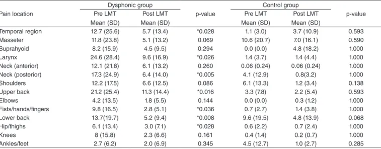
Table 1 shows the results regarding the location and inten-sity of pain reported before and after LMT. In the DG, pain was reduced after LMT in the following regions: temporal, lar-ynx, posterior region of the neck, ists, hands, ingers, upper and lower back, hips, and thighs. In the CG, there was no pain reduction in any body part.
ranged from 50 to 70% for both groups. The intraevaluator agreement was 30% for the vowel “a” and 70% for spontane-ous speech, for both groups.
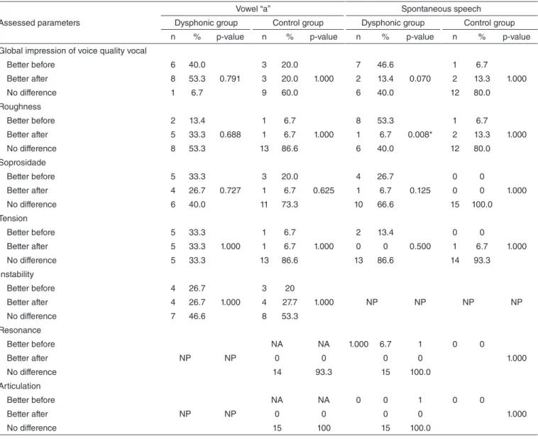
The results presented in Table 2 refer to the similar judg-ment of at least two of the three judges for each parameter analyzed. The perceptual-auditory analysis of the vowel “a” did not present differences after LMT was applied in all of the assessed parameters of both groups. It is important to mention that, even though there are no signiicant differ-ences, 55.6% of the dysphonic individuals presented a “bet-ter” global impression of voice quality after LMT. Regarding spontaneous speech, the perceptual-auditory analysis showed “worse” roughness after LMT in the DG. For the other param-eters, there was no difference after LMT in both analyzed groups. It is important to mention that no differences were observed in DG in more than 70% of the sample after LMT considering the parameters roughness, tension, resonance, and articulation.
Table 3 shows the results of acoustic parameters before and after LMT. The analysis of fundamental frequency (f0) was made separately for men and women. Therefore, in DG, f0 before LMT was 209 Hz for women and 126 Hz for men, and, after LMT, f0 was 213 Hz and 127 Hz, respectively. For the CG, f0 before LMT was 212 Hz for women and 97 Hz for men, and, after LMT, 216 Hz and 100 Hz, respectively. No differences were observed between the moments pre- and postLMT in both groups and genders. As to the other parameters, it was observed that only the CG demonstrated reduced jitter values after LMT.
Table 4 reveals the sensations reported by individuals after LMT. The DG showed positive sensations regarding the larynx and the articulation. The other investigated sensations, despite not presenting signiicant positive perceptions, showed that most individuals reported positive sensations in these param-eters after LMT. In the CG, no signiicant sensations were observed after LMT.
DISCUSSION
This study aimed at approaching dysphonias based on abu-sive vocal behavior, classiied in literature as functional or orga-nofunctoinal dysphonia(1). These types of dysphonia may be
associated with imbalance in cervical and perilaryngeal mus-cles, and speech-language treatment may use manual therapies associated with vocal techniques addressed to voice training, observed in clinical practice. However, this study tried to observe the immediate effects of a manual technique called LMT(11), in
a single therapy session, without association with a vocal train-ing, lasting 20 minutes. LMT is irst described(11) as a massage
in sternocleidomastoid muscles, going to the other muscles in the suprahyoid and laryngeal regions. Despite the different time of application, this study followed these described criteria.
This article observed reduced pain intensity on the temporal regions, larynx, posterior part of the neck, and lower and upper back after LMT in dysphonic individuals (Table 1). Pain can be present in behavioral dysphonia situations(23), once laryngeal
mus-cles in dysphonic people are more rigid(2,11). These results reinforce
the idea that it is possible to bring back balance to muscles that are distal and proximal to the larynx by using LMT, improving blood irrigation in the applied region, making it less resistant.
Regarding the reduced pain intensity on the lower back, hips, and thighs (Table 1), it is possible to relate it to the fact that the person was sitting during LMT, and the posture was corrected by the therapist, at rest, therefore being relaxed. Maybe, this fact is not associated to the dysphonia; however, some authors in the physical therapy ield consider questions related to imbalance in muscle chains(24,25). The authors state(24,25)
that body movement and postural adaptation result from the action of muscle chains constituted by gravitational muscles working in synergy in the same chain. They are characterized as a set of muscles in the same direction, usually polyarticular, with associated biomechanical function. The adequate balance
Table 1. Values, in millimeters, of the intensity of musculoskeletal pain reported by individuals in the dysphonic and in the control groups, before and after Laryngeal Manual Therapy
Wilcoxon Test (*p≤0.05)
Caption: LMT = Laryngeal Manual Therapy; SD = standard deviation Pain location
Dysphonic group
p-value
Control group
p-value Pre LMT
Mean (SD)
Post LMT Mean (SD)
Pre LMT Mean (SD)
Post LMT Mean (SD)
Temporal region 12.7 (25.6) 5.7 (13.4) *0.028 1.1 (3.0) 3.7 (10.9) 0.593
Masseter 11.8 (23.8) 5.1 (13.2) 0.069 10.6 (20.7) 7.0 (16.1) 0.590
Suprahyoid 8.2 (15.9) 4.5 (9.5) 0.294 0.0 (0.0) 4.8 (18.2) 1.000
Larynx 24.6 (28.4) 9.6 (16.9) *0.026 1.4 (3.7) 1.4 (4.4) 1.000
Neck (anterior) 12.1 (21.8) 6.1 (13.2) 0.260 0.06 (0.24) 0.06 (0.24) 1.000
Neck (posterior) 17.3 (24.9) 6.4 (14.0) *0.005 4.1 (12.9) 0.8(3.2) 1.000
Shoulders 12.2 (17.5) 6.6 (12.5) 0.086 6.1 (13.3) 1.2 (3.4) 0.138
Upper back 21.2 (25.4) 11.3 (14.4) *0.016 3.3 (7.8) 2.2 (5.4) 0.593
Elbows 4.2 (13.5) 1.8 (5.5) 0.144 0.0 (0.0) 0.3 (1.2) 1.000
Fists/hands/fingers 9.8 (16.5) 2.8 (5.1) *0.036 0.7 (2.7) 1.4 (3.8) 1.000
Lower back 13.7(19.7) 5.2 (9.4) *0.008 9.6 (19.5) 4.8 (13.9) 0.068
Hip/thighs 6.1 (13.4) 3.0 (7.1) *0.028 0.6 (2.2) 0.7 (2.4) 1.000
Knees 8 (15.8) 2.3 (6.6) 0.161 0.4 (1.4) 0.2 (0.7) 1.000
Table 2. Auditory-perceptual judgment regarding the best vowel emission and spontaneous speech of individuals in the dysphonic and in the control groups, before and after Laryngeal Manual Therapy
Assessed parameters
Vowel “a” Spontaneous speech
Dysphonic group Control group Dysphonic group Control group
n % p-value n % p-value n % p-value n % p-value
Global impression of voice quality vocal
Better before 6 40.0
0.791
3 20.0
1.000
7 46.6
0.070
1 6.7
1.000
Better after 8 53.3 3 20.0 2 13.4 2 13.3
No difference 1 6.7 9 60.0 6 40.0 12 80.0
Roughness
Better before 2 13.4
0.688
1 6.7
1.000
8 53.3
0.008*
1 6.7
1.000
Better after 5 33.3 1 6.7 1 6.7 2 13.3
No difference 8 53.3 13 86.6 6 40.0 12 80.0
Soprosidade
Better before 5 33.3
0.727
3 20.0
0.625
4 26.7
0.125
0 0
1.000
Better after 4 26.7 1 6.7 1 6.7 0 0
No difference 6 40.0 11 73.3 10 66.6 15 100.0
Tension
Better before 5 33.3
1.000
1 6.7
1.000
2 13.4
0.500
0 0
1.000
Better after 5 33.3 1 6.7 0 0 1 6.7
No difference 5 33.3 13 86.6 13 86.6 14 93.3
Instability
Better before 4 26.7
1.000
3 20
1.000 NP NP NP NP
Better after 4 26.7 4 27.7
No difference 7 46.6 8 53.3
Resonance
Better before
NP NP
NA NA 1.000 6.7 1 0 0
1.000
Better after 0 0 0 0
No difference 14 93.3 15 100.0
Articulation
Better before
NP NP
NA NA 0 0 1 0 0
1.000
Better after 0 0 0 0
No difference 15 100 15 100.0
Signals Test (*p<0.05).
Caption: NP = nonassessed parameter
Table 3. Values of acoustic parameters, before and after Laryngeal Manual Therapy, of the individuals in the dysphonic and in the control groups
Acoustic parameters
Dysphonic group
p-value
Control group
p-value Pre LMT
Mean (SD)
Post LMT Mean (SD)
Pre LMT Mean (SD)
Post LMT Mean (SD)
Jitter 1.05 (0.80) 1.12 (0.78) 0.800 1.59 (1.53) 0.77 (0.67) *0.033
Shimmer 3.13 (0.80) 3.17 (1.24) 0.845 3.92 (1.92) 3.06 (1.67) 0.098
NHR 0.12 (0.02) 0.12 (0.02) 0.783 0.12 (0.02) 0.13 (0.02) 0.566
Wilcoxon test(*p≤0.05).
Caption: NHR = noise-to-harmonics ratio; LMT = Laryngeal Manual Therapy; SD = standard deviation
Table 4. Numeric distribution of immediate sensations reported by individuals in the dysphonic and in the control groups, after Laryngeal Manual Therapy
Signals Test *p<0.05. Sensations
Voice Larynx Articulation Breathing
Dysphonic
group Control group
Dysphonic
group Control group
Dysphonic
group Control group
Dysphonic
group Control group
Positive 10 7 10 6 7 2 3 6
No difference 2 8 3 6 8 13 10 4
Negative 3 1 2 3 0 0 2 5
control relects on appropriate muscle synergies and produces effective motor response, which minimize and restore the dis-placement of the center of gravity(24). On the other hand, at the
presence of postural changes, the body reorganizes itself in chains of compensation, searching for an adaptive response. Therefore, when there is imbalance, postural changes are estab-lished, and, in some cases, they lead to pain. So, the LMT may have contributed with the improvement of pain in the lower back, hip and thighs, even if not focused on these regions, pro-viding improvement in the muscle chain. Further interdisciplin-ary studies are required to understand the effect of massage on the head and neck regions and its effects on other parts of the body, which are not in the control and study ield of Speech-Language Pathology and Audiology.
Regarding voice quality, the worsened roughness of dys-phonic individuals (Table 2) may be related to muscle rebalance in the perilaryngeal region achieved after LMT. Dysphonic indi-viduals make compensatory and inadequate muscle adjustments in a scenario of hyperfunctional dysphonia(26).
These adjust-ments along the voice system are made to improve dificulties related to voice production, such as the presence of rough-ness and breathirough-ness, in order to reach better voice quality in a vicious, repetitive, and constant cycle of muscle imbal-ance(11). Probably, after LMT, the voice tension was decreased,
even if not demonstrated by the auditory-perceptual evalua-tion of the judges, which might have led to worse roughness. On the other hand, in a study assessing the effects of one type of LMT in 25 sessions, the authors observed improved voice quality, especially regarding tension(10). In clinical practice,
usually the application of voice techniques indicated to relax laryngeal muscles may lead to worsened voice quality, mak-ing it rougher; however, they provide a softer voice, fulillmak-ing the objective of the exercise. This fact may have happened in this study after LMT.
Regarding the acoustic analysis, LMT only changed the jitter of individuals without vocal complaints (Table 3), which indicates that LMT improved the stability in the frequency of vocal emission. It is worth to mention that, even though the jitter is correlated with auditory-perceptual characteristics of roughness and breathiness(27,28), this study did not show changes
related to these parameters in the sustained vowel after LMT. The authors who assessed the effects of LMT(11) mentioned
there was no improvement in voice quality immediately after LMT, which is different from the indings of this analysis. However, they found improved acoustic parameters of jitter relative average perturbation 1 week after the LMT was con-ducted, which did not happen immediately after the procedure. In studies conducted with other types of laryngeal massage(10,20),
the authors observed differences after the manual therapy, with improved voice quality and better jitter and shimmer parame-ters, which was not found in this study. On the other hand, in a study assessing the effects of 12 sessions of LMT in dysphonic women(19), no signiicant changes were observed in the
acous-tic parameters, and reported that the 20-minute LMT may have contributed with less favorable results(19). The same may
have occurred in this article, because most parameters did not show differences after 20 minutes of LMT.
One of the limitations in this study is that no evaluations were made after a period of time, for instance, one week, which would allow comparisons with other studies in literature with this control(11).
The immediate sensations regarding voice, larynx, articu-lation, and breathing show that, in the dysphonic group, indi-viduals were able to see signiicant improvement, especially in the larynx and articulation, which did not happen in the con-trol group (Table 4). Sensations such as “it is easier to speak”, “clearer speech”, “lighter throat”, “more relaxed”, “more loosen”, and “softer throat” show that LMT causes laryngeal comfort and relaxation, thus improving the speech articulation. Reports regarding voice, even if without statistical signiicance, were “cleaner voice”, “less rough”, and “softer voice.” Some nega-tive reports were also observed regarding voice, larynx, and breathing, both in the DG and CG: “rougher voice”, “burning throat”, and “loss of breath”, indicating that not all individu-als beneit from this technique, as expected. Mathieson et al.(11)
also observed improvement in symptoms such as “dry throat”, “itch”, “pain”, “tightness”, and “tension in the throat” after one week of LMT; however, they reported a tendency to the recur-rence of “tightness in the throat.”
There is evidence that manual therapy, in its various forms, may be useful in a primary intervention, in cases when muscle tension is present in dysphonias, even though this statement is based on a few studies(10,11,18,19). Therefore, more controlled,
randomized, and blind studies are required to understand LMT better and to investigate the role of this kind of treatment associ-ated with other interventions, in individuals with different types of dysphonia. This is necessary because the application of mas-sages on the head and neck is frequent in voice clinic, however, little is known about their immediate and long-term effects.
CONCLUSION
LMT could decrease the intensity of musculoskeletal pain in the following regions: temporal, larynx, posterior part of the neck, ists/hands/ingers, lower back, and hips/thighs in dys-phonic individuals, which did not occur for individuals with-out vocal changes.
As to voice quality after LMT, the roughness parameter became worse in the dysphonic group. Besides, positive sen-sations were reported in the larynx and in the articulation by dysphonic individuals after LMT.
ACKNOWLEDGMENTS
To Fundação de Amparo à Pesquisa do Estado de São Paulo – FAPESP, process n. 2012/02901-2.
REFERENCES
1. Behlau M, Madazio G, Pontes P. Disfonias organofuncionais. In: Azevedo R, Pontes PAL. Voz: o livro do especialista. Rio de Janeiro: Revinter. 2001; p. 296. 2. Menoncin LCM, Jurkiewicz AL, Silvério KCA, Camargo PM, Wolff NMM.
Alterações musculares e esqueléticas cervicais em mulheres disfônicas. Intl Arch Otorhinolaryngol. 2010; 14(4): 461-66.
3. Bigaton DR, Silvério KCA, Berni KCS, Distefano G, Forti F, Guirro RRJ. Postura craniocervical em mulheres disfônicas. Rev Soc Bras Fonoaudiol. 2010;15(3):329-34.
4. Angsuwarangsee T, Morrison M. Extrinsic laryngeal muscular tension in patients with voice disorders. J Voice. 2002;16(3):333-43.
5. Hsiung MW, Hsiao YC. The characteristic features of muscle tension dysphonia before and after surgery in benign lesions of the vocal fold. ORL J Otolaryngol Relat Spec. 2004;66(5):246-54.
6. Kooijman PG, de Jong FI, Oudes MJ, Huinck W, van Acht H, Graamans K. Muscular tension and body posture in relation to voice handicap and voice quality in teachers with persistent voice complaints. Folia Phoniatr Logop. 2005;57(3):134-47.
7. Behlau M, Gama ACC, Cielo CA. Técnicas Vocais. In: Marchesam IQ, Silva HJ, Tomé MC. Tratado das Especialidades em Fonoaudiologia. São Paulo: Guanabara-Koogan; 2014. p. 127-152.
8. Roy N, Bless DM, Heisey D, Ford CN. Manual circumlaryngeal therapy for functional dysphonia: an evaluation of short- and long-term treatment outcomes. J Voice. 1997;11(3):321-31.
9. Ternström S, Andersson M, Bergman U. An effect of body massage on voice loudness and phonation frequency in reading. Logoped Phoniatr Vocol. 2000; 25:146-50.
10. Van Lierde KM, De Ley S, Clement G, Bodt De, Van Cauwenberge P. Outcome of laryngeal manual therapy in four Dutch adults with persistent moderate-to-severe vocal hyperfunction: a pilot study. J Voice. 2004;18(4):467-74.
11. Mathieson L, Hirani SP, Epstein R, Baken RJ, Wood G, Rubin JS. Laryngeal manual therapy: a preliminary study to examine its treatment effects in the management of muscle tension dysphonia. J Voice. 2009;23(3):353-66. 12. Marszalek S, Niebudek-Bogusz W, Zebryk-Stopa A, Woz´nicka E,
Malin´ska J, Golusin´ski W et al. Assessment of the inluence of osteopathic myofascial techniques on normalization of the vocal tract functions in patients with occupational dysphonia. Int J Occup Med Environ Health. 2012;25(3):225-35.
13. Marszalek S, Zebryk-Stopa A, Wojnowki W, Wiskirska-Woznica B, Golusin´ski W. The application of the manual physiotherapy treatment for patient with after trauma dysphonia. Otolaryngol Pol. 2011;65(4):285-8.
14. Schneider SL, Sataloff RT. Voice therapy for the professional voice. Otolaryngol Clin North Am. 2007;40(5):1133-49.
15. Rubin JS, Blake E, Mathieson L. Musculoskeletal patterns in patients with voice disorders. J Voice. 2007;21(4):477-84.
16. Roy N, Nissen SL, Dromey C, Sapir S. Articulatory changes in muscle tension dysphonia: evidence of vowel space expansion following manual circumlaryngeal therapy. J Commun Disord. 2009;42(2):124-35. 17. Van Lierde KM, De Bodt M, Dhaeseleer E, Wuyts F, Claeys S. The
treatment of muscle tension dysphonia: a comparison of two treatment techniques by means of an objective multiparameter approach. J Voice. 2010;24(3):294-301.
18. Mathieson L. The evidence for laryngeal manual therapies in the treatment of muscle tension dysphonia. Curr Opin Otolaryngol Head Neck Surg. 2011;19(3):171-6.
19. Silverio KCA, Brasolotto AG, Siqueira LTD, Carneiro CG, Fukushiro AP, Guirro RRJ. Effect of application of transcutaneous electrical nerve stimulation and laryngeal manual therapy in dysphonic women: clinical trial. J Voice. 2015;29(2):200-8.
20. Aronson AE. Clinical voice disorders: an interdisciplinary approach. 3rd ed. New York, NY: Thieme; 1990.
21. Shipp T. Vertical laryngeal position: research indings and applications for singers. J Voice. 1987;1(3):217-19.
22. Sonninen A. The external frame function in the control of pitch in the human voice. Ann N Y Acad Sci. 1968;155(1):68-90
23. Silverio, KCA, Siqueira, LTD, Lauris, JRP, Brasolotto AG. Dor musculoesquelética em mulheres disfônicas. CoDAS. 2014;26(5):374-81. 24. Yoshitomi, SK, Tanaka C, Duarte M, Lima F, Morya E, Hazime F. Respostas posturais à perturbação externa inesperada em judocas de diferentes níveis de habilidade. Rev Brasil de Medicina do Esporte. 2006;12(3):159-63. 25. Rosário JLP, Sousa A, Cabral CMN, João SMA, Marques AP. Reeducação
postural global e alongamento estático segmentar na melhora da lexibilidade, força muscular e amplitude de movimento: um estudo comparativo. Fisioterapia e Pesquisa. 2008;15(1):12-8.
26. Yamasaki R, Behlau M, do Brasil Ode O, Yamashita H. MRI anatomical and morphological differences in the vocal tract between dysphonic and normal adult women. J Voice. 2011;25(6):743-50.
27. Dejonckere P, Remacle M, Fresnel-Elbaz E, Woisard V, Crevier-Buchman L, Milliet B. Differentiated perceptual evaluation of pathological voice quality: reliability and correlations with acoustic measurements. Rev Laryngol Oto Rhinol (Bord). 1996;117(3):219-24.