Physiopathology and clinical management of one-lung
ventilation*
HALINA CIDRINI FERREIRA, WALTER ARAÚJO ZIN, PATRÍCIA RIEKEN MACEDO ROCCO
J Bras Pneumol 2004; 30(5) 566-73.
*Study carried out in the Laboratory of Respiratory Physiology and the Laboratory of Pulmonary Investigation, Carlos Chagas Filho Institute of Biophysics, Rio de Janeiro Federal University. Sponsors: Programa de Núcleos de Excelência – Ministério de Ciência e Tecnologia (PRONEX-MCT, Program for the Development of Centers of Excellence – Ministry of Science and Technology), Conselho Nacional de Desenvolvimento Científico e Tecnológico (CNPq, Brazilian Council for Scientific and Technological Development), Fundação Carlos Chagas Filho de Amparo à Pesquisa do Estado do Rio de Janeiro (FAPERJ, Carlos Chagas Filho Foundation for the Support of Research in the state of Rio de Janeiro).
Correspondence to: Patricia Rieken Macêdo Rocco, MD, Ph.D. Universidade Federal do Rio de Janeiro. Instituto de Biofísica Carlos Chagas Filho. Centro de Ciências da Saúde. CEP 21949-900 – Rio de Janeiro – RJ – Brasil.
Phone number: 55 21 2562 6557, Fax number: 55 21 2280 8193 E-mail: [email protected]
Submitted: 14 January 2004. Accepted, after review: 25 June 2004.
Key words: tidal volume, hypoxemia, ventilatory management, mechanical ventilation
DEFINITION
One-lung ventilation consists of mechanical ventilation of the selected lung and exposure or intentional airway blocking of the other. This technique facilitates viewing of intrathoracic structures, thereby providing optimal surgical conditions, since adequate pulmonary exposure facilitates resection and reduces surgical time(1,2).
HISTORY
Selective intubation was described for the first time in 1932 by Gale and Waters, who aimed to open the thorax and surgically manipulate the lungs. The authors used a single-light tube that was inserted into the right or left mainstem bronchus(3). Since then, various alternative methods have been proposed in order to make this technique safer and facilitate its practice.
The lung separation techniques previously described involve the use of bronchial tubes, bronchial blockers, or double-lumen tubes (the devices most commonly used in current surgical practice)(4).
INDICATIONS FOR USE
Situations in which one-lung ventilation are indicated may be divided into two groups: indications and relative indications. Indications include hemothorax, massive hemorrhage, unilateral cysts, bronchopleural fistulae and unilateral pulmonary diseases. Relative indications i n c l u d e p n e u m o n e c t o m i e s , l o b e c t o m i e s , esophageal resection and thoracoscopies(5).
COMPLICATIONS
The principal complications that arise from one-lung ventilation are hypoxemia, hemorrhage, hemodynamic instability, bronchial rupture caused by excessive inflation of the balloon on the tip of the double-lumen tube, and alveolar lesions caused by the use of fractions of inspired oxygen (FiO2) of 1.0(1).
PHYSIOLOGICAL REPERCUSSIONS OF
ONE-LUNG VENTILATION
Atelectasis of the nonventilated lung
Alveolar collapse results from the action of opposite forces on the pulmonary parenchyma. These include elastic recoil pressure and surface
forces of the alveolar air-liquid interface versus positive transpulmonary pressures. Positive basal alveolar pressure is fundamental to alveolar stabilization. When there is total collapse, as is the case for the nonventilated lung during one-lung ventilation, reversal of this state is not easily achieved and requires higher pressures to produce alveolar reopening. The term “time constant” cannot be applied to units that are in a state of collapse since these units are totally excluded from the process of distribution of the inspired air. This means that the air diffuses through an area that is less compliant, as compared with the lung as a whole.
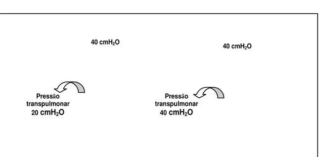
Egan et al. demonstrated that the size of a pulmonary lobe may increase from three to four times, thereby maintaining the same inspiratory pressure(6). When atelectasis of various pulmonary lobes is induced, the remaining lobe absorbs all the stress imposed by the high airway pressure. This is due to the fact that the resulting pleural pressure does not rise significantly. Since the other lobes cease to contribute to the increase in gas volume, the pleural pressures around the remaining lobe remain practically unaltered, thereby generating a high transpulmonary pressure over this lobe alone(6) (Figure 1).
Blood flow distribution during one-lung ventilation
lung and thereby prevent the drop in PaO2. Passive mechanisms include gravity and aggressive surgical compression (direct compression of the pulmonary vessels)(7). Gravity is the determining factor in the regional distribution of the blood flow. This distribution depends on airway pressures and the local relationship between arterial and venous pressures. Furthermore, gravity creates a vertical gradient in the distribution of the flow. Therefore, the blood flow in the nondependent lung is lower than that in the dependent lung (8).
The main active mechanism is hypoxic pulmonary vasoconstriction (HPV). The normal response of the pulmonary vasculature to atelectasis is that of increasing pulmonary vascular resistance, which diverts the blood flow from the nondependent to the dependent lung, thereby minimizing the shunt. Subsequently, the HPV mechanism, occurring predominantly in arterioles and inferior pulmonary veins, has a protective effect. The main stimulus to HPV is the decrease in PaO2 and in venous oxygen tension (PvO2).
W h e n P v O2 i s n o r m a l , t h e r e i s m a x i m u m vasoconstriction response, which diminishes when PvO2 is increased or reduced. Cardiac output may also influence HPV, since changes in the former cause alterations in lung vascular pressures(9). It is of note that reduced FiO2 in the dependent lung increases the vascular resistance of this lung, thereby attenuating the diversion of the blood flow coming from the nondependent lung(10,11).
Several factors, such as anesthetic agents(12), vasodilators(13), arterial carbon dioxide tension(9), m a n i p u l a t i o n o f t h e l u n g(7) a n d e p i d u r a l anesthesia(14), can affect HPV. These factors may either minimize or maximize its effects.
Arterial oxygenation
Hypoxemia is a complication that affects from 9% to 27% of patients undergoing one- lung ventilation and is influenced by several factors. Initially, soon after the beginning of one-lung ventilation, the blood flow in the nonventilated lung becomes deoxygenated. This reduces the
Figure 1 – Simplified diagram showing that, when the entire left lung is insufflated with a pressure of 40 cmH2O, the rib cage absorbs the stress generated by the applied pressure. The resulting transpulmonary pressure is 20 cmH2O. In the right lung, atelectasis was provoked in several pulmonary lobes by progressive bronchial obstructions. The sole remaining lobe absorbs all the stress imposed by the high airway pressure. This occurs because the pleural pressure does not increase significantly. Therefore, the other lobes cease to contribute to the increase in gas volume. This results in high transpulmonary pressure. (Modified from Egan EA. Lung inflation, lung solute permeability, and alveolar edema. J Appl Physiol 1982;53:121-5)
Pressão
transpulmonar 40 cmH2O
Pressão
transpulmonar 20 cmH2O
40 cmH2O
surface area available for gas exchange and consequently decreases PaO2(11).
Patient positioning during thoracic surgery is another significant factor affecting oxygenation. Studies show that oxygenation is more satisfactory when the patient is placed in the lateral decubitus, rather than the dorsal decubitus, position. Watanabe et al. demonstrated that alveolar oxygen tension decreases more rapidly in the dorsal decubitus position than in the lateral decubitus position, thereby leading to more accentuated hypoxemia(15). Therefore, the lateral decubitus position is used in most studies aiming to minimize the effects of patient position on arterial oxygenation(7,15).
EXECUTION OF ONE-LUNG
VENTILATION
Choosing the size of the double-lumen tube and confirming its correct positioning
Currently, the technique most frequently used for separating the two lungs is that which employs the double-lumen tube. The main advantages of this technique are rapidity and ease of use. The double-lumen tube offers, if necessary, the possibility of shifting to traditional bilateral ventilation. In addition, it permits the concomitant use of several techniques to correct any possible hypoxemia (continuous positive airway pressure, insufflation of oxygen, etc)(16).
The size of the tube must be proportional to the weight and height of the patient. In addition, it must permit free passage through the mainstem bronchus, with no resistance. Occlusion of the tube by inflation of the balloon must be taken into account in order to prevent air leaks(17).
Correct positioning of the double-lumen tube is confirmed by fiberoptic bronchoscopy and pulmonary auscultation. The use of the fiberoptic bronchoscope was first described in 1982 by Watson et al.(18) and still seems to best meet the needs of surgeons(17,19).
Improper positioning of the tube causes complications in 20% to 30% of patients undergoing thoracic surgery. Hypoxemia and hypercapnia, as well as elevations in the plateau and apical pressures, are indicative of incorrect positioning of the tube. The three most important studies on the monitoring of airway pressures during one-lung ventilation were carried out in
1995. Cohen fixed 40 cmH2O as the upper pressure limit during one-lung ventilation(20). Slinger fixed 45 cmH2O as the highest apical pressure(21). Ovassapian proposed that the apical pressure not surpass 150% of the basal value of traditional bilateral ventilation(22). In a more recent study conducted by Szedi et al., it was demonstrated that there are quantifiable increases in the plateau and apical pressures, but that these values should not be considered in isolation since they present low sensitivity and poor diagnostic precision(23).
In a retrospective study of 234 one-lung intubations, arterial oxygen saturation of less than 90% was observed in 9% of the patients, apical pressure above 40 cmH2O in another 9%, an intubation error during isolation of one of the lungs in 7%, and airway trapping in 2%. This demonstrates that, although fiberoptic bronchoscopy had initially been performed, thus ensuring the correct positioning of the tube, a shift during surgical manipulation may alter its position, resulting in airway obstruction and increased inspiratory pressure(24).
Tidal volume (VT) similar to that used during traditional mechanical ventilation
During one-lung ventilation, it is recommended that the dependent lung be ventilated with a tidal volume (VT) similar to that used when ventilating both lungs during traditional mechanical ventilation(2,25). High V
Tsare used in order to maintain arterial oxygenation(26). Katz et al. have demonstrated that VT levels of 8 to 15 ml/kg do not significantly affect either the pulmonary shunt or the PaO2(27). In addition, the authors reported that VT levels of less than 8ml/kg resulted in a reduction in the residual functional capacity, thereby causing atelectasis in the dependent lung and consequent gas exchange impairment. It is clear that the choice of the VT is solely based on improving oxygenation. However, little attention is given to the potential deleterious effects of high VTs on patients undergoing one-lung ventilation.
Recent studies have demonstrated that mechanical ventilation alone may initiate or aggravate lung injury(28-30). High V
alveolar-capillary membrane, a functional alteration of the cells, liberation of proinflammatory cytokines, an alteration in the ionic transport and a reduction in the surfactant secretion, constituting pulmonary injury(28-31).
Although most of the patients undergoing one-lung ventilation do not present a previous history of lung disease, the administration of high VTs may lead to lung parenchymal injury(26,32). A reduction in V
T, in combination with the use of positive end-expiratory pressure (PEEP), may minimize these alterations(26,33).
Use of high fractions of inspired oxygen In addition to the use of high VTs, an FiO2 of 1.0 is used in order to maintain satisfactory arterial oxygenation. This concentration of oxygen causes vasodilatation in the dependent lung, thereby increasing its capacity to accommodate the blood flow coming from the nondependent lung. Capan et al. showed that the use of an FiO2 of 1.0 resulted in shunt fractions of 25% to 30% and PaO2 values between 150 mmHg and 210 mmHg during one-lung ventilation(34). More recently, Bardoczky et al. analyzed the effects of different concentrations of oxygen combined with positioning patients in the dorsal decubitus or lateral decubitus position and concluded that all of them presented an increase in the alveolar-arterial oxygen difference, regardless of the FiO2 values used. However, with an FiO2 of 1.0, the reduction in PaO2 was smaller and therefore did not cause hypoxemia. It is noteworthy that the reduction in PaO2 was greater in patients in the dorsal decubitus position than in those in the lateral decubitus position(35).
High FiO2 values, on the other hand, cause several complications, such as absorption atelectasis, as well as alterations in vital capacity, respiratory rate, pH, PaO2 and in the pulmonary diffusing capacity for carbon monoxide. This leads to a later increase in the shunt fraction(36).
Positive end-expiratory pressure
During one-lung ventilation, especially when in the lateral decubitus position, the functional residual capacity of the dependent lung is reduced due to factors related to the induction of general anesthesia, such as compression of the abdominal contents and the mediastinum(2,25).
The application of PEEP to the dependent lung prevents alveolar collapse and increases functional residual capacity, thus improving the
ventilation-perfusion ratio and the compliance of this lung(25). However, PEEP efficacy depends on the levels used. High PEEP levels may have deleterious effects on arterial oxygenation. This is due to the compression of the intra-alveolar vessels resulting from increased lung volume, thereby leading to an increase in pulmonary vascular resistance and a reduction in cardiac output(37).
There is much controversy in the literature over the efficacy of PEEP in controlling PaO2 during one-lung ventilation. Cohen et al. demonstrated that the application of a PEEP level of 10 cmH2O to patients with low PaO2 increased the functional residual capacity to normal values, thereby reducing the pulmonary vascular resistance, improving the ventilation-perfusion ratio and increasing PaO2(38). On the other hand, Capan et al. provided evidence that oxygenation did not improve in the presence of PEEP(34).More recently, Inomata et al. proposed an explanation for the conflicting results regarding the administration of PEEP during one-lung ventilation(39). They ascertained that there was an increase in airway resistance and, as a result, patients presented auto-PEEP. In patients with no previous history of lung disease, arterial oxygenation, shunt fraction and cardiac rate were satisfactory when PEEP levels similar to those of auto-PEEP were used. Therefore, in order to apply an ideal PEEP level, it is recommended that auto-PEEP levels be determined(39).
ANESTHESIA
In vitro studies have shown that volatile anesthetics directly reduce the action of hypoxic vasoconstriction and may theoretically lead to increased perfusion of the nonventilated lung, thus increasing the shunt fraction and, as a result, reducing PaO2(40).
The effects of propofol on PaO2, shunt fraction, pulmonary perfusion and cardiac output have been analyzed in comparison to volatile anesthetics previously studied, and it was demonstrated that PaO2 values, pulmonary perfusion and cardiac output were higher when propofol was used, and that the shunt fraction was significantly lower than t h a t o b s e r v e d w i t h t h e o t h e r v o l a t i l e anesthetics(43,44).
ALTERNATIVE METHODS FOR
MINIMIZING HYPOXEMIA DURING
ONE-LUNG VENTILATION
Continuous positive airway pressure
Continuous positive airway pressure is administered to the nondependent lung in order to maintain airway and alveolar patency, thereby making gas exchange possible, since the alveoli are not in total collapse. The pressure values for continuous positive airway pressure must be adjusted in order to maintain sufficient arterial oxygenation. Generally, the combination of continuous positive airway pressure with PEEP in the dependent lung produces more satisfactory results(45-47).
Nitric oxide
Nitric oxide (NO) is a key factor in endothelium-dependent relaxation. Endothelial NO release is i n d u c e d b y L - a r g i n i n e a n d b y i n c r e a s e d concentrations of calcium in the cytoplasm. It diffuses through the intracellular site of the smooth muscle cells and binds the heme group that is present in the guanylate cyclase. This activation leads to smooth muscle relaxation by the synthesis of the 3’,5’-cyclic guanosine monophosphate, which causes relaxation and vasodilatation. Its half-life ranges from 110 msec to 130 msec(48).
Inhaled NO (5 to 80 ppm) selectively reduces pulmonary vascular resistance, that is, its effects are restricted to the pulmonary circulation, and, since it is deactivated by hemoglobin immediately after entering the circulation, it has no effect on systemic circulation(48). This concept that pulmonary circulation may be modulated by the administration of NO had led to its utilization in attempts to reduce hypoxemia during one-lung ventilation. It is notable that NO inhalation only causes vasodilatation in ventilated areas, not affecting HPV in nonventilated areas. Administration of NO during one-lung
ventilation could theoretically improve oxygenation since it selectively reduces pulmonary vascular resistance and increases the blood flow into the ventilated lung(49).
However, there is controversy over the results obtained by NO use during one-lung ventilation. Booth et al. administered NO (40 ppm) to nine patients, and reported improved oxygenation(50). However, the authors of two other studies found no evidence of improved PaO2 resulting from the use of NO(51,52).
There are several theories that attempt to explain the lack of NO efficacy during one-lung ventilation. One is based on the fact that, in the lateral decubitus position, 75% to 80% of the cardiac output flows through the ventilated lung. This suggests that the pulmonary vessels in the ventilated lung are already dilated sufficiently to accommodate this increased flow, and additional dilation would therefore be impossible(52).
HIGH-FREQUENCY VENTILATION
High-frequency ventilation was initially developed in the 1970s and 1980s with the aim of ventilating patients with acute lung injury. The principal characteristics of high-frequency ventilation are the use of low tidal volumes (approximately 1.0 ml/kg), high respiratory rate, and low airway pressures – analogous to PEEP, although without the need to maintain high volumes in order to adequately eliminate carbon dioxide(53).
As a result of the convection created by low levels and molecular diffusion (reduction in the carbon dioxide gradient from the alveolus to the conducting airways), carbon dioxide elimination during high-frequency ventilation is efficacious and is increased by the turbulence created by the convective flow (Taylor dispersion). These mechanisms make it possible to effectively eliminate carbon dioxide despite the use of tidal volumes lower than that of the pulmonary dead space(53).
with the aim of improving arterial oxygenation. Some studies have demonstrated its efficacy by supporting the hypothesis that PaO2 normalizes, thereby improving surgical success rates(55,56). Dikmen et al. analyzed PaO2, carbon dioxide tension, arterial pressure and cardiac rate in 15 patients undergoing elective thoracic surgery in the lateral decubitus position. High-frequency ventilation was applied to the nondependent lung. The authors concluded that this technique was effective in establishing normal values for the parameters analyzed(55).
ALVEOLAR RECRUITMENT
It is known that general anesthesia causes atelectasis in dependent pulmonary regions(57). Based on that fact, Tusman et al. demonstrated, in 1999, that the alveolar recruitment maneuver improves oxygenation, reverses alveolar collapse and increases compliance during anesthesia(58).
In 2002, the same group analyzed the effects of the alveolar recruitment maneuver during one-lung ventilation. They concluded that the use of this technique in the dependent lung promotes proper oxygenation, since it optimizes the ventilation-perfusion ratio and totally reverses the shunt(59). However, further studies are needed in order to elucidate which mechanisms are involved in alveolar and capillary recruitment during one-lung ventilation.
CONCLUSION
The execution of one-lung ventilation still constitutes a challenge in clinical and surgical practice. Many techniques have been developed w i t h t h e a i m o f m i n i m i z i n g t h e r e l a t e d complications. However, further studies are warranted in order to find the ideal way to employ and monitor this technique.
REFERENCES
1. Plummer S, Hartley M, Vaughan RS. Anaesthesia for telescopic procedures in the thorax. Br J Anaesth 1998;80:223-34.
2. Szegedi LL. Pathophysiology of one-lung ventilation. Anesthesiol Clin North America 2001;19(3):435-53. 3. Gale JW, Waters RM. Closed endobronchial anesthesia
in thoracic surgery: preliminary report. J Thorac Surg 1932;1:432-7.
4. Nazari S, Trazzi R, Moncalvo A, Zonta A, Campani M. Selective bronchial intubation for one lung anaesthesia in thoracic surgery. A new method. Anaesthesia 1986;41:519-26.
5. Ost D. Independent lung ventilation. Clin Chest Med 1996;17:591-601.
6. Egan EA. Lung inflation, lung solute permeability, and alveolar edema. J Appl Physiol 1982;53:121-5. 7. Ishikawa S, Nakazawa K, Makita K. Progressive changes
in arterial oxygenation during one-lung anaesthesia are related to the response to compression of the non-dependent lung. Br J Anaesth 2003;90:21-6. 8. Benumof JL. One-lung ventilation and hypoxic
pulmonary vasoconstriction: implications for anesthetic management. Anesth Analg 1985;64:821-33. 9. B e n u m o f J L , W a h r e n b r o c k E A . B l u n t e d h y p o x i c
pulmonary vasoconstriction by increased lung vascular pressures. J Appl Physiol 1975;38:846-50.
1 0 . Benumof JL. Intermittent hypoxia increases lobar hypoxic pulmonary vasoconstriction. Anesthesiology 1983;58:399-404.
11 . Schwarzkopf K; Schreiber T; Preussler NP, Gaser E, Hüter L, Bauer R, Schubert H, Karzai W. Lung perfusion, shunt fraction, and oxygenation during one lung ventilation in pigs: the effects of desflurane, isoflurane, and propofol. J Cardiothorac Vasc Anesth 2003;17:73-5. 1 2 . Slinger P, Scott WA. Arterial oxygenation during
one-lung ventilation. A comparison of enflurane and isoflurane. Anesthesiology 1995;82:940-6.
1 3 . Fradj K, Samain E, Delefosse D, Farah E, Marty J. Placebo-controlled study of inhaled nitric oxide to treat hypoxaemia during one-lung ventilation. Br J Anaesth 1999; 82:208-12.
14. Ishibe Y, Shiokawa Y, Umeda T, Uno H, Nakamura M, Izumi T. The effect of thoracic epidural anesthesia on hypoxic pulmonary vasoconstriction in dogs: an analysis of the pressure-flow curve. Anesth Analg 1996;82:1049-55. 1 5 . Watanabe S, Noguchi E, Yamada S, Hamada N, Kano T.
Sequential changes of arterial oxygen tension in the supine position during one-lung ventilation. Anesth Analg 2000;90:28-34 .
1 6 . Tobias JD. Variations on one-lung ventilation. J Clin Anesth 2001; 13: 35-39.
1 7 . Slinger P. A view of and through double – lumen tubes. J Cardiothorac Vasc Anesth 2003;17:287-8.
1 8 . Watson CB, Bowe EA, Burk W. One-lung anesthesia for pediatric thoracic surgery: a new use for the fiberoptic bronchoscope. Anesthesiology 1982;56:314-5. 1 9 . Malik S, Shapiro WA, Jablons D, Katz JA. Contralateral
tension pneumothorax during one-lung ventilation for l o b e c t o m y : d i a g n o s i s a i d e d b y f i b e r o p t i c bronchoscopy. Anesth Analg 2002;95:570-2. 2 0 . C o h e n E . A n e s t h e t i c m a n a g e m e n t o f o n e - l u n g
ventilation. In: Cohen E, 1st ed. The practice of thoracic anesthesia. Philadelphia: Lippincott, 1995;308-40. 21. Slinger P. New trends in anesthesia for thoracic surgery
including thoracoscopy. Can J Anaesth 1995;42:77-84. 2 2 . Ovassapian A. Flexible bronchoscopic positioning of right – sided double-lumen endobronchial tubes. J Bronchol 1995;2:12-9.
2 3 . Szegedi LL, Bardoczky GI, Engelman EE, d’Hollander A A . A i r w a y p r e s s u r e c h a n g e s d u r i n g o n e - l u n g ventilation. Anesth Analg 1997;84:1034-7.
25. Campos JH. Effects on oxygenation during selective lobar versus total lung collapse with or without continuous positive airway pressure. Anesth Analg 1997;85:583-6. 26.Gama de Abreu M, Heintz M, Heller A, Széchényi R,
Albrecht DM, Koch T. One Lung ventilation with high tidal volumes and zero positive end-expiratory pressure is injurious in the isolated rabbit lung model. Anesth Analg 2003;96:220-8.
2 7 . Katz JA, Laverne RG, Fairley B, Thomas AN. Pulmonary oxygen exchange during endobronchial anesthesia: effect of tidal volume and PEEP. Anesthesiology 1982;56:164-71.
2 8 . Dreyfuss D, Saumon G. Ventilator-induced lung injury. Lessons from experimental studies. Am J Respir Crit Care Med 1998;157:294-323.
2 9 . Berg JT, Fu Z, Breen EC, Tran HC, Mathieu-Costello O, West J. High lung inflation increases mRNA levels of E C M c o m p o n e n t s a n d g r o w t h f a c t o r s i n l u n g parenchyma. J Appl Physiol 1997;83:120-8.
3 0 . Carlton DP, Cummings JJ, Scheerer RG, Poulain FR, Bland RD. Lung overexpansion increases pulmonary microvascular protein permeability in young lambs. J Appl Physiol 1990;69:577-83.
31. Wilson MR, Choudhury S, Goddard ME, O’Dea K, Nicholson AG, Takata M. High tidal volume upregulates intrapulmonary cytokines in an in vivo mouse of ve n t i l a t o r - i n d u c e d l u n g i n j u r y. J A p p l P h ys i o l 2003;95:1385-93.
3 2 . Zin WA, Ferreira HC, Momesso DP, Boechem NT, Nascimento CS, Prota LFM et al. Which is the best tidal volume in one-lung ventilation [abstract] ? Eur Respir J 2002;20: 284.
33. Frank JA, Gutierrez JA, Jones KD, Allen L, Dobbs L, Matthay MA. Low tidal volume reduces epithelial and endothelial injury in acid-injured rat lungs. Am J Respir Crit Care Med 2002;165:242-9.
34. Capan LM, Turndorf H, Patel C, Ramanathan S, Acinapura A, Chalon J. Optimization of Arterial oxygenation during one-lung anesthesia. 1980;59:847-51.
3 5 . Bardoczky GI, Szegedi LL, d’Hollander AA, Moures JM, de Francquen P, Yernault JC. Two-Lung and one-lung v e n t i l a t i o n i n p a t i e n t s w i t h c h r o n i c o b s t r u c t i v e pulmonary disease: The effects of position and FiO2. Anesth Analg 2000;90:35-41.
3 6 . W i n t e r P M , S m i t h G . T h e t o x i c i t y o f o x y g e n . Anesthesiology 1972;37:210-41.
3 7 . Benumof JL. One-lung ventilation: which lung should be PEEPed? Anesthesiology 1982;56:161-3.
38. Cohen E, Thys DM. PEEP during one-lung anesthesia improves oxygenation in patients with low PaO2. Anesth Analg 1985;64:200.
39. Inomata S, Nishikawa T, Saito S, Kihara S. “Best” PEEP during one-lung ventilation. Br J Anaesth 1997;78:754-6. 4 0 . Marshall C, Lindgren L, Marshall BE: Effects of h a l o t h a n e , e n f l u r a n e a n d i s o f l u r a n e o n h y p o x i c pulmonary vasoconstriction in rat lungs in vitro. Anesthesiology 1993;79:1348-53.
41. Bjernaes LJ. Hypoxia induced pulmonary vasoconstriction in man: inhibition due to diethyl ether and halothane anaesthesia. Acta Anaesthesiol Scan 1978;22:578. 42. Groh J. Effects of isoflurane on regional pulmonary
blood flow during one-lung ventilation. Br J Anaesth 1995; 74: 209-16.
43. Kazuo Abe, Shimizu T. The effects of propofol, isoflurane, and sevoflurane on oxygenation and shunt fraction during one-lung ventilation. Anesth Analg 1998;87:1164-9. 4 4 . Konrad Schwarzkopf. Lung perfusion, shunt fraction,
and oxygenation during one-lung ventilation in pigs: the effects of desflurane, isoflurane, and propofol. J Card Thoracic and Vascular Anesth 2003;17(1):73-5. 4 5 . Hogue CW. Effectiveness of low levels of nonventilated
lung continuous positive airway pressure in improving arterial oxygenation during one-lung ventilation. Anesth Analg 1994;79:364-7.
4 6 . Hughes SA, Benumof J. Operative lung continuous positive airway pressure to minimize FiO2 during one-lung ventilation. Anesth Analg 1990;71:92-5. 47. Campos JH. Effects on oxygenation during selective lobar
versus total lung collapse with or without continuous positive airway pressure. Anesth Analg 1997;85:583-6. 4 8 . Steudel W, Hurford WE, Zapol WM. Inhaled Nitric Oxide:
basic biology and clinical applications. Anesthesiology 1999;91:1090-121.
4 9 . Rich GF, Lowson SM, Johns RA, Daugherty MO, Uncles DR. Inhaled nitric oxide selectively decreases pulmonary vascular resistance without impairing oxygenation during one-lung ventilation in patients undergoing cardiac surgery. Anesthesiology 1994;80:57-62. 5 0 . Booth J. Effect of unilateral inhaled NO during selective
ventilation in anesthetized humans. Anesthesiology 1994;81:1457.
51. Moutafis M, Liu N, Dalibon N, Kuhlman G, Ducros L, Castelain MH, Fischler M. The effects of inhaled nitric oxide and its combination with intravenous almitrine on PaO2 during one-lung ventilation in patients undergoing thoracoscopic procedures. Anesth Analg 1997;85:1130-5.
5 2 . Schwarzkopf K, Klein U, Schreiber T, Preussetaler NP, Bloos F, Helfritsh H, Sauer F, Karzai W. Oxygenation during one lung ventilation: the effect of inhaled nitric oxide and increasing levels of inspired fraction of oxygen. Anesth Analg 2001;92:842-7.
5 3 . McRae K. Anesthesia for airway surgery. Anesthesiol Clin North America 2001;19:497-541.
5 4 . Wood B, Karna P, Adams A. Specific compliance and g a s e x c h a n g e d u r i n g h i g h - f r e q u e n c y o s c i l l a t o r y ventilation. Crit Care Med 2002;30:1523-7.
5 5 . Dikmen Y, Aykac B, Erocay H. Unilateral high frequency jet ventilation during one-lung ventilation. Eur J Anesthesiol 1997;14:239-43.
5 6 . den Hoed PT, Leendertse-Verloop K, Bruining HA, Bonjer HJ. Comparison of one-lung ventilation and high-frequency ventilation in thoracoscopic surgery. Eur J Surg 1999;165:1031-4.
5 7 . Brismar B, Hedenstierna G, Lundquist H, Strandberg A, Svensson L, Tokics L. Pulmonary densities during anesthesia with muscular relaxation: a proposal of atelectasis. Anesthesiology 1985;62:422-8.
5 8 . Tusman G, Böhm SH, Vazquez de Anda GF, do Campo JL, Lachmann B. Alveolar recruitment strategy improves arterial oxygenation during general anaesthesia. Br J Anaesth 1999;82:8-13.