Recebido em 7/12/2010 Aprovado em
9/12/2010
individual analysis using SPECT among an
obsessive-compulsive population
Perfusão cerebral e análise individual automatizada utilizando SPECT
em uma população com transtorno obsessivo-compulsivo
Euclides Timóteo da Rocha
1, Carlos Alberto Buchpiguel
2, Euripedes Constantino Miguel
3,
Stela Verzinhase Peres
4,
Geraldo Busatto Filho
5ABSTRACT
Objective: To make individual assessments using automated quantiication methodology in
order to screen for perfusion abnormalities in cerebral SPECT examinations among a sample of subjects with OCD. Methods: Statistical parametric mapping (SPM) was used to compare 26 brain SPECT images from patients with OCD individually with an image bank of 32 normal subjects, using the statistical threshold of p < 0.05 (corrected for multiple comparisons at the level of individual voxels or clusters). The maps were analyzed, and regions presenting voxels that remained above this threshold were sought. Results: Six patients from a sample of 26 OCD images showed abnormalities at cluster or voxel level, considering the criteria described above, which represented 23.07%. However, seven images from the normal group of 32 were also indicated as cases of perfusional abnormality, representing 21.8% of the sample.
Con-clusion: The automated quantiication method was not considered to be a useful tool for
clinical practice, for analyses complementary to visual inspection.
RESUMO
Objetivo: Avaliar uma amostra de pacientes com transtorno obsessivo-compulsivo (TOC), in-dividualmente, empregando uma metodologia de quantiicação automatizada para rastrear anormalidades de perfusão em exames de SPECT cerebral. Métodos: Foi utilizado o Statistical Parametric Mapping (SPM) para comparar 26 imagens de SPECT cerebral de pacientes com TOC, individualmente, com um banco de 32 imagens de voluntários normais, usando o limiar estatístico de p < 0,05 (corrigido para comparações múltiplas ao nível do voxel individual ou clusters). Os mapas foram analisados procurando por regiões que apresentassem voxels acima desse limiar.
Resultados: Seis pacientes da amostra de 26 imagens com TOC mostraram anormalidades ao nível do cluster ou voxel, considerando os critérios descritos acima, os quais representaram 23,07%. Contudo, sete imagens do grupo de 32 voluntários normais também foram apontadas com anor-malidades de perfusão, que representou 21,8% da amostra.Conclusão: O método de quantiica-ção automatizada não foi considerado como uma ferramenta útil na prática clínica, como forma de análise complementar à inspeção visual.
1 Hospital de Câncer – Fundação Pio XII, Department of Nuclear Medicine, Barretos, SP, Brazil; São Paulo State University (Unesp), Medical School, Blood Transfusion Center, Botucatu, SP, Brazil.
2 University of São Paulo (USP), School of Medicine, Department of Radiology, Nuclear Medicine Service. 3 USP, School of Medicine, Institute of Psychiatry.
4 Hospital de Câncer – Fundação Pio XII, Research Support Group.
5 USP, School of Medicine, Institute of Psychiatry; Department of Radiology, Nuclear Medicine Service.
Endereço para correspondência: Euclides Timóteo da Rocha
Departamento de Medicina Nuclear, Hospital de Câncer – Fundação Pio XII
Av. Antenor Duarte Vilela, 1331, Bairro Dr. Paulo Prata – 14784-400 – Barretos, São Paulo, Brasil E-mail: [email protected]
Keywords
Brain SPECT, SPM, OCD, individual analysis.
Palavras-chave
INTRODUCTION
The neurobiology of obsessive-compulsive disorder (OCD) has been widely investigated over the past decades. Functio-nal neuroimaging studies using positron emission tomogra-phy (PET), single-photon emission computed tomogratomogra-phy (SPECT) and functional magnetic resonance imaging (fMRI) have sought to investigate the pathophysiology of OCD, and these have mainly implicated circuits involving fronto-sub-cortical connections. A series of published papers has indica-ted functional brain abnormalities in group comparisons of OCD patients relative to healthy controls, particularly invol-ving the orbitofrontal cortex, anterior cingulate cortex, basal ganglia and thalamus1-4. Cortical regions such as the orbito-frontal cortex and the cingulate gyrus have been recognized to be heterogeneous among humans, with evidence of the existence of subregions that possibly perform speciic roles in the pathophysiology of OCD5,6.
Despite the great impact that neuroimaging studies have had with regard to elucidating the pathophysiolo-gy of the so-called functional psychiatric disorders such as major depressive disorder, OCD and psychoses, the results from such investigations have generally come from statis-tical comparisons of means from cerebral measurements, between groups of patients and normal controls. On the other hand, when brain functional images of any type are inspected individually, abnormalities are only detected in a certain proportion of patients with psychiatric disorders, and with considerable variability regarding the nature and cerebral location. In this respect, there has been a scarcity of systematic studies which suggest that such neuroimaging examinations could be useful for practical clinical diagnostic purposes in the individual evaluation of subjects with func-tional psychiatric disorders. This is in sharp contrast with the well-established practical applications for functional neuroi-maging methods in the diferential diagnosis of neurological diseases that presumably present well-deined cerebral pa-thological indings7-9.
In recent years, automated image analysis methods have been developed to allow voxelwise quantiications of in-dings in PET, SPECT and MRI studies. Classically, these me-thods have been used to make statistical comparisons of mean signal intensities in each voxel of the cerebral volume between groups of patients and normal controls, or alter-natively, to compare images from the same group under two diferent conditions10,11. The possibility of using SPECT in association with some form of automated quantiication for evaluating individual patients with functional psychiatric disorders is of great interest, but there have been few publi-shed papers using this approach. Nevertheless, automated voxel-based image analysis methods have been applied in case-by-case evaluations for investigating epileptic foci12, for comparative assessments with visual analysis among
pa-tients with Alzheimer’s disease, and even for assessments of normal subjects13,14.
Conventional methods using regions-of-interest to quan-tify brain perfusion abnormalities are not appropriate for stu-dying subregions within functionally heterogeneous brain structures of relevance to the pathophysiology of OCD, such as the orbitofrontal and anterior cingulate cortices. Thus, au-tomated voxel-based quantiication methods may aford a better strategy for assessing subtle and highly circumscribed changes in cerebral SPECT examinations in individual OCD cases. Hence, the aim of the present study was to evalua-te whether the voxel-based statistical parametric mapping (SPM) approach might have a complementary role in investi-gating possible perfusional abnormalities among a group of patients with OCD (n = 26) assessed individually with SPECT. We predicted that the SPM-based approach could enable the detection of subtle brain perfusion abnormalities in a proportion of OCD patients, in a frequency greater than that found in the group of healthy control subjects (n = 32) eva-luated with exactly the same imaging methods.
MATERIAL AND METHODS
A sample of 26 patients (15 males/11 females, 23 right-han-ded) fulilling DMS-IV criteria for OCD15 was selected. Their mean age was 32.1 years (SD 7.8). Eleven of the patients had never previously undergone any kind of treatment. Four pa-tients agreed to remain free from medication for three we-eks in order to undergo washout from previous treatments. The remaining patients had taken medications in the past, but had been free from all OCD treatments over the four--year period preceding the start of this study. The patients were recruited at the Institute of Psychiatry of Hospital das Clínicas, School of Medicine of the University of São Paulo (IPq-HC-FMUSP). All of the patients underwent a structured clinical interview in accordance with DSM-IV- Patient Edition (SCID-I/P)16. This group of OCD patients was recruited for a controlled study on cerebral SPECT and structural MRI17. The intensity of obsessive-compulsive symptoms was measured moments before performing the cerebral SPECT examina-tion, with the Yale-Brown-Obsessive-Compulsive Scale18 (mean = 26.8; SD = 5.9).
abnormalities as assessed by structural magnetic resonance scans, visually inspected by two radiologists independently from each other. The study received institutional review bo-ard approval.
Protocol for acquisition and processing of cerebral SPECT images
The protocols used for acquisition and processing of cere-bral SPECT data were the same for all subjects in the above groups. After venous puncture in a supericial vein of the arm, the individuals remained at rest for 20 minutes and then received a dose of 740 MBq of 99mTc-ECD. After a further 20 minutes, image acquisition was started. All the examina-tions were performed using a device with two detectors and high-resolution collimators (OPTIMA NX, General Electric, Milwaukee, USA). The images were acquired using the step--and-shoot method. A matrix of 128 X 128 was used, and 128 projections were acquired (20 seconds per projection).
The orbital-meatal line was used for reconstructing the SPECT images, which was done by using the iltered back--projection method with attenuation correction in accor-dance with Chang’s algorithm (µ = 0.12 cm-1). All the images were reconstructed with pixels of 2.25 mm and a tenth-order Butterworth ilter with Nyquist frequency of 0.57. Transverse sections through the reconstructed images were selected for the subsequent stages of the analysis.
Generation of individualized statistical parametric maps showing rCBF abnormalities Individualized statistical parametric maps for each subject were produced using the SPM program, version 1999, exe-cuted in MATLAB 4.2. Firstly, SPECT images of all patients with OCD and controls were spatially normalized, with li-near 12-point aine transformations, to an anatomical tem-plate provided by the SPM program, which approximates the stereotaxic space of the Talairach-Tournoux atlas20. Subsequently, images were re-sliced using bi-linear inter-polation to a inal voxel size of 2x2x2 mm3, and smoothed with an isotropic Gaussian ilter (12 mm full-width at half maximum) in order to improve the signal-to-noise ratio and to reduce errors attributed to inter-individual variations in gyral and sulcal anatomy. For each of the subjects with obsessive-compulsive disorder (n = 26), a voxel-by-voxel t--test map was obtained by comparison against the pool of controls (n = 32). With the purpose of accounting for inter--individual diferences in overall cerebral blood low, the regional 99mTc-ECD uptake was standardized to the mean overall uptake using proportional scaling. The measure of total brain radioactive uptake was obtained automatically by the SPM program, given by the mean counts of all voxels included in the SPECT volume of each subject, after the spa-tial transformations described above. In order to reduce the number of statistical comparisons, only voxels with signal
intensities above 50% of the mean overall value were ente-red in each analysis. The individualized SPM{t} maps for each subject were transformed to the unit’s normal distribution (Z scores), thresholded at 3.09 (corresponding to ρ < 0.001, uncorrected for multiple comparisons), and displayed as statistical parametric maps into standard space. The same procedure was carried out to provide individualized statis-tical parametric maps of rCBF abnormalities for the control subjects, by comparing each control individual against the remaining thirty-one subjects.
Clusters of rCBF abnormalities in the individualized maps were then examined in terms of size (k) and peak height (u), and were considered signiicant only if retaining statistical signiicance after correction for multiple comparisons based on Gaussian random ield theory (p < 0.05), either at the level of individual voxel height or at the level of clusters21.
RESULTS
Table 1 shows the results from the SPM maps relating to the individual comparisons of each of the 26 patients with OCD against the pool of healthy controls, with voxels that were screened at the signiicance threshold of p < 0.05, corrected for multiple comparisons. Signiicant abnormalities due to hypoperfusion were found in one patient at voxel level and in another subject at the cluster level; with regard to hyper-perfusion, there were three patients with signiicant indings at the voxel level and one subject at cluster level (Figure 1). Thus among the 26 images analyzed, only six cases showed abnormalities, i.e. 23.07% of the full sample. Such abnormali-ties were considerably variable in their location, involving the frontal, temporal and occipital cortices (Table 1).
Among the 32 images from healthy controls analyzed for hypoperfusion, two abnormal examinations were found at voxel level and two at cluster level, of which one presented signiicance at both levels, i.e. three healthy subjects with ab-normalities (Table 2). The abab-normalities were located in the right occipital cortex and right inferior frontal cortex (Figure 2). In evaluating the presence of hyperperfusion, two examina-tions with voxels above the threshold were found at voxel level, and four at cluster level. However, one examination at voxel level and another at cluster level had already been described with hypoperfusion contrast. Thus, four subjects were deemed to present perfusion abnormalities, thus total seven healthy subjects (21.8%) with abnormal examinations considering both contrasts.
Table 1. rCBF indings of hypoperfusion and hyperperfusion in OCD group evaluated using SPM
Age Hypoperfusion Hyperperfusion
Z max ρ voxel level ρ cluster level No. of voxels Location Z max ρ voxel level ρ cluster level No. of voxels Location
32 Zero Vx 4.86 0.02 0.09 199 Left
posterior occipital
34 3.71 0.714 0.940 18 5.03 0.009 0.000 1,813 Right
superior temporal
28 Zero Vx 4.36 0.131 0.004 467 Left
cerebellum
24 4.47 0.08 0.008 409 Right
inferior frontal
3.66 0.762 0.792 40
32 Zero Vx 4.61 0.05 0.01 343 Left
temporal
38 5.62 0.001 0.028 300 Left medial frontal
4.50 0.07 0.207 144 Left
cerebellum
Z max: Z score attained; p voxel level: signiicance level at voxel level; p cluster level: signiicance level at cluster level.
Table 2. rCBF indings of hypoperfusion and hyperperfusion in normal volunteers evaluated using SPM
age Hypoperfusion Hyperperfusion
Z max ρ voxel level ρ cluster level No. of voxels Location Z max ρ voxel level ρ cluster level No. of voxels Location
26 4.80 0.02 0.001 615 Right
Occipital
Zero Vx
38 3.61 0.813 0.815 37 4.15 0.05 0.001 665 Left inferior
frontal
41 4.12 0.28 0.01 348 Left
ventricle
Zero Vx
47 5.02 0.01 0.000 791 Right
inferior frontal
5.03 0.004 0.029 284 Anterior cingulate
22 Zero Vx 4.21 0.219 0.05 249 Left medial
frontal
20 3.71 0.723 0.617 62 4.39 0.124 0.029 290 Right
superior frontal
32 3.83 0.574 0.04 272 Left
ventricle
3.91 0.493
Z max: Z score attained; p voxel level: signiicance level at voxel level; p cluster level: signiicance level at cluster level.
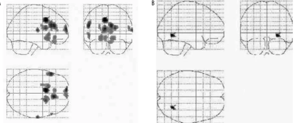
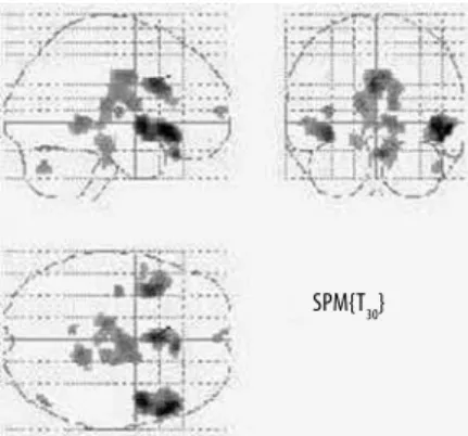
Figure 1. (A-B) Individual SPM maps of 38(A) and 55(B) years OCD patients. Y-BOCS score were 26 and 24, respectively. SPM maps (A) showed hypoperfusion in the inferior frontal cortex and anterior cingulate gyrus which were signiicant after correction for multiple comparison with p < 0.02 and 0.01; SPM map in (B) shows a small group of voxels, not signiicant.
DISCUSSION
A group of patients with OCD was evaluated with the aim of measuring the eiciency of automated, SPM-based quan-tiications for detecting brain perfusional abnormalities in-dividually, as assessed with SPECT22. Even considering the examinations with abnormalities that reached statistical sig-niicance either at the voxel level or at the cluster level, the number of patients whose abnormalities were mapped out and correlated with the diagnosis of OCD was very small. Mo-reover, contrary to our prediction, similar numbers of abnor-mal results were found in both the OCD and control groups. These results indicate that the evaluation of threshold per-fusion changes using an automated voxelwise analysis me-thod is not clinically useful and does not add anything to the routine clinical evaluation.
Despite this, it’s important to note that OCD patients showed a wide spectrum of severity of symptoms with Y-BOCS mean 26,77 ± 5,89, ranging 16-36. The majority (53%) of patients diagnosed with OCD were categorized as seri-ous, while a smaller portion was identiied as moderate or extreme as to severity of symptoms. In this sample eight pa-tients met DMS-IV criteria for major depressive episode and 10 met criteria for other anxiety disorders such as phobias and generalized anxiety disorder.
Moreover, all the healthy subjects recruited to the con-trol group had undergone an assessment, which included a general medical anamnesis and the structured clinical inter-view for the DSM-IV diagnosis of psychiatric disorders. They also underwent structural magnetic resonance scan, that vi-sually did not show any abnormality.
At this point it’s important to take into account that single subject analysis is a very interesting tool for clinical practice and it has been successfully applied to neurological disorders characterized by precise regional deicit in brain function14,20. On the contrary, OCD is a really heterogeneous psychiatric disease where functional abnormalities are dii-cult to detect in resting condition and particularly at single subject level21.
Based on these results, we concluded that SPM-based analyses of individual cases could be a valuable complemen-tary tool within routine clinical practice for the assessment of brain abnormalities in SPECT datasets of patients with neu-rological conditions. Our present results, using exactly the same methods, indicate that the same clinical applicability cannot be reached for the diagnostic evaluation of patients with OCD.
However, caution is warranted in the evaluation of the present negative results for a number of reasons. Firstly, the low anatomical resolution of SPECT has to be born in mind, along with inter-individual diferences in anatomy and meta-bolism and mood variations at the time of administering the radiopharmaceutical, thus leading to increased variability of brain perfusion patterns in both groups. Also, although the diagnoses of OCD were based on widely validate diagnostic criteria (DSM-IV), OCD is characterized by heterogeneity in its clinical presentation, and this may also add variability to rCBF patterns as assessed by SPECT.
Finally, it should be noted that the construction of in-dividual statistical maps using SPM is not the only metho-dological strategy for using automated methods to analyze cerebral SPECT data in clinical practice. For instance, alter-native analysis methods have been developed to support the diagnosis of neuropsychiatric conditions, enabling fully automated categorization of individual structural or func-tional brain images based on machine-learning techniques, such as support vector machines (SVMs)23,24. Recent studies have demonstrated the reliability and validity of SVM-based techniques, as well as their good diagnostic performance in discriminating, for instance, between Alzheimer’s disease pa-tients and healthy control individuals24. Therefore, replication of the current negative results are warranted using SVM-ba-sed methods, with the aim of studying whether these more sophisticated forms of analysis might lead to similar results or, instead, show greater promise for clinical applications for automated evaluations of rCBF SPECT data from OCD sub-jects. For the time being, we conclude that automated, SPM--based voxelwise analysis methods have very limited
appli-Table 3. Severity of symptoms in OCD patients
Age Comp. Obs. Y-BOCS
32 13 16 29
34 12 15 27
28 17 17 34
24 15 16 31
32 12 13 25
38 15 11 26
Comp.: compulsion; Obs.: obsession;
Y-BOCS: Yale-Brown Obsessive Compulsive Scale.
Figure 2. Individual SPM maps of 47 years old healthy volunteer. SPM map shows hypoperfusion in the inferior frontal cortex signiicant after correction for multiple comparison with p < 0.01.
cability in clinical practice to aid in the diagnostic evaluation of OCD patients.
REFERENCES
1. Jang JH, Kim JH, Jung WH, Choi JS, Jung MH, Lee JM, et al. Functional connectivity in fronto-subcortical circuitry during the resting state in obsessive-compulsive disorder. Neurosci Lett. 2010;474:158-62.
2. Schilman EA, Klavir O, Winter C, Sohr R, Joel D. The role of the striatum in compulsive behavior in intact and orbitofrontal-cortex-lesioned rats: possible involvement of the se-rotonergic system. Neuropsychopharmacology. 2010;35:1026-39.
3. Pena-Garijo J, Ruipérez-Rodríguez MA, Barros-Loscertales A. The neurobiology of obses-sive-compulsive disorder: new indings from functional magnetic resonance imaging (I). Rev Neurol. 2010;50:477-85.
4. Black DW, Shaw M, Blum N. Pathological gambling and compulsive buying: do they fall within an obsessive-compulsive spectrum? Dialogues Clin Neurosci. 2010;12:175-85.
5. Rotge JY, Langbour N, Jaafari N, Guehl D, Bioulac B, Aouizerate B, et al. Anatomical al-terations and symptom-related functional activity in obsessive-compulsive disorder are correlated in the lateral orbitofrontal cortex. Biol Psychiatry. 2010;67:37-8.
6. DeLong M, Wichmann T. Changing views of basal ganglia circuits and circuit disorders. Clin EEG Neurosci. 2010;41:61-7.
7. Aso K, Ogasawara K, Sasaki M, Kobayashi M, Suga Y, Chida K, et al. Preoperative cerebro-vascular reactivity to acetazolamide measured by brain perfusion SPECT predicts develop-ment of cerebral ischemic lesions caused by microemboli during carotid endarterectomy. Eur J Nucl Med Mol Imaging. 2009;36:294-301.
8. Ayalon L, Peterson S. Functional central nervous system imaging in the investigation of obstructive sleep apnea. Curr Opin Pulm Med. 2007;13:479-83.
9. Maehara T. Neuroimaging of epilepsy. Neuropathology. 2007;27(6):585-93.
10. Friston KJ, Frith CD, Liddle PF, Frackowiak RS. Comparing functional (PET) images: the as-sessment of signiicant change. J Cereb Blood Flow Metab. 1991;11:690-9.
11. Friston KJ, Poline JB, Holmes AP, Price CJ, Frith CD. Detecting activations in PET and FMRI: levels of inference and power. Neuroimage, 1996;4:223-35.
12. Lee JD, Kim HJ, Lee BI, Kim OJ, Jeon, TJ, Kim MJ. Evaluation of ictal brain SPECT using sta-tistical parametric mapping in temporal lobe epilepsy. Eur J Nucl Med. 2000;27:1658-65.
13. Imran MB, Kawashima R, Sato K, Kinomura S, Ono S, Qureshy A, et al. Detection of CBF deicit in neuropsychiatric by an expert system: a 99mTc-HMPAO brain SPECT study using automated image registration. Nucl Med Commun. 1999;20:25-32.
14. Rocha ET, Buchpiguel CA, Nitrini R, Tazima S, Peres SV, Busatto Filho G. Diagnosis of re-gional cerebral blood low abnormalities using spect: agreement between individualized statistical parametric maps and visual inspection by nuclear medicine physicians with diferent levels of expertise in nuclear neurology. Clinics. 2009;64:1145-53.
15. American Psychiatric Association, 1994. DSM-IV: Diagnostic and Statistical Manual of Mental Disorders, 4th ed. American Psychiatric Press, Washington, DC.
16. First MB, Spitzer RL, Willians JBW, Gibbons M. Structures Clinical Interview for DSM-IV – Patients Edition (SCIDP). Washington, DC: American Psychiatric Press; 1995.
17. Busatto GF, Zamignani DR, Buchpiguel CA, Garrido GE, Glabus MF, Rocha ET, et al. A voxel--based investigation of regional cerebral blood low abnormalities in obsessive-compulsi-ve disorder using single photon emission computed tomography (SPECT). Psychiatry Res. 2000;99:15-27.
18. Goodman WK, Price LH, Rasmussen SA Mazure C, Fleischmann RL, Hill CL, et al. The Yale--Brown Obsessive-Compulsive scale I: development, use and reliability. Arch Gen Psychia-try. 1989;46:1006-11.
19. Talairach J, Tournoux P. Coplanar stereotaxic atlas of the human brain. New Y, NY: Thieme Medical; 1998.
20. Nishimiya M, Matsuda H, Imabayashi E, Kuji I, Sato N. Comparison of SPM and NEUROSTAT in voxelwise statistical analysis of brain SPECT and MRI at the early stage of Alzheimer’s disease. Ann Nucl Med. 2008;22:921-7.
21. Dougall N, Nobili F, Ebmeier KP. Predicting the accuracy of a diagnosis of Alzheimer’s di-sease with 99mTc HMPAO single photon emission computed tomography. Psychiatry Res. 2004;131:157-68.
22. Signorini M, Paulesu E, Friston K, Perani D, Colleluori A, Lucignani G, et al. Rapid asses-sment of regional cerebral metabolic abnormalities in single subjects with quantitative and non-quantitative 18F-FDG PET: a clinical validation of statistical parametric mapping. Neuroimage. 1999;9:63-80.
23. Kalatzis I, Pappas D, Piliouras N, Cayouras D. Support vector machines based analysis of brain SPECT images for determining cerebral abnormalities in asymptomatic diabetic pa-tients. Med Inform Internet Med. 2003;28:221-30.