Nicotine Inhibits Cisplatin-Induced Apoptosis
via Regulating
α
5-nAChR/AKT Signaling in
Human Gastric Cancer Cells
Yanfei Jia1☯, Haiji Sun2☯, Hongqiao Wu1, Huilin Zhang1, Xiuping Zhang1, Dongjie Xiao1, Xiaoli Ma1*, Yunshan Wang1*
1Central Laboratory, Jinan Central Hospital Affiliated to Shandong University, Jinan, China,2College of Life Science, Shandong Normal University, Jinan, China
☯These authors contributed equally to this work.
*mxl7125@126.com(XM);w_yunshan@126.com(YSW)
Abstract
Gastric cancer incidence demonstrates a strong etiologic association with smoking. Nico-tine, the major component in tobacco, is a survival agonist that inhibits apoptosis induced by certain chemotherapeutic agents, but the precise mechanisms involved remain largely unknown. Recently studies have indicated thatα5-nicotinic acetylcholine receptor (α 5-nAChR) is highly associated with lung cancer risk and nicotine dependence. Nevertheless, no information has been available about whether nicotine also affects proliferation of human gastric cancer cells through regulation ofα5-nAChR. To evaluate the hypothesis thatα5-nAChR may play a role in gastric cancer, we investigated its expression in gastric cancer tissues and cell lines. The expression ofα5-nAChR increased in gastric cancer tis-sue compared with para-carcinoma tistis-sues. In view of the results, we proceeded to investi-gate whether nicotine inhibits cisplatin-induced apoptosis via regulatingα5-nAChR in gastric cancer cell. The results showed that nicotine significantly promoted cell proliferation in a dose and time-dependent manner throughα5-nAChR activation in human gastric cells. Furthermore, nicotine inhibited apoptosis induced by cisplatin. Silence ofα5-nAChR ablated the protective effects of nicotine. However, when co-administrating LY294002, an inhibitor of PI3K/AKT pathway, an increased apoptosis was observed. This effect correlated with the induction of Bcl-2, Bax, Survivin and Caspase-3 by nicotine in gastric cell lines. These results suggest that exposure to nicotine might negatively impact the apoptotic potential of chemotherapeutic drugs and thatα5-nAChR/AKT signaling plays a key role in the anti-apoptotic activity of nicotine induced by cisplatin.
Introduction
Gastric cancer is one of the major causes of cancer deaths in the world. Apparently, both genetic and environmental factors are involved in gastric carcinogenesis. Previous findings have unraveled the strong association between cigarette smoke and gastric cancer incidence [1–3], however, the detailed mechanism has not been fully studied.
OPEN ACCESS
Citation:Jia Y, Sun H, Wu H, Zhang H, Zhang X, Xiao D, et al. (2016) Nicotine Inhibits Cisplatin-Induced Apoptosis via Regulatingα5-nAChR/AKT Signaling in Human Gastric Cancer Cells. PLoS ONE 11(2): e0149120. doi:10.1371/journal.pone.0149120
Editor:Salvatore V Pizzo, Duke University Medical Center, UNITED STATES
Received:June 1, 2015
Accepted:January 15, 2016
Published:February 24, 2016
Copyright:© 2016 Jia et al. This is an open access article distributed under the terms of theCreative Commons Attribution License, which permits unrestricted use, distribution, and reproduction in any medium, provided the original author and source are credited.
Data Availability Statement:Due to restrictions imposed by Jinan Central Hospital, data are available upon request. Interested researchers may contact Dr. Xiaoli Ma (mxl7125@126.com) to request data access.
Funding:This work is supported by the National Natural Science Foundation of China (No. 81272588) and Shandong Provincial Natural Science Foundation of China (No. ZR2012HM061and ZR2013CM010).
Nicotine, a major component of cigarette smoke, has been shown to be involved in the initi-ation, promotion, and even progression of several tumors including gastric cancer. Several lines of evidence suggest that nicotine exerts its cellular functions through nicotinic acetylcho-line receptors (nAChRs). Different epithelial cells, not only neuron cells, express nAChRs and the structure of nAChRs is a homo-(α7orα9) or heteropentamer (α2–α10; b2–b4). Studies have demonstrated that nicotine stimulated the proliferation of human gastric cancer cells through its interaction withα7-nAChR [4,5]. While different members of nAChR family may regulate converging signaling pathways, they often have diverse and even opposing actions. Recently, genome wide association studies have indicated thatα5-nAChR is highly associated with lung cancer risk and nicotine dependence [6,7]. Nevertheless, no information has been available about whether nicotine also affects proliferation of human gastric cancer cells through regulation ofα5-nAChR.
The delivery of chemotherapeutic agent cisplatin following surgical resection currently defines the standard treatment for gastric cancer [8–10]. Unfortunately, acquired resistance to cisplatin is common and evasion of cell apoptosis is recognized as one of the major mecha-nisms responsible for cisplatin resistance [11,12]. In particular, nicotine could inhibit apopto-sis induced by cisplatin in lung cancer cells [13–15] and oral cancers [16]. The inhibitory effect of nicotine on apoptosis has been attributed to its ability to activate anti-apoptotic proteins like Bcl-2 and Survivin, as well as inactivation of proapoptotic proteins like Bax and Caspase-3, through the activation of both PI3K/AKT and PKC/ERK signaling pathways in cancer cells [13–15]. This effect of nicotine on cell apoptosis is also mediated by nAChRs, but in addition toα5-nAChR, other subunits seem to be involved [17,18].
Although many of these mechanisms have been observed in lung cancer [19–21], there is no evidence of the anti-apoptotic effect and the mechanism exerted by nicotine on gastric cancer cells. The aim of the present study was to investigate nicotine inhibits cisplatin-induced apo-ptosis via regulatingα5-nAChR in gastric cancer cell. Moreover, we propose the involvement of pro-survival factors, such as Bcl-2 and Survivin, activated by AKT pathways, respectively.
Materials and Methods
Ethics Statement
The study protocol was approved by the Medical Ethics and Human Clinical Trial Committee of the Jinan Central Hospital. Written informed consent was obtained from all patients.
Tissue specimens, Cell culture and drug treatment
Fifty formalin-fixed, paraffin-embedded samples containing 40 specimens of gastric cancer and 10 para-carcinoma tissues were retrospectively and randomly selected from the files of the Jinan Central Hospital after the protocol was approved by the local research ethics committee. According to the record of smoking (or not) in the case history of the patients, 32 cases had no smoking intake history, 8 had smoking intake history. All the samples were evaluated for diag-nosis by two experienced pathologists for diagdiag-nosis.
Immunohistochemistry
Immunohistochemical staining using the streptavidin peroxidase method (S-P method) was per-formed on 4μm sections of paraffin-embedded specimens to detectα5-nAChR expression in gas-tric cancer and para-carcinoma tissues. In brief, after deparaffinization and hydration, the slides were treated with endogenous peroxidase in 0.3% H2O2for 30 min and blocked for 2 h at room temperature with 1.5% blocking serum in phosphate-buffered saline (PBS). The sections were then incubated with anti-α5-nAChR antibody (Abcam, Inc. Cambridge, MA) (1:100 dilution) at 4°C overnight, washed with PBS, and incubated with secondary anti-mouse biotinylated antibody (KIT-5010, Max Vision, Maixin.Bio, China) (1:2000) in PBS for 30 min at 37°C. The antibody binding was detected using the streptavidin–biotin–peroxidase complex/HRP (Code K0377; Dako) with 3, 3- diaminobenzidine for 3 min as a chromogenic substrate. The slides were then lightly counterstained with hematoxylin. As a negative control, duplicate sections were stained without exposure to the primary antibodies. The results were observed under a microscope.
Quantitative Real-time RT-PCR
Total RNA was isolated using the Trizol reagent (Invitrogen) according to the manufacturer’s instructions. Twenty-five nanogram total RNA per sample was reverse transcribed by using the Reverse Transcription Reaction Kit (Takara Code: DRR061S) according to the manufacturer's instructions. Quantitative real-time PCR was performed analyzed on the Applied Biosystems 7300 Real-Time PCR System to determine the relative amounts ofα5-nAChR andβ-actin (internal con-trol) mRNAs expressed. The SYBR Green Supermix was used for all real-time PCR reactions. PCR primers used in this study are as follows:α5-nAChR Forward: GAAACTGAGAGTGGTAGTGGA CCAA, Reverse: GGGCTATGAATTTCC AATCTTCAAC; glyceraldehyde 3-phosphate dehydro-genase (GAPDH) forward, 5’-CATGAGAAGTATGACAACAGCCT-3’and reverse, 5’-AGTCCT TCCACG ATACCAAAGT-3’. The quantitative real-time PCR parameters were 95°C for 10s as a pre-denature step followed by 40 PCR cycles of 95°C for 5 s, 60°C for 30 s and 72°C for 10 min. All the samples were performed in triplicates in each experiment. The relative amount mRNA was cal-culated using the comparative CT method after normalization toβ-actin mRNA levels.
Western blotting analysis
Cell pellets were homogenized in extraction buffer (50 mmol/L Tris-HCl, pH 6.8, 0.1% SDS, 150μmol/L NaCl, 100 mg/L phenylmethylsulfonyl fluoride, 1 mg/L aprotinin, 1% NP-40 and 0.5% sodium orthovanadate), incubated at 4°C for 30 min, and centrifuged for 20 min at 12000 g/min. Total protein in the cell lysate was measured with use of the Bio-Rad colorimetric kit (Bio-Rad, Hercules, CA, USA). For western blot analysis, total protein was separated on 10% SDS-PAGE and transferred onto nitrocellulose membranes (0.45μm, Millipore, Billerica, MA, USA), which were incubated for 24 h at 4°C with the antibodies forα5-nAChR (1:500, ab41173 or ab166718), AKT(1:500, Epitomics Cat no:1085–1), P-AKT(1:500, Epitomics Cat no:2118– 1), Caspase-3 (1:500, Epitomics Cat no:1087–1), Bcl-2 (1:500, Epitomics Cat no:1017–1), Survi-vin (1:500, Epitomics Cat no:2463–1) and GAPDH (1:1000; ab37168), then horseradish peroxi-dase-conjugated anti-mouse/rabbit IgG antibody (Santa Cruz Biotechnology) after a final wash. Signals were detected with use of an enhanced chemiluminescence kit (Amersham Phar-macia, Buckinghamshire, UK). GAPDH level was an internal standard.
RNA interference
was synthesized by Shanghai Genepharma Co. Ltd. (China). A pair of negative control siRNA was also designed with sequences different from siRNA-CHRNA5 and not homologous to any sequences found in gene bank (sense: UUCUCCGAACGUGUCACGUTT-3’, antisense: 5’-ACGUGACACGUUCGGAGA-3’). The cells were plated in six-well plates. When cells reached 30–50% confluence, the siRNAs were added to a final concentration of 50 nM with lipofectamine 2000 (Invitrogen) according to the manufacturer's instruction.
Cell Viability Assay
Cell viability was determined by the CCK8 assay (Dojindo, Tokyo, Japan). Briefly, cells plated in 96-well plates (1500 cells/well) were treated with nicotine at the indicated doses. The cell proliferation assay was performed by the addition of 10μl CCK8 solution to each well, followed by incubation at 37°C for 2 h. Absorbance was measured at a wavelength of 450 nm using a microplate reader (Synergy 2 Multi-Mode Microplate Reader; BioTek, Winooski, VT, USA).
Annexin V/7-AAD staining
BGC823 cells were cultured at confluence into 6-well tissue culture plates (Falcon, Becton Dickinson Labware) in a complete medium. Then the medium was replaced by fresh complete medium or by serum free medium; the cells were then stimulated with 100μM nicotine alone or in combination with 20 mM cisplatin for 24 h based on our previous data, trypsinized and washed twice with PBS. The cells were stained with PE labeled annexin V/7-AAD (7-aminoac-tinomycine-D) according to the instructions of the manufacturer (annexin V/7-AAD kit; Bec-ton, Dickinson and Company). Briefly, a washed cell pellet was resuspended in 500μl binding buffer. Next, 5μl Annexin-V-PE and 5μl 7-AAD were added. Flow cytometric analysis was per-formed immediately after supravital staining. Data acquisition and analysis were perper-formed in a Becton Dickinson FACSCalibur flow cytometer using CellQuest software. The cells in early stages of apoptosis were AnnexinV positive and 7-AAD negative, whereas the cells in the late stages of apoptosis were both AnnexinV and 7-AAD positive.
Hoechst 33342 staining
Cells were seeded in 12-well tissue culture plates and treated with the indicated concentrations of nicotine and cisplatin. At the end of each incubation, cells were fixed with 4% paraformalde-hyde for 20 min, washed with PBS, and then incubated with Hoechst 33342 (1μg/ml) for 10 min. After washing with PBS, cells were observed using a fluorescent microscope (Olympus, Japan). At least 400 cells from 12 randomly selected fields per dish were counted, and each treatment was performed in triplicate.
Statistics analysis
The result of the immunohistochemistry was analyzed using theχ2test. All results were pre-sented as means ± S.D. from triplicate experiments performed in a parallel manner unless oth-erwise indicated. The significance of difference between control groups and nicotine treatment groups was determined by a two-tailed Student’s t-test. Differences were considered significant at P<0.05 or P<0.01.
Results
α
5-nAChR expressions in gastric cancer tissue specimens and cell lines
some cytoplasm staining was also observed (Fig 1A). Expression ofα5-nAChR was higher in gastric cancer tissues (77.5%, 31/40) than that in para-carcinoma tissues (20%, 2/10). The results showed a trend that the positive rate ofα5-nAchR expression increased in patients with nicotine intake history (87.5%, 7/8) compared with patients without nicotine intake history (75.0%, 24/32).
α5-nAChR mRNA and protein were detectable in a panel of gastric cancer cell lines. The cell lines expressed different levels ofα5-nAChR protein, where cell lines with a higher expres-sion were BGC823, AGS and SGC7901 normalized to that of GAPDH(Fig 1B). The levels of
α5-nAChR mRNA expression detected by RT-PCR correlated with the levels of protein expres-sion (Fig 1C). BGC823 expresses higher levels ofα5-nAChR than that of other cell lines. For further functional experiments, BGC823 cell line was chosen due to its higher expression (suit-able for induction by nicotine or silencing by siRNA).
Nicotine promoted gastric cancer cell proliferation through
α
5-nAChR
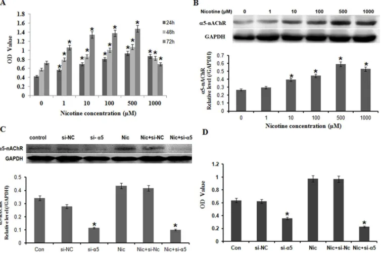
To determine whether nicotine may regulate the proliferation of gastric cells, we examined the effect of nicotine with various concentrations on cell viability in BGC823 by a CCK8 analysis (Fig 2A). BGC823 were exposed to nicotine (0, 1, 10, 100, 500, 1000μM) for 24, 48, and 72h, respectively. As shown inFig 2A, nicotine promoted cell proliferation in a time-dependent and concentration-dependent relation. Nicotine significantly stimulated cell proliferation at lower concentration (1, 10, 100, 500μM) and time (24–72 h), but a noticeable decrease in cell number was observed in nicotine-treated cultures at higher concentration (1000μM) and time (72 h). Studies showed levels of nicotine in the body vary widely among individuals even when smok-ing the same number of identical cigarettes [22–24].The concentration of nicotine (100μM) used in the present study mimicked the daily intake of cigarettes in moderate smokers [25,26]. The concentration of nicotine (100μM) is equivalent to concentration in the saliva in smoker who intake 25 cigarettes/day [27]. For further functional experiments, the concentration of nic-otine (100μM) was chosen to treat gastric cells for 24h.
To determine whether the nicotine-mediated promotion of cell proliferation in gastric cells is mediated throughα5-nAChR signaling, we analyzed the effect of nicotine on the expression ofα5-nAChR protein in BGC823 cells by Western blot. As shown inFig 2B, treatment of nico-tine at the dose of niconico-tine (0, 1, 10, 100, 500, 1000μM) for 24 hours promoted the expression ofα5-nAChR protein in a dose-dependent manner. To further confirm the involvement of
α5-nAChR signaling pathway in the nicotine-mediated promotion of gastric cancer cell prolif-eration, we blockedα5-nAChR protein expression by transfecting BC823 cells with a
α5-nAChR-specific siRNA (si-α5-nAChR) (Fig 2C) and evaluated its effects on nicotine-medi-ated promotion of cell proliferation by CCK assay (Fig 2D). By comparison with the scrambled nonspecific control siRNA (si-NC), transfection with si-α5-nAChR inhibited cell proliferation. Moreover, si-α5-nAChR transfection significantly reduced the nicotine-mediated promotion of cell proliferation in BGC823 cells (Fig 2D).
Nicotine inhibition of cisplatin-induced apoptosis
Fig 1. The expression ofα5-nAChR in gastric cancer tissues and cell lines.A: Expression ofα5-nAChR in gastric para-carcinoma tissue (Left) and
cancer Tissues (Right). (IHC 200×); B: Protein expression ofα5-nAChR in gastric cancer cell lines (N87, MKN28, MGC803, BGC823, HGC27, SGC7901,
MKN45, AGS); C: The expression ofα5-nAChR mRNA in the above cells in (A) was analyzed by RT-PCR, and normalized to that of GAPDH.
Cisplatin induced apoptosis was further assayed by AnnexinV/7-AAD staining. Flow cytome-try plots showed that the proportions of total apoptotic cells (AnnexinV+/7-AAD−) decreased
from43.2±2.32% to29.9 ± 1.26%, when exposed to cisplatin combined with 100μM nicotine (Fig 3B). These results suggested that nicotine can suppress apoptosis induced by cisplatin.
α
5-nAChR/AKT signaling pathway involved in anti- apoptotic effects of
nicotine in cisplatin-induced apoptosis of BGC823 cells
To determine the role of AKT in mediating the effects of nicotine in these cells, we determined whether nicotine induces activation of AKT in BGC823 cells. InFig 4A, 20 mM cisplatin strongly suppressed activity of AKT (lane 2) but nicotine increased AKT expression (lane 3) in the presence of cisplatin. It suggested that AKT was activated after exposure to 100μM nicotine in BGC823 cells as reported in various papers [29,30]. The down-regulation ofα5-nAChR expression decreased P-AKT expression (lane 4). Treatment with 10 mM LY294002, a phos-phatidylinositol 3-kinase (PI3K)/AKT pathway inhibitor, decreased the anti-apoptotic effects of nicotine in BGC823 cells (lane 5). In addition, treatment with LY294002 combined with si-CHRNA5 transfection significantly repressed the nicotine induced P-AKT protein levels (lane
Fig 2. Nicotine promoted BGC823 cell proliferation throughα5-nAChR.A: BGC823 were exposed to nicotine (0, 1, 10, 100, 500, 1000μM) for 24, 48,
and 72h, respectively. Nicotine promoted cell proliferation in a time-dependent and concentration-dependent relation; B: Treatment of nicotine at the dose of nicotine (0, 1, 10, 100, 500, 1000μM) for 24 hours promoted the expression ofα5-nAChR protein in a dose-dependent manner; C: Cells were transfected with α5-nAChR specific siRNA (si-α5) and then treated with nicotine at 100μM for 24 hours.α5-nAChR expressions were significantly decreased compared with
scrambled nonspecific control siRNA (si-NC) and Nic (Nicotine) + si-NC group; D: By comparison with the si-NC, transfection with si-α5 considerably
inhibited cell proliferation.*P<0.05 vs. the untreated control; each experiment was performed in triplicate.
6). These data suggest thatα5-nAChR /AKT pathways play a critical role in mediating the effects of nicotine and chemotherapy in gastric cancer cells.
We further examined the effects of nicotine on cisplatin-induced caspase-3 activation in BGC823 cells by Western blot assay. As shown inFig 4B, cisplatin induced an increase in cas-pase-3 activation, whereas nicotine blocked cisplatin-induced cascas-pase-3 activation in BGC823 cells. Moreover, exposure of BGC823 cells to cisplatin caused the expression of apoptotic pro-tein Bax increased while the prosurvival propro-tein Bcl-2 and Survivin decreased, which were inhibited by treatment with nicotine.
Meantime, as shown inFig 4C, the anti-apoptotic effect of nicotine was obviously blocked by si-α5-nAChR in BGC823 cells. Flow cytometry plots showed that the proportion of total apo-ptotic cells (AnnexinV+/7-AAD−) increased from 18.5 ± 0.92% to 34.9 ± 1.74% after si-5
treat-ment compared with si-NC group. Addition of AKT inhibitor LY294002 with si-α5-nAChR significantly promoted the apoptotic effect of cisplatin from 31.5 ± 1.57% to 61.2 ± 3.06% com-pared with LY294002 group. These results indicate that nicotine plays an important role in the prevention of cisplatin-induced apoptosis viaα5-nAChR/AKT signaling pathway.
Discussion
This study is the first to demonstrate a vital role ofα5-nAChR in nicotine anti-apoptosis of cisplatin in human gastric cancer cells. The analysis of clinical specimens indicated that
α5-nAChR expression is generally higher in tumor cells compared with para-carcinoma cells.
Fig 3. Nicotine inhibition of cisplatin-induced apoptosis.A: Cisplatin induced cells apoptosis compared with the control. In contrast, the cells co-treated with 100μM nicotine and 20 mM cisplatin showed little nuclear fragmentation and few apoptotic bodies by fluorescence microscopy (200 X); B: Cisplatin
induced apoptosis was assayed by AnnexinV/7-AAD staining. Flow cytometry plots showed that the proportion of total apoptotic cells was decreased when exposed to 20 mM cisplatin combined with 100μM nicotine.
The results showed a trend that the positive rate ofα5-nAchR expression was higher in patients with nicotine intake history than patients without nicotine intake history. The in vitro results showed that nicotine up-regulated the expression ofα5-nAChR protein and inhibited cis-platin-induced apoptosis by regulatingα5-nAChR/AKT signaling pathway in gastric cancer cells. Nicotine prevented cisplatin-induced activation of caspase-3 and Bax, and up-regulated the expression of anti-apoptotic proteins, Bcl-2 and Survivin. Furthermore, activation of AKT was found to play a role in mediating cisplatin-induced apoptosis, as well as anti-apoptotic effects of nicotine in BGC823 cells. It may be interesting to pay attention to the relationship between cigarette smoke (second smoking) and gastric cancer incidence.
Fig 4.α5-nAChR/AKT signaling involved in anti-apoptotic effects of nicotine in cisplatin-induced apoptosis of BGC823 cells.A: P-AKT was
activated after exposure to 100μM nicotine in BGC823 cells (lane 2 and lane3). Cisplatin strongly suppressed activity of P-AKT (lane 1 and lane 2) but
nicotine also induced P-AKT in the presence of cisplatin (lane 2 and lane 3). Down-regulation ofα5-nAChR expression decreased the level of P-AKT (lane 4
and lane 5). Treatment with LY294002 downregulated P-AKT expression (lane 3 and lane 5). Combination LY294002 with si-CHRNA5 transfection significantly repressed the nicotine induced P-AKT protein levels (lane 3 and lane 6);*p<0.05; B: Cisplatin induced an increase in caspase-3 and Bax
activation in BGC823 cells a decrease in Bcl-2 and Survivin expressions, whereas nicotine blocked cisplatin- induced Bcl-2, Bax, caspase-3 and Survivin expressions; With silence ofα5-nAChR co-administrated LY294002, an increased apoptosis was observed with the induction of Bcl-2, Bax, Survivin and
Caspase-3 by nicotine in BGC823 cells.*p<0.05; C: Assessment of apoptosis by AnnexinV/7-AAD staining in each group. BGC823 cells were pre-treated with 100μM nicotine and cisplatin for 24 h, and/or si-α5-nAChR for 48h, and/or AKT inhibitor LY294002 for 24h, and harvested.
Previous findings have unraveled the strong association between cigarette smoke and gastric cancer [31,32]. Nicotine was shown to increase the proliferation and migration of gastric can-cer cells by inducing cyclooxgenase-2 (COX-2), prostaglandin E2, VEGF,β-adrenoceptors, protein kinase C [26,33,34]. These effects appear to be mediated through the homopentameric
α7-nAChRs [35]. However, recent studies have demonstrated an unexpected effect of the het-eropentamericα5-nAChRs on nicotine intake [36–38]. Our previous works reported that the nicotine/α5-nAChR pathway is critical for lung cancer cell viability [21,39]. Nevertheless, no information has been available about whether nicotine also affects proliferation of human gas-tric cancer cells through regulation ofα5-nAChR. Here, we studied the role ofα5-nAChR in human gastric cancer. Our results demonstrated that expression ofα5-nAChR was higher in gastric cancer tissues compared to para-carcinoma tissue, which suggested thatα5-nAChR may play a role in gastric carcinogenesis. Furthermore, nicotine promotedα5-nAChR protein expression and blocking the activation ofα5-nAChR by siRNA attenuated nicotine-induced
Fig 5. Schematic diagram of nicotine-mediatedα5-nAChR/AKT signaling pathway in the prevention of cisplatin-induced apoptosis in gastric
cancer.Nicotine may interact withα5-nAChR on the surface of gastric cancer cells, then activate AKT signaling pathway and up-regulate Survivin and Bcl-2
expressions to prevent cisplatin-induced apoptosis.
gastric cancer cell proliferation. It suggested thatα5-nAChR is one of the major molecules to mediate cell proliferation stimulated by nicotine in gastric cancer cells.
The current standard chemotherapy for gastric cancer is cisplatin, but the success rate of this treatment is poor. The efficacy of cisplatin depends on the ability to induce DNA damage [40,41]. Hence, how cancer cells respond to cisplatin- induced apoptosis plays a critical role in cisplatin sensitivity. Recent reports show that nicotine inhibits cisplatin induced apoptosis in lung cancer cells, oral cancer, Raw264.7 and El4 cells [13,16,18], which may suggest that nicotine has the abil-ity not only to promote cancer development by activating cell growth pathways, but also to reduce the efficacy of chemotherapeutic agents by stimulating survival pathways. In agreement with the previous findings, results from the present study showed that co-treatment of the cells with cis-platin and nicotine, a significant activation of caspase-3 was observed, indicating that nicotine is able to determine an inhibition of the apoptotic potential of the cisplatin in gastric cancer.
The signaling pathways that regulate cell processes, including cell proliferation, cell cycle progression and cell apoptosis, have significant impact on deciding cellular response to cis-platin. Previous studies have shown that activation of AKT pathway may lead to resistance to cisplatin [42,43]. An over-expression of serine phosphorylated AKT has been detected in a wide range of human cancers, including gastric cancer [44,45]. As reported in various papers, nicotine by binding to nAChR leads to downstream activation of tumor promoting proteins including activation of the AKT pathways [46–48]. Exposure to nicotine might negatively impact on the apoptotic potential of cisplatin in human oral cancer cells, and the AKT pathway was required for nicotine function [16]. However, AKT pathway of nicotinic downstream sig-naling and the anti-apoptotic effects of nicotine in gastric cancer cells were unknown. There-fore, we investigated whether P-AKT may account for the antagonizing effect of nicotine on the cytotoxicity of cisplatin. We found that co-treated with siRNA-α5-nAChR and LY294002, a PI3k/AKT pathway inhibitor, increased cisplatin-induced apoptosis and attenuated the effects of nicotine in BGC823 cells. Several studies highlighted nicotine activated AKT signal-ing pathways is in modulatsignal-ing cell proliferation and survival through activation of the prosur-vival protein Bcl-2 and Survivin [15,49,50]. Our research also demonstrated that role of nicotine on cell apoptosis induced by cisplatin throughα5-nAChR/AKT is confirmed by induction of Survivin and Bcl-2, as final effectors of the pathways above.
In summary, we demonstrated that nicotine activatedα5-nAChR/AKT signaling and is involved in the resistance of cisplatin in gastric cancer. These findings provide new insights into the possible molecular mechanisms of nicotine inhibition of cisplatin-induced apoptosis in human gastric cancer cells (Fig 5). Nicotine present in cigarette smoke may interfere with gastric cancer pharmacological treatment by inhibiting chemotherapeutic drug-induced apo-ptosis. Strategies aimed at understanding nicotine-mediated signaling may facilitate the devel-opment of improved therapies in gastric cancer.
Supporting Information
S1 Fig. Expression ofα5-nAChR andα7-nAChR without or withα5-siRNA treatment. (TIF)
S2 Fig. Down-regulation ofα5-nAChR expression decreased the level of P-AKT. (TIF)
S3 Fig. Expression ofα5-nAChR in BGC823 cells without or with si-α5-nAChR treatment. (TIF)
S5 Fig. Nicotine promoted SGC7901 cell proliferation throughα5-nAChR. (TIF)
S6 Fig. Nicotine inhibition of cisplatin-induced apoptosis of SCG7901. (TIF)
Author Contributions
Conceived and designed the experiments: XLM YSW. Performed the experiments: YFJ HJS HQW HLZ XPZ DJX. Analyzed the data: YFJ HJS XLM YSW. Contributed reagents/materials/ analysis tools: YFJ HJS XLM YSW. Wrote the paper: YFJ XLM YSW.
References
1. Ladeiras-Lopes R, Pereira AK, Nogueira A, Pinheiro-Torres T, Pinto I, Santos-Pereira R, et al. Smoking and gastric cancer: systematic review and meta-analysis of cohort studies. Cancer Causes Control. 2008; 19(7):689–701. doi:10.1007/s10552-008-9132-yPMID:18293090.
2. Tredaniel J, Boffetta P, Buiatti E, Saracci R, Hirsch A. Tobacco smoking and gastric cancer: review and meta-analysis. Int J Cancer. 1997; 72(4):565–73. PMID:9259392.
3. Nishino Y, Inoue M, Tsuji I, Wakai K, Nagata C, Mizoue T, et al. Tobacco smoking and gastric cancer risk: an evaluation based on a systematic review of epidemiologic evidence among the Japanese popu-lation. Jpn J Clin Oncol. 2006; 36(12):800–7. doi:10.1093/jjco/hyl112PMID:17210611.
4. Wang W, Chin-Sheng H, Kuo LJ, Wei PL, Lien YC, Lin FY, et al. NNK enhances cell migration through alpha7-nicotinic acetylcholine receptor accompanied by increased of fibronectin expression in gastric cancer. Ann Surg Oncol. 2012; 19 Suppl 3:S580–8. Epub 2011/10/05. doi: 10.1245/s10434-011-2064-xPMID:21969082.
5. Shin VY, Liu ES, Ye YN, Koo MW, Chu KM, Cho CH. A mechanistic study of cigarette smoke and cyclo-oxygenase-2 on proliferation of gastric cancer cells. Toxicol Appl Pharmacol. 2004; 195(1):103–12. doi: 10.1016/j.taap.2003.10.009PMID:14962510.
6. Macqueen DA, Heckman BW, Blank MD, Janse Van Rensburg K, Park JY, Drobes DJ, et al. Variation in the alpha 5 nicotinic acetylcholine receptor subunit gene predicts cigarette smoking intensity as a function of nicotine content. Pharmacogenomics J. 2014; 14(1):70–6. doi:10.1038/tpj.2012.50PMID: 23358500; PubMed Central PMCID: PMC3778124.
7. Boezen HM. Genome-wide association studies: what do they teach us about asthma and chronic obstructive pulmonary disease? Proc Am Thorac Soc. 2009; 6(8):701–3. doi: 10.1513/pats.200907-058DPPMID:20008879.
8. Pasini F, Fraccon AP, G DEM. The role of chemotherapy in metastatic gastric cancer. Anticancer Res. 2011; 31(10):3543–54. PMID:21965776.
9. De Dosso S, Zanellato E, Nucifora M, Boldorini R, Sonzogni A, Biffi R, et al. ERCC1 predicts outcome in patients with gastric cancer treated with adjuvant cisplatin-based chemotherapy. Cancer Chemother Pharmacol. 2013; 72(1):159–65. doi:10.1007/s00280-013-2181-2PMID:23645290.
10. Takahari D, Hamaguchi T, Yoshimura K, Katai H, Ito S, Fuse N, et al. Feasibility study of adjuvant che-motherapy with S-1 plus cisplatin for gastric cancer. Cancer Chemother Pharmacol. 2011; 67(6):1423–
8. doi:10.1007/s00280-010-1432-8PMID:20809123.
11. Galluzzi L, Senovilla L, Vitale I, Michels J, Martins I, Kepp O, et al. Molecular mechanisms of cisplatin resistance. Oncogene. 2012; 31(15):1869–83. doi:10.1038/onc.2011.384PMID:21892204.
12. Sun XP, Dong X, Lin L, Jiang X, Wei Z, Zhai B, et al. Up-regulation of survivin by AKT and hypoxia-inducible factor 1alpha contributes to cisplatin resistance in gastric cancer. FEBS J. 2014; 281(1):115–
28. doi:10.1111/febs.12577PMID:24165223.
13. Zeng F, Li YC, Chen G, Zhang YK, Wang YK, Zhou SQ, et al. Nicotine inhibits cisplatin-induced apopto-sis in NCI-H446 cells. Med Oncol. 2012; 29(1):364–73. doi:10.1007/s12032-010-9792-9PMID: 21267677.
14. Zhang J, Kamdar O, Le W, Rosen GD, Upadhyay D. Nicotine induces resistance to chemotherapy by modulating mitochondrial signaling in lung cancer. Am J Respir Cell Mol Biol. 2009; 40(2):135–46. doi: 10.1165/rcmb.2007-0277OCPMID:18676776; PubMed Central PMCID: PMC2633138.
2006; 103(16):6332–7. doi:10.1073/pnas.0509313103PMID:16601104; PubMed Central PMCID:
PMC1458878.
16. Xu J, Huang H, Pan C, Zhang B, Liu X, Zhang L. Nicotine inhibits apoptosis induced by cisplatin in human oral cancer cells. Int J Oral Maxillofac Surg. 2007; 36(8):739–44. doi:10.1016/j.ijom.2007.05. 016PMID:17611077.
17. Marrero MB, Bencherif M. Convergence of alpha 7 nicotinic acetylcholine receptor-activated pathways for anti-apoptosis and anti-inflammation: central role for JAK2 activation of STAT3 and NF-kappaB. Brain Res. 2009; 1256:1–7. doi:10.1016/j.brainres.2008.11.053PMID:19063868.
18. Wang YY, Liu Y, Ni XY, Bai ZH, Chen QY, Zhang Y, et al. Nicotine promotes cell proliferation and induces resistance to cisplatin by alpha7 nicotinic acetylcholine receptormediated activation in Raw264.7 and El4 cells. Oncol Rep. 2014; 31(3):1480–8. doi:10.3892/or.2013.2962PMID:24399025.
19. Zheng Y, Ritzenthaler JD, Roman J, Han S. Nicotine stimulates human lung cancer cell growth by inducing fibronectin expression. Am J Respir Cell Mol Biol. 2007; 37(6):681–90. doi:10.1165/rcmb. 2007-0051OCPMID:17600315.
20. Zovko A, Viktorsson K, Lewensohn R, Kolosa K, Filipic M, Xing H, et al. APS8, a polymeric alkylpyridi-nium salt blocks alpha7 nAChR and induces apoptosis in non-small cell lung carcinoma. Mar Drugs. 2013; 11(7):2574–94. doi:10.3390/md11072574PMID:23880932; PubMed Central PMCID:
PMC3736439.
21. Ma X, Jia Y, Zu S, Li R, Jia Y, Zhao Y, et al. alpha5 Nicotinic acetylcholine receptor mediates nicotine-induced HIF-1alpha and VEGF expression in non-small cell lung cancer. Toxicol Appl Pharmacol. 2014; 278(2):172–9. doi:10.1016/j.taap.2014.04.023PMID:24793809.
22. Mayer B. How much nicotine kills a human? Tracing back the generally accepted lethal dose to dubious self-experiments in the nineteenth century. Arch Toxicol. 2014; 88(1):5–7. doi: 10.1007/s00204-013-1127-0PMID:24091634; PubMed Central PMCID: PMC3880486.
23. Grando SA. Connections of nicotine to cancer. Nat Rev Cancer. 2014; 14(6):419–29. doi:10.1038/ nrc3725PMID:24827506.
24. Gourlay SG, Benowitz NL, Forbes A, McNeil JJ. Determinants of plasma concentrations of nicotine and cotinine during cigarette smoking and transdermal nicotine treatment. Eur J Clin Pharmacol. 1997; 51 (5):407–14. PMID:9049583.
25. Lawson GM, Hurt RD, Dale LC, Offord KP, Croghan IT, Schroeder DR, et al. Application of urine nico-tine and cotinine excretion rates to assessment of niconico-tine replacement in light, moderate, and heavy smokers undergoing transdermal therapy. J Clin Pharmacol. 1998; 38(6):510–6. PMID:9650540.
26. Shin VY, Jin HC, Ng EK, Yu J, Leung WK, Cho CH, et al. Nicotine and 4-(methylnitrosamino)-1-(3-pyri-dyl)-1-butanone induce cyclooxygenase-2 activity in human gastric cancer cells: Involvement of nico-tinic acetylcholine receptor (nAChR) and beta-adrenergic receptor signaling pathways. Toxicol Appl Pharmacol. 2008; 233(2):254–61. doi:10.1016/j.taap.2008.08.012PMID:18805435.
27. Cheng YA, Tsai CC. Nicotine- and arecoline-induced interleukin-1 secretion and intercellular adhesion molecular-1 expression in human oral epidermoid carcinoma cells in vitro. Arch Oral Biol. 1999; 44 (10):843–51. Epub 1999/10/26. PMID:10530917.
28. Kanetaka K, Enjoji A, Furui J, Nagata Y, Fujioka H, Shiogama T, et al. Effects of intermittent 5-fluoroura-cil and low-dose cisplatin therapy on advanced and recurrent gastric cancer. Anticancer Res. 2012; 32 (8):3495–9. PMID:22843936.
29. Egleton RD, Brown KC, Dasgupta P. Nicotinic acetylcholine receptors in cancer: multiple roles in prolif-eration and inhibition of apoptosis. Trends Pharmacol Sci. 2008; 29(3):151–8. doi:10.1016/j.tips.2007. 12.006PMID:18262664.
30. Arredondo J, Chernyavsky AI, Jolkovsky DL, Pinkerton KE, Grando SA. Receptor-mediated tobacco toxicity: cooperation of the Ras/Raf-1/MEK1/ERK and JAK-2/STAT-3 pathways downstream of alpha7 nicotinic receptor in oral keratinocytes. FASEB J. 2006; 20(12):2093–101. doi:10.1096/fj.06-6191com
PMID:17012261.
31. Shin VY, Cho CH. Nicotine and gastric cancer. Alcohol. 2005; 35(3):259–64. doi:10.1016/j.alcohol. 2005.04.007PMID:16054988.
32. Liu Y, Liu BA. Enhanced proliferation, invasion, and epithelial-mesenchymal transition of nicotine-pro-moted gastric cancer by periostin. World J Gastroenterol. 2011; 17(21):2674–80. doi:10.3748/wjg.v17. i21.2674PMID:21677839; PubMed Central PMCID: PMC3110933.
34. Huang RY, Chen GG. Cigarette smoking, cyclooxygenase-2 pathway and cancer. Biochim Biophys Acta. 2011; 1815(2):158–69. doi:10.1016/j.bbcan.2010.11.005PMID:21147199.
35. Lien YC, Wang W, Kuo LJ, Liu JJ, Wei PL, Ho YS, et al. Nicotine promotes cell migration through alpha7 nicotinic acetylcholine receptor in gastric cancer cells. Ann Surg Oncol. 2011; 18(9):2671–9.
doi:10.1245/s10434-011-1598-2PMID:21347787.
36. Spitz MR, Amos CI, Dong Q, Lin J, Wu X. The CHRNA5-A3 region on chromosome 15q24-25.1 is a risk factor both for nicotine dependence and for lung cancer. J Natl Cancer Inst. 2008; 100(21):1552–6. doi: 10.1093/jnci/djn363PMID:18957677; PubMed Central PMCID: PMC2720751.
37. Improgo MR, Scofield MD, Tapper AR, Gardner PD. The nicotinic acetylcholine receptor CHRNA5/A3/ B4 gene cluster: dual role in nicotine addiction and lung cancer. Prog Neurobiol. 2010; 92(2):212–26.
doi:10.1016/j.pneurobio.2010.05.003PMID:20685379; PubMed Central PMCID: PMC2939268. 38. Wojas-Krawczyk K, Krawczyk P, Biernacka B, Grzybek M, Kolodziej P, Kucharczyk T, et al. The
poly-morphism of the CHRNA5 gene and the strength of nicotine addiction in lung cancer and COPD patients. Eur J Cancer Prev. 2012; 21(2):111–7. doi:10.1097/CEJ.0b013e32834c9b40PMID: 21955800.
39. Sun H, Ma X. alpha5-nAChR modulates nicotine-induced cell migration and invasion in A549 lung can-cer cells. Exp Toxicol Pathol. 2015; 67(9):477–82. doi:10.1016/j.etp.2015.07.001PMID:26205096.
40. Tanida S, Mizoshita T, Ozeki K, Tsukamoto H, Kamiya T, Kataoka H, et al. Mechanisms of Cisplatin-Induced Apoptosis and of Cisplatin Sensitivity: Potential of BIN1 to Act as a Potent Predictor of Cisplatin Sensitivity in Gastric Cancer Treatment. Int J Surg Oncol. 2012; 2012:862879. doi:10.1155/2012/ 862879PMID:22778941; PubMed Central PMCID: PMC3384945.
41. Checinska A, Hoogeland BS, Rodriguez JA, Giaccone G, Kruyt FA. Role of XIAP in inhibiting cisplatin-induced caspase activation in non-small cell lung cancer cells: a small molecule Smac mimic sensitizes for chemotherapy-induced apoptosis by enhancing caspase-3 activation. Exp Cell Res. 2007; 313 (6):1215–24. doi:10.1016/j.yexcr.2006.12.011PMID:17291493.
42. Falasca M. PI3K/Akt signalling pathway specific inhibitors: a novel strategy to sensitize cancer cells to anti-cancer drugs. Curr Pharm Des. 2010; 16(12):1410–6. PMID:20166984.
43. Zhang LL, Zhang J, Shen L, Xu XM, Yu HG. Overexpression of AKT decreases the chemosensitivity of gastric cancer cells to cisplatin in vitro and in vivo. Mol Med Rep. 2013; 7(5):1387–90. doi:10.3892/ mmr.2013.1400PMID:23546174.
44. Zhou W, Fu XQ, Liu J, Yu HG. RNAi knockdown of the Akt1 gene increases the chemosensitivity of gastric cancer cells to cisplatin both in vitro and in vivo. Regul Pept. 2012; 176(1–3):13–21. doi:10. 1016/j.regpep.2012.02.003PMID:22387880.
45. Yu LL, Dai N, Yu HG, Sun LM, Si JM. Akt associates with nuclear factor kappaB and plays an important role in chemoresistance of gastric cancer cells. Oncol Rep. 2010; 24(1):113–9. PMID:20514451.
46. Nishioka T, Guo J, Yamamoto D, Chen L, Huppi P, Chen CY. Nicotine, through upregulating pro-sur-vival signaling, cooperates with NNK to promote transformation. J Cell Biochem. 2010; 109(1):152–61.
doi:10.1002/jcb.22392PMID:19911375; PubMed Central PMCID: PMC3683296.
47. Nakada T, Kiyotani K, Iwano S, Uno T, Yokohira M, Yamakawa K, et al. Lung tumorigenesis promoted by anti-apoptotic effects of cotinine, a nicotine metabolite through activation of PI3K/Akt pathway. J Toxicol Sci. 2012; 37(3):555–63. PMID:22687995.
48. Schaal C, Chellappan SP. Nicotine-mediated cell proliferation and tumor progression in smoking-related cancers. Mol Cancer Res. 2014; 12(1):14–23. doi:10.1158/1541-7786.MCR-13-0541PMID: 24398389; PubMed Central PMCID: PMC3915512.
49. Cucina A, Dinicola S, Coluccia P, Proietti S, D'Anselmi F, Pasqualato A, et al. Nicotine stimulates prolif-eration and inhibits apoptosis in colon cancer cell lines through activation of survival pathways. J Surg Res. 2012; 178(1):233–41. doi:10.1016/j.jss.2011.12.029PMID:22520577.
50. Jin Q, Menter DG, Mao L, Hong WK, Lee HY. Survivin expression in normal human bronchial epithelial cells: an early and critical step in tumorigenesis induced by tobacco exposure. Carcinogenesis. 2008; 29(8):1614–22. doi:10.1093/carcin/bgm234PMID:18635526; PubMed Central PMCID: