www.bjournal.com.br
Volume 44 (2) 84-181 February 2011
doi:
Braz J Med Biol Res, February 2011, Volume 44(2) 105-111
10.1590/S0100-879X2010007500142
E3 ubiquitin ligase Cbl-b potentiates the apoptotic action of
arsenic trioxide by inhibiting the PI3K/Akt pathway
Yingchun Li, Xiujuan Qu, Jinglei Qu, Ye Zhang, Jing Liu, Yuee Teng, Xuejun Hu, Kezuo Hou
and Yunpeng Liu
Institutional Sponsors
The Brazilian Journal of Medical and Biological Research is partially financed by
E3 ubiquitin ligase Cbl-b potentiates
the apoptotic action of arsenic trioxide
by inhibiting the PI3K/Akt pathway
Yingchun Li
1*, Xiujuan Qu
1*, Jinglei Qu
1, Ye Zhang
1, Jing Liu
1, Yuee Teng
1,
Xuejun Hu
2, Kezuo Hou
1and Yunpeng Liu
11Department of Medical Oncology, 2Department of Medical Respiratory, The First Hospital,
China Medical University, Shenyang, China
Abstract
Arsenic trioxide (ATO) is a strong inducer of apoptosis in malignant hematological cells. Inducible phosphatidyl inositol 3 ki-nase (PI3K)-Akt activation promotes resistance to ATO. In the present study, we evaluated whether E3 ubiquitin ligase Cbl-b, a negative regulator of PI3K activation, is involved in the action of ATO. The effect of ATO on cell viability was measured by the Trypan blue exclusion assay or by the 3-(4,5-dimethylthiazol-2-yl)-2,5-diphenyltetrazolium bromide (MTT) assay.
Apopto-sis was determined by flow cytometry and protein expression was assayed by Western blotting. ATO decreased the viability
of HL60 cells and induced cellular apoptosis, which was accompanied by transient activation of Akt. The PI3K/Akt inhibitor,
LY294002, significantly increased ATO-induced apoptosis (P < 0.05). In addition, ATO up-regulated the expression of Cbl-b
proteins. Furthermore, ATO inhibited cell viability with an IC50 of 18.54 μM at 24 h in rat basophilic leukemia-2H3 cells. ATO induced cellular apoptosis with transient activation of Akt and Cbl-b was also up-regulated. Rat basophilic leukemia-2H3 cells transfected with a dominant negative (DN) Cbl-b mutation showed overexpression of Cbl-b (DN) and enhanced Akt activation. Compared with cells transfected with vector, ATO-induced apoptosis was decreased and G2/M phase cells were increased at
the same concentration (P < 0.05). The PI3K/Akt inhibitor, LY294002, re-sensitized Cbl-b (DN) overexpressing cells to ATO and reversed G2/M arrest (P < 0.05). Taken together, these results suggest that Cbl-b potentiates the apoptotic action of ATO
by inhibition of the PI3K/Akt pathway.
Key words: Arsenic trioxide; Apoptosis; Cbl-b; PI3K/Akt pathway
Introduction
Correspondence: Yunpeng Liu, Department of Medical Oncology, The First Hospital, China Medical University, 155 North Nanjing Street, Heping District, Shenyang City, 110001, China. Fax: +86-24-8328-2543. E-mail: [email protected]
*These authors contributed equally to this study.
Received August 2, 2010. Accepted December 6, 2010. Available online December 17, 2010. Published February 7, 2011.
Arsenic trioxide (As2O3, ATO) is very effective for the treatment of acute promyelocytic leukemia, with very little toxicity (1,2). ATO is also a potent inducer of apoptosis in a number of other cell types such as acute myeloid leukemia (AML) (3), multiple myeloma (4), and lymphocytic leukemia (5) cells. Several mechanisms have been proposed to ex-plain ATO-induced apoptosis, including the down-regulation
of the PML-RARα fusion protein (6), the involvement of a
mitochondrial pathway (7-9), production of superoxides (8,10), triggering of apoptosis-associated factors (3), and signal transduction (4,10). However, the sensitivity of dif-ferent types of cells to ATO differs, and the low sensitivity of some cells has restricted its clinical application.
Phosphatidyl inositol 3 kinase (PI3K)/Akt signaling is
frequently activated in blast cells in AML patients, and contributes strongly to the proliferation, survival and drug resistance of these cells (11-14). Constitutive and inducible Akt activity promotes resistance to ATO in AML blasts and in several cell lines, such as NB4, U937, HL60, and K562 cells (14-17). The combination of small molecule inhibitors of the PI3K/Akt pathway and standard chemotherapy has been successful in attenuating chemotherapeutic resis-tance, but further study of the molecules modulating PI3K/ Akt signaling is necessary.
106 Yingchun Li et al.
investigations showed that Cbl-b sensitizes both leukemia and gastric cancer cells to anthracyclines by modulating the ERK and Akt survival pathways (25). Another investigation also demonstrated that inhibition of PI3K/Akt signaling by Cbl (Cbl-b and c-Cbl) may be involved in both ATO-induced apoptosis of NB4 cells and ATO-induced G2/M phase arrest of gastric cancer cells (26). Since this result was derived from indirect evidence with pharmacologic inhibitors of PI3K/ Akt and proteasome, it is still unclear whether or not Cbl-b is involved in the action of ATO on AML cells.
In the present study, we investigated the effects of Cbl-b on the apoptotic action of ATO in leukemic cell lines. The re-sults showed that ATO induces apoptosis and up-regulation of Cbl-b in HL60 and rat basophilic leukemia (RBL)-2H3 cells. Loss of Cbl-b function increases Akt activation, and subsequently suppresses ATO-induced apoptosis. These results indicate that Cbl-b facilitates ATO-induced apoptosis by the inhibition of PI3K/Akt signaling.
Material and Methods
Reagents and antibodies
Anti-tubulin and anti-Cbl-b antibodies were purchased from Santa Cruz Biotechnology (USA). Anti-phospho-Akt (Ser-473) and anti-Akt antibodies were purchased from Cell Signaling Technology (USA). Arsenic trioxide and LY294002 were purchased from Sigma-Aldrich (USA).
Cell culture
The human promyelocytic cell line HL60 was grown in RPMI 1640 medium containing 10% heat-inactivated fetal calf serum (FCS), 5 U/mL penicillin and 50 mg/mL streptomycin under a 95% air/5% CO2 atmosphere. Rat basophilic leukemia RBL-2H3 cells and cells overexpress-ing a dominant negative (DN) Cbl-b mutation (27) were
maintained as monolayer cultures in Dulbecco’s modified
Eagle’s medium (DMEM) containing 100 U/mL penicillin and 10% heat-inactivated FBS (27). The transfected cells were cultured in the presence of 0.4 mg/mL G418 (USA).
Cell viability assay
The effect of ATO on HL60 cell viability was measured by the Trypan blue exclusion assay (26). At assay time, HL60 cells were collected, mixed with an equal volume of PBS containing 0.4% Trypan blue dye, and manually counted. Actual cell numbers were calculated by multiplying diluted times compared with initial cell numbers. Cell viability % = viable cell numbers / total (viable + dead) cell numbers x 100 (6). The in vitro cell viability effect of ATO on RBL-2H3 cells was determined by measuring 3-(4,5-dimethylthiazol-2-yl)-2,5-diphenyltetrazolium bromide (MTT) dye absorbance
of living cells as described previously (28,29). Briefly, cells
were seeded at 5 x 10³ cells/well in 96-well plates in the presence of the designated doses of ATO. After exposure
to the drugs for the indicated time, 20 μL MTT solution (5
mg/mL in PBS) was added to each well and the plates were incubated for an additional 4 h at 37°C. MTT solu-tion in medium was aspirated off. To achieve solubilizasolu-tion
of the formazan crystals formed in viable cells, 150 μL
dimethylsulfoxide (DMSO) was added to each well and absorbance at 570 nm was measured using a microplate reader (Bio-Rad, USA).
Flow cytometric assay of apoptosis
To evaluate the induction of apoptosis, samples of
cells treated with ATO were taken at relevant times, fixed
in ice-cold 70% ethanol for 12 h, and then incubated with
20 μg/mL RNase A and 10 μg/mL propidium iodide for 30 min in the dark. Finally, samples were evaluated by flow
cytometry and data were analyzed using the CellQuest software (Becton Dickinson, USA). The experiment was repeated three times.
Western blot analysis
Western blotting was performed using standard tech
-niques (26). Briefly, cells were washed twice with PBS
and lysed in 1% Triton lysis buffer (1% Triton X-100, 50 mM Tris-Cl, pH 7.4, 150 mM NaCl, 10 mM EDTA, 100 mM NaF, 1 mM Na3VO4, 1 mM PMSF, and 2 μg/mL aprotinin) on ice. Protein concentration was determined by the Lowry
method. Total proteins (30-50 μg) were subjected to so -dium dodecyl sulfate-polyacrylamide gel electrophoresis (SDS-PAGE) and transferred to nitrocellulose membranes (Immoblin-P, USA). Membranes were blocked with 5% skim milk in Tris-buffered saline Tween-20 (TBST; 10 mM Tris, pH 7.4, 150 mM NaCl, and 0.1% Tween 20) at room tem-perature for 2 h and incubated with the indicated primary antibodies at 4°C overnight. After washing with TBST, the membrane was reacted with the appropriate horseradish peroxidase-conjugated secondary antibodies for 30 min at room temperature. After extensive washing with TBST, proteins were visualized using the enhanced
chemilumines-cence reagent (SuperSignal Western Pico Chemilunescent
Substrate; Pierce, USA).
Statistical analysis
Data are reported as means ± SD. Differences between two groups were determined by the Student t-test. P < 0.05 was considered to be statistically significant.
Results
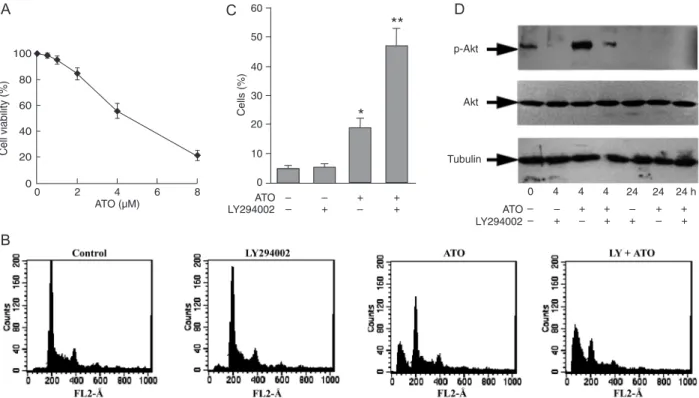
The PI3K/Akt inhibitor LY294002 enhanced ATO-induced apoptosis of HL60 cells
Akt remained unchanged upon treatment with ATO. Basal phospho-Akt (p-Akt) expression was strongly increased after 4 h of ATO treatment, but decreased to below the basal level after 24 h (Figure 1D). Treatment for 24 h with
25 μM LY294002 alone had no effect. However, when used
in combination, LY294002 greatly potentiated the induction
of apoptosis by ATO (P < 0.05; Figure 1B, C) and signifi -cantly decreased the expression of p-Akt at 24 h (Figure 1D), and reduced the transient increase at 4 h. These data indicate that ATO-induced apoptosis of HL60 cells involves the inhibition of PI3K/Akt signaling by ATO itself, and that this action is potentiated by the addition of LY294002.
Up-regulation of E3 ubiquitin ligase Cbl-b by ATO
Previous reports demonstrated that Cbl-b inhibits the PI3K/Akt signaling pathway by down-regulating PI3K
ac-tivation. We examined the expression levels of Cbl-b and
p-Akt in HL60 cells in order to further clarify the correlation between Cbl-b and p-Akt as well as their important roles.
Treatment with ATO significantly increased the expression
of Cbl-b starting at 4 h and then gradually increased to the maximal level at 24 h. And p-Akt expression strongly
increased at 4 h and then gradually decreased to below the basal level. The results indicate that there is a correlation between Cbl-b and p-Akt (Figure 2).
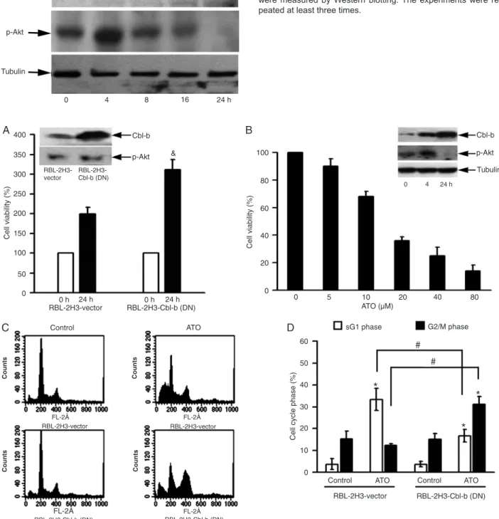
Cbl-b enhanced ATO-induced apoptosis
RBL-2H3 cells overexpressing DN Cbl-b have been used in previous studies (27,30). It was observed that p-Akt ex-pression is slightly increased in Cbl-b (DN) overexpressing cells. Consistently, overexpression of Cbl-b (DN) strongly accelerates cell proliferation by about 1.5 times compared to RBL-2H3 cells transfected with vector (RBL-2H3-vector; Figure 3A). After treatment with ATO, dose-dependent inhibition of cell growth was observed in RBL-2H3 cells with an IC50 at 24 h of 18.54 μM. p-Akt up-regulation was observed at 4 h, followed by down-regulation at 24 h, and Cbl-b was also up-regulated by ATO (Figure 3B). Next,
we analyzed the ability of 25 μM ATO to induce apoptosis
in RBL-2H3-vector cells and in 2H3 cells overexpressing DN Cbl-b. As shown in Figure 3C and D, ATO strongly induced apoptosis in RBL-2H3-vector cells at 12 h. The sub-G1 population was approximately 34.5%, but cells in the G2/M phase were not increased. In contrast, DN Cbl-b
Figure 1. Stimulation of apoptosis by arsenic trioxide (ATO) and phosphatidyl inositol 3 kinase (PI3K)/Akt inhibitors in HL60 cells. A, Cell viability was assessed by the Trypan blue exclusion assay after treatment with ATO for 24 h. B, Frequency of apoptosis in HL60
cell cultures treated for 24 h with 4 μM ATO, either alone or in combination with 25 μM PI3K/Akt inhibitor LY294002. C, Percentage of apoptotic cells in response to ATO and LY294002. D, Total proteins or the proteins in the cytosolic fractions were isolated and
phospho-Akt (p-phospho-Akt), phospho-Akt and β-tubulin levels were measured by Western blotting, as described in the Material and Methods section. The experi -ments were repeated at least three times. Differences between two groups were determined by the Student t-test. *P < 0.05 compared
to control cells. **P < 0.05 compared to cells treated with ATO alone.
A
B
108 Yingchun Li et al.
Figure 2. Up-regulation of Cbl-b by arsenic trioxide. Total proteins or the proteins in the cytosolic fractions were isolated and E3
ubiquitin ligase Cbl-b, phospho-Akt (p-Akt) and β-tubulin levels were measured by Western blotting. The experiments were re -peated at least three times.
Figure 3. Modulation of Cbl-b level and its relationship to apoptosis. A, Rat basophilic leukemia (RBL)-2H3 cells overexpressing domi-nant negative Cbl-b and the control cells were grown for 24 h, and cell viability was then assessed by the 3-(4,5-dimethylthiazol-2-yl)-2,5-diphenyltetrazolium bromide (MTT) assay. The inset represents the relative levels of Cbl-b and phospho-Akt (p-Akt) proteins in both kinds of cells. B, Rat basophilic leukemia (RBL)-2H3 cells were treated with the indicated concentrations of arsenic trioxide (ATO) for 24 h, and cell viability was assessed by the MTT assay. The inset represents the relative levels of Cbl-b and p-Akt proteins in total cell
extracts obtained from cells treated with 25 μM ATO for the indicated number of hours. C, D, Frequency of apoptotic cells and G2/M
phase cells in untreated cultures, and in cultures treated with 25 μM ATO for 12 h. The experiments were repeated at least three times.
Differences between two groups were determined by the Student t-test. &P < 0.05 compared to RBL-2H3 cells transfected with vector
overexpression significantly decreased the percentage of apoptotic cells and increased G2/M phase cells (P < 0.05).
These results indicate that loss of function of Cbl-b and sub-sequent deregulation of p-Akt may attenuate ATO-induced apoptosis and change the phase of the cell cycle.
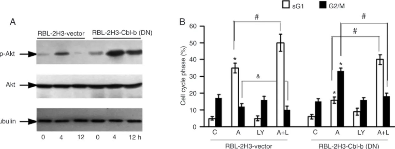
Effect of Cbl-b on Akt activity
The effect of Cbl-b was confirmed by measuring the
levels of p-Akt in transfected and parental cells after ATO treatment (Figure 4A). DN Cbl-b overexpression in RBL-2H3 cells slightly enhanced basal Akt activation compared
to control cells. After treatment with 25 μM ATO, the level of
p-Akt expression in DN Cbl-b overexpressing cells was much higher than that in control cells. These results indicate that DN Cbl-b attenuation of ATO-induced apoptosis may occur through the enhancement of Akt activation. Furthermore,
the PI3K/Akt inhibitor LY294002 significantly enhanced
ATO-induced apoptosis and reversed ATO-induced the
G2/M phase arrest in DN Cbl-b overexpressing cells (P < 0.05). LY294002 also significantly enhanced ATO-induced apoptosis in parental cells (P < 0.05; Figure 4B).
Discussion
Previous studies have demonstrated that ATO could induce apoptosis in AML cell lines (3,6,7,9,10), and that ATO-induced apoptosis is accompanied by decreased Akt
activity (15-17). In the present study, we have confirmed
that ATO induces apoptosis of AML HL60 cells. In the pro-cess of apoptosis, Akt activity falls sharply after a transient strong increase. The PI3K/Akt inhibitor LY294002 greatly enhanced ATO-induced apoptosis. Chemotherapeutic
agents such as anthracyclines and cisplatin have also been reported to increase PI3K/Akt activity during drug-induced apoptosis (12,25). Activated Akt can regulate cell survival by phosphorylation of downstream substrates such as BAD, ASK1, IKK, and CREB, which indirectly or directly control the apoptotic machinery (12-14). Therefore, Akt activation might be a negative feedback response to chemotherapy and
stress, and could in turn influence drug-induced apoptosis.
The potentiation of ATO-induced apoptosis by LY294002
indicates that Akt inhibition is necessary and sufficient for
ATO-induced apoptosis.
The PI3K/Akt pathway could be modulated by several factors (12). The E3 ubiquitin ligase Cbl-b functions as a negative regulator of PI3K activation. Ubiquitination of the p85-regulatory subunit of PI3K by Cbl-b affects its phosphorylation of downstream substrates, including Akt
(18-22). T lymphocytes from Cbl-b-deficient mice show
enhanced proliferation and cytokine production in response to the triggering of T-cell receptors (22,23). Zhang et al.
(24) recently reported that Cbl-b-deficient T cells show
deregulated proliferation, a process resulting in part from the failure of Akt inhibition. Our recent study demonstrated that Cbl-b sensitizes both leukemia and gastric cancer cells to anthracyclines by modulating the ERK and Akt survival pathways (25). The present results demonstrate that ATO up-regulates Cbl-b expression in the process of apoptosis in both HL60 and RBL-2H3 cells. These events imply the importance of Cbl-b up-regulation by ATO, which is consistent with our previous assumption regarding NB4 cells (26). The NB4 cell line is characterized by the PML/
RARα fusion protein and ATO strongly promotes apopto -sis in NB4 cells by modulating and degrading the fusion
Figure 4. Modulation of phospho-Akt (p-Akt) level by arsenic trioxide. A, Total proteins or the proteins in the cytosolic
frac-tions were isolated and p-Akt and Akt levels were measured by Western blotting. B, Frequency of apoptotic cells and G2/M
phase cells in untreated cultures, and in cultures treated with 25 μM arsenic trioxide (A) and/or 25 μM LY294002 (LY) for 12
h. The experiments were repeated at least three times. Differences between two groups were determined by the Student
t-test. *P < 0.05 compared to control (C) cells; #P < 0.05 for comparison between the two groups indicated. &P = 0.057 for
110 Yingchun Li et al.
References
1. Hu J, Shen ZX, Sun GL, Chen SJ, Wang ZY, Chen Z.
Long-term survival and prognostic study in acute promyelocytic leukemia treated with all-trans-retinoic acid, chemotherapy, and As2O3: an experience of 120 patients at a single
institu-tion. Int J Hematol 1999; 70: 248-260.
2. Soignet SL, Maslak P, Wang ZG, Jhanwar S, Calleja E,
Dardashti LJ, et al. Complete remission after treatment of acute promyelocytic leukemia with arsenic trioxide. N Engl J Med 1998; 339: 1341-1348.
3. Perkins C, Kim CN, Fang G, Bhalla KN. Arsenic induces apoptosis of multidrug-resistant human myeloid leukemia cells that express Bcr-Abl or overexpress MDR, MRP, Bcl-2, or Bcl-x(L). Blood 2000; 95: 1014-1022.
4. Lunghi P, Giuliani N, Mazzera L, Lombardi G, Ricca M, Cor-radi A, et al. Targeting MEK/MAPK signal transduction mod-ule potentiates ATO-induced apoptosis in multiple myeloma cells through multiple signaling pathways. Blood 2008; 112: 2450-2462.
5. Merkel O, Heyder C, Asslaber D, Hamacher F, Tinhofer I, Holler C, et al. Arsenic trioxide induces apoptosis preferen-tially in B-CLL cells of patients with unfavourable prognostic factors including del17p13. J Mol Med 2008; 86: 541-552. 6. Chen GQ, Zhu J, Shi XG, Ni JH, Zhong HJ, Si GY, et al. In
vitro studies on cellular and molecular mechanisms of arse-nic trioxide (As2O3) in the treatment of acute promyelocytic
leukemia: As2O3 induces NB4 cell apoptosis with
down-regulation of Bcl-2 expression and modulation of PML-RAR alpha/PML proteins. Blood 1996; 88: 1052-1061.
7. Cai X, Shen YL, Zhu Q, Jia PM, Yu Y, Zhou L, et al. Arsenic trioxide-induced apoptosis and differentiation are associated
respectively with mitochondrial transmembrane potential collapse and retinoic acid signaling pathways in acute pro-myelocytic leukemia. Leukemia 2000; 14: 262-270.
8. Woo SH, Park IC, Park MJ, Lee HC, Lee SJ, Chun YJ, et al.
Arsenic trioxide induces apoptosis through a reactive oxy-gen species-dependent pathway and loss of mitochondrial membrane potential in HeLa cells. Int J Oncol 2002; 21: 57-63.
9. Larochette N, Decaudin D, Jacotot E, Brenner C, Marzo I, Susin SA, et al. Arsenite induces apoptosis via a direct effect on the mitochondrial permeability transition pore. Exp Cell Res 1999; 249: 413-421.
10. Choi YJ, Park JW, Suh SI, Mun KC, Bae JH, Song DK, et
al. Arsenic trioxide-induced apoptosis in U937 cells involve generation of reactive oxygen species and inhibition of Akt.
Int J Oncol 2002; 21: 603-610.
11. Franke TF, Hornik CP, Segev L, Shostak GA, Sugimoto C. PI3K/Akt and apoptosis: size matters. Oncogene 2003; 22: 8983-8998.
12. West KA, Castillo SS, Dennis PA. Activation of the PI3K/
Akt pathway and chemotherapeutic resistance. Drug Resist Updat 2002; 5: 234-248.
13. Tazzari PL, Cappellini A, Ricci F, Evangelisti C, Papa V, Grafone T, et al. Multidrug resistance-associated protein 1 expression is under the control of the phosphoinositide 3 kinase/Akt signal transduction network in human acute my-elogenous leukemia blasts. Leukemia 2007; 21: 427-438. 14. Martelli AM, Nyakern M, Tabellini G, Bortul R, Tazzari PL,
Evangelisti C, et al. Phosphoinositide 3-kinase/Akt signaling pathway and its therapeutical implications for human acute protein (6). However, ATO also drives HL60 cells lacking
the PML/RARα fusion protein to apoptosis, and the pres
-ent evidence proved that Cbl was modified by ATO in the
process regardless of whether the cells carried the PML/
RARα fusion protein. It is suggested that Cbl involvement
in ATO-induced apoptosis is an important mechanism. To
confirm this hypothesis, we used RBL-2H3 cells transfected
with DN Cbl-b. Consistent with previously reported results (24,25), DN Cbl-b strongly accelerated cell proliferation by about 1.5 times compared to control cells. Further results showed that the loss of function of Cbl-b by overexpression of DN Cbl-b strongly increases Akt activation after treatment and thereby desensitization of the cells. This suggests that Cbl-b might play a positive role in ATO-induced leukemic cell apoptosis. Since Cbl involvement in the antitumor activity of ATO via its regulation of the PI3K/Akt pathway is cur-rently thought to be independent of the primary mechanism
of action of the drug, further study is worthwhile. We also
noted that the blot of Cbl-b showed double bands in HL60 cells, and a single band in RBL-2H3 cells. This difference in the patterns of Cbl proteins depending on cell type needs further investigation. In addition, DN Cbl-b overexpression
significantly induced G2/M phase arrest in the process of apoptosis by ATO. Higher Akt activation might influence
the vital substrates, such as p53, which is the determinant in the action of ATO, apoptosis or G2/M phase arrest (26). Consistent with this explanation, the PI3K/Akt inhibitor
LY294002 significantly enhanced ATO-induced apoptosis in
both cells, and also markedly reversed ATO-induced G2/M phase arrest in DN Cbl-b overexpressing cells.
In summary, our results suggest that PI3K/Akt pathway activation antagonizes ATO-induced leukemic cell apopto-sis. Ubiquitin ligase Cbl-b potentiates the apoptotic action of ATO by inhibition of the PI3K/Akt pathway.
Recent reports have shown that mutations in the Cbl
family RING finger domain are not only associated with
preleukemic chronic myelomonocytic leukemia, juvenile myelomonocytic leukemia, and other myeloproliferative neoplasms, but also with progression to AML (31,32). It is well known that the inhibition of PI3K/Akt activation by
Cbl-b is dependent on a functional Cbl RING finger (18-22).
myeloid leukemia. Leukemia 2006; 20: 911-928.
15. Tabellini G, Tazzari PL, Bortul R, Evangelisti C, Billi AM, Grafone T, et al. Phosphoinositide 3-kinase/Akt inhibition increases arsenic trioxide-induced apoptosis of acute pro-myelocytic and T-cell leukaemias. Br J Haematol 2005; 130: 716-725.
16. Tabellini G, Cappellini A, Tazzari PL, Fala F, Billi AM, Man-zoli L, et al. Phosphoinositide 3-kinase/Akt involvement in arsenic trioxide resistance of human leukemia cells. J Cell Physiol 2005; 202: 623-634.
17. Ramos AM, Fernandez C, Amran D, Sancho P, de Blas E, Aller P. Pharmacologic inhibitors of PI3K/Akt potentiate the apoptotic action of the antileukemic drug arsenic trioxide via glutathione depletion and increased peroxide accumulation in myeloid leukemia cells. Blood 2005; 105: 4013-4020.
18. Fang D, Wang HY, Fang N, Altman Y, Elly C, Liu YC. Cbl-b,
a RING-type E3 ubiquitin ligase, targets phosphatidylinositol 3-kinase for ubiquitination in T cells. J Biol Chem 2001; 276: 4872-4878.
19. Dikic I, Szymkiewicz I, Soubeyran P. Cbl signaling networks in the regulation of cell function. Cell Mol Life Sci 2003; 60: 1805-1827.
20. Guenou H, Kaabeche K, Dufour C, Miraoui H, Marie PJ. Down-regulation of ubiquitin ligase Cbl induced by twist
haploinsufficiency in Saethre-Chotzen syndrome results in
increased PI3K/Akt signaling and osteoblast proliferation.
Am J Pathol 2006; 169: 1303-1311.
21. Lin AE, Mak TW. The role of E3 ligases in autoimmunity and
the regulation of autoreactive T cells. Curr Opin Immunol
2007; 19: 665-673.
22. Jeon MS, Atfield A, Venuprasad K, Krawczyk C, Sarao R,
Elly C, et al. Essential role of the E3 ubiquitin ligase Cbl-b in T cell anergy induction. Immunity 2004; 21: 167-177. 23. Bachmaier K, Krawczyk C, Kozieradzki I, Kong YY, Sasaki T,
Oliveira-dos-Santos A, et al. Negative regulation of lympho-cyte activation and autoimmunity by the molecular adaptor Cbl-b. Nature 2000; 403: 211-216.
24. Zhang R, Zhang N, Mueller DL. Casitas B-lineage lymphoma b inhibits antigen recognition and slows cell cycle progres-sion at late times during CD4+ T cell clonal expanprogres-sion. J Immunol 2008; 181: 5331-5339.
25. Qu X, Zhang Y, Li Y, Hu X, Xu Y, Xu L, et al. Ubiquitin li-gase Cbl-b sensitizes leukemia and gastric cancer cells to anthracyclines by activating the mitochondrial pathway and modulating Akt and ERK survival signals. FEBS Lett 2009; 583: 2255-2262.
26. Li Y, Qu X, Qu J, Zhang Y, Liu J, Teng Y, et al. Arsenic trioxide induces apoptosis and G2/M phase arrest by inducing Cbl to inhibit PI3K/Akt signaling and thereby regulate p53 activa-tion. Cancer Lett 2009; 284: 208-215.
27. Qu X, Sada K, Kyo S, Maeno K, Miah SM, Yamamura H. Negative regulation of FcepsilonRI-mediated mast cell acti-vation by a ubiquitin-protein ligase Cbl-b. Blood 2004; 103: 1779-1786.
28. Ji C, Ren F, Xu M. Caspase-8 and p38MAPK in DATS-induced apoptosis of human CNE2 cells. Braz J Med Biol Res 2010; 43: 821-827.
29. Vistica DT, Skehan P, Scudiero D, Monks A, Pittman A, Boyd MR. Tetrazolium-based assays for cellular viability: a criti-cal examination of selected parameters affecting formazan production. Cancer Res 1991; 51: 2515-2520.
30. Qu X, Liu Y, Ma Y, Zhang Y, Li Y, Hou K. Up-regulation of the Cbl family of ubiquitin ligases is involved in ATRA and bufalin-induced cell adhesion but not cell differentiation.
Biochem Biophys Res Commun 2008; 367: 183-189.
31. Caligiuri MA, Briesewitz R, Yu J, Wang L, Wei M, Arnoczky
KJ, et al. Novel c-CBL and CBL-b ubiquitin ligase mutations in human acute myeloid leukemia. Blood 2007; 110: 1022-1024.