AKT inhibitor suppresses hyperthermia-induced
Ndrg2 phosphorylation in gastric cancer cells
Yurong Tao
1*, Yan Guo
1*, Wenchao Liu
1*, Jian Zhang
2, Xia Li
2, Lan Shen
2, Yi Ru
2, Yan Xue
1,
Jin Zheng
3, Xinping Liu
2, Jing Zhang
2and Libo Yao
2 1Department of Oncology, State Key Discipline of Cell Biology, Xijing Hospital, The Fourth Military Medical University, Xi’an, Shaanxi, China 2State Key Laboratory of Cancer Biology and Department of Biochemistry and Molecular Biology, The Fourth Military Medical University, Xi’an, Shaanxi, China 3Department of Traditional Chinese and Western Medicine of Oncology, Tangdu Hospital, The Fourth Military Medical University, Xi’an, Shaanxi, ChinaAbstract
Hyperthermia is one of the most effective adjuvant treatments for various cancers with few side effects. However, the underlying molecular mechanisms still are not known. N-myc downstream-regulated gene 2 (NDRG2), a tumor suppressor, has been shown to be involved in diverse cellular stresses including hypoxia, lipotoxicity, etc. In addition, Ndrg2 has been reported to be related to progression of gastric cancer. In the current study, our data showed that the apoptosis rate of MKN28 cells increased relatively rapidly to 13.4% by 24 h after treatment with hyperthermia (426C for 1 h) compared to 5.1% in control cells (P,0.05). Nevertheless, there was no obvious change in the expression level of total Ndrg2 during this process. Further investigation demonstrated that the relative phosphorylation levels of Ndrg2 at Ser332, Thr348 increased up to 3.2- and 1.9-fold (hyperthermia groupvscontrol group) at 3 h in MKN28 cells, respectively (P,0.05). We also found that heat treatment significantly increased AKT phosphorylation. AKT inhibitor VIII (10mM) decreased the phosphorylation level of Ndrg2 induced by hyperthermia. Accordingly, the apoptosis rate rose significantly in MKN28 cells (16.4%) treated with a combination of AKT inhibitor VIII and hyperthermia compared to that (6.8%) of cells treated with hyperthermia alone (P,0.05). Taken together, these data demonstrated that Ndrg2 phosphorylation could be induced by hyperthermia in an AKT-dependent manner in gastric cancer cells. Furthermore, AKT inhibitor VIII suppressed Ndrg2 phosphorylation and rendered gastric cancer cells susceptible to apoptosis induced by hyperthermia.
Key words: Hyperthermia; Gastric cancer; Ndrg2; Apoptosis; AKT inhibitor
Introduction
Gastric cancer is the second most common cause of cancer-related death in the world, and it remains difficult to cure. Over the last decades, surgical resection has been used as the main treatment to cure local gastric cancer. Recently, postoperative hyperthermia has been used as a strong adjuvant therapy to improve the prognosis of cancer (1).
Mild hyperthermia is a type of hyperthermia, which treats the cancer by exposing the malignant tumor to controlled temperature up to 40-436C. Apart from cell necrosis resulting from severe hyperthermia (.456C), the main function of mild hyperthermia is to induce apoptosis of tumor cells. In addition, the anti-tumor effect of mild hyperthermia also includes disrupting cytoskeleton components, changing
membrane permeability, and inhibiting tumor cell growth. However, despite the multiple positive therapeutic effects of hyperthermia, the detailed molecular mechanisms under-lying this process have not been elucidated.
The N-myc downstream-regulated gene 2 (NDRG2) belongs to the NDRG family, a new family of differentiation-related genes consisting of four members namedNDRG1, NDRG2, NDRG3, and NDRG4 (2). Compared to other family members, highly structural homology and functional redundancy have been revealed for Ndrg2 and Ndrg1 (3,4). Besides contributing to cellular differentiation, Ndrg2 was extensively studied as a tumor suppressor in various types of cancer (5-9). Recent accumulating evidence has
Correspondence: Jing Zhang, State Key Laboratory of Cancer Biology and Department of Biochemistry and Molecular Biology, The Fourth Military Medical University, 17 Changle Western Rd, Xi’an, Shaanxi 710032, China. Fax: ++86-29-8477-3947. E-mail: [email protected] (Jing Zhang) or [email protected] (Yurong Tao)
*These authors contributed equally to this study.
demonstrated that Ndrg2 played an important role in cellular stress. For instance, Ndrg2 was involved in AKT-mediated protection ofbcells against lipotoxicity (10). The phosphorylation of Ndrg2 could be induced by different stress conditions in skeletal muscle (11). Moreover, expression of Ndrg2 was markedly up-regulated in tumor cell lines that were exposed to hypoxic conditions. Further investigation indicated that Ndrg2 contributed to hypoxia-induced radioresistance of HeLa cells (12). Although several studies have shown that NDRG2 is a stress-related gene, little information is available regarding the role of Ndrg2 in hyperthermia-induced heat stress.
In the present study, we found that AKT mediated phosphorylation of Ndrg2 induced by hyperthermia in gastric cancer cells. Further study revealed that AKT inhibitor VIII suppressed Ndrg2 phosphorylation and enhanced hyperthermia-induced apoptosis. The effects of the AKT inhibitor on heat sensitivity are discussed in relation to the phosphorylation of Ndrg2 in gastric cancer cells.
Material and Methods
Chemicals
AKT inhibitor VIII, glycogen synthase kinase 3 (GSK-3) inhibitor CT99021 and phosphoinositide 3-kinase (PI3K) inhibitor LY294002 were obtained from Calbiochem (EMD Biosciences Inc., Germany). 3-(4,5-Dimethylthiazol-2-yl)-2,5-diphenyltetrazolium bromide (MTT) and trypan blue were purchased from Sigma-Aldrich, Inc. (USA). The 5-ethynyl-2-deoxyuridine (EdU) Cell Proliferation Assay Kit was obtained from Rui BO Bio Co. (China).
Cell culture and treatment
The human gastric cancer cell lines MKN45 and MKN28 (Riken Cell Bank, Japan) were cultured in RPMI 1640 medium (Invitrogen, USA) supplemented with 10% fetal bovine serum. Cell cultures were maintained at 376C in a 5% CO2/95% air atmosphere. For heat treatment, the
cells were seeded onto 24- or 6-well plates for 24 h to allow exponential growth and were subjected to heat shock at 426C in a precision water bath for 1 h. The cells were then re-incubated at 376C for the indicated times.
Cell growth analysis
Cells were seeded at an initial density of 26104cells/
mL on 96-well plates for 24 h and exposed to hyperthermia. Six wells from each group were selected for the MTT assay. MTT was added to each well at a final concentration of 0.5 mg/mL at the indicated times and dissolved in 150mL
dimethylsulfoxide and measured using a multiscanner reader (TECAN-spectra mini Grodig, Austria) at a wave-length of 490 nm. Cell growth curves were drawn by using average absorbance at 490 nm from three independent experiments. Percent inhibition was calculated using the formula: % inhibition = 1 - (absorbance test / absorbance control)6100%.
EdU analysis
The cells were seeded on 96-well plates and exposed to the media. All cells were treated with 50mM EdU for 4 h
at 376C. After fixation with 4% paraformaldehyde for 15 min, the cells were treated with 0.5% Triton X-100 for 20 min and rinsed with PBS three times. The cells were then exposed to 100mL 1X ApolloHreaction cocktail for
30 min and incubated with 5mg/mL Hoechst 33342 to
stain cell nuclei for 30 min. Images were observed under a microscope. The percentage of EdU-positive cells was calculated from 5 random fields per well each seeded in triplicate. About 200 cells were observed in each well. The EdU-labeled (red) and unlabeled cells (blue) were counted. EdU-labeling index (%) = labeled (red) cells / [labeled (red) cells ++ unlabeled cells (blue)] 6 100%.
Three independent experiments were performed.
Analysis of apoptosis
The cells were collected at the indicated times: washed twice with PBS and incubated in the dark for 15 min with binding buffer (10 mM HEPES/NaOH, pH 7.4, 140 mM NaCl and 2.5 mM CaCl2), Annexin V-FITC
(200 mg/mL; BD Pharmingen, USA) and propidium iodide (PI, 1 mg/mL; Sigma-Aldrich Co.). The fluorescence of Annexin V-FITC and PI was measured by flow cytometry using an Epics Elite apparatus (USA). Data were analyzed using the CellQuest software (Becton Dickinson, USA).
Trypan blue exclusion test
The cells were collected at the indicated times: 0.5 mL of a suitable cell suspension (cells diluted in medium without serum to an approximate concentration of 16105
to 26105cells/mL) was placed in a screw-cap test tube.
A 0.4% solution of trypan blue was prepared in buffered isotonic salt solution and 0.1 mL 0.4% trypan blue was added to the tube. After 5 min at 156 to 306C (room temperature), the stained cell suspension was loaded into a hemocytometer and examined immediately under a microscope at low magnification. The number of blue-staining cells and the number of total cells were counted. Non-viable cells were stained and viable cells were excluded. Percentage of trypan blue-stained cells (%) was calculated as (number of blue cells / number of total cells)6100.
Western blot analysis
The cell lysates were prepared with RIPA buffer (50 mM Tris, pH 7.5, 150 mM NaCl, 1% NP-40, 0.5% sodium deoxycholate, 0.1% SDS) containing 2mg/mL
polyviny-lidene difluoride membranes. After blocking with 5% non-fat dry milk, the membranes were probed with the various primary antibodies overnight at 46C followed by the primary antibody for 1 h. The secondary antibodies were previously conjugated to IRDyeTM 800 (1:20,000 dilution;
Rockland Inc., USA) and detected using the Odyssey infrared imaging system (LI-COR Inc., USA). The primary antibodies were used against the following antigens: Ndrg2, Caspase-3 and a-tubulin antibody (Santa Cruz Biotechnology, USA), pSer332 and Ndrg2-pThr348 antibodies (Kinasource, UK), AKT and pAKT (Cell Signaling, USA). The intensity of individual bands was quantified by image analysis using NIH Image 1.62. The amount of Western product for Ndrg2-pSer332 and Ndrg2-pThr348 was normalized according to the corre-sponding total Ndrg2 Western product.
Immunofluorescence
This procedure was adapted from Ref. 7. Briefly, for Ndrg2 subcellular localization, the cells were fixed in a freshly prepared solution of 4% paraformaldehyde, rinsed, and permeabilized with 0.1% Triton X-100 in PBS. After blockage with goat serum, the cells were incubated overnight at 46C with the anti-Ndrg2 antibody (diluted 1:100; Abnova, USA) followed by the Cy3-conjugated anti-mouse antibody (diluted 1:400; Sigma) at room temperature for 1 h. The nuclei were visualized using DAPI and fluorescence was detected using an Olympus fluorescence microscope.
Statistical analysis
All experiments were performed in triplicate. Data are reported as means ± SD. The statistical significance of differences between groups was determined by one-way analysis of variance (ANOVA) followed by the Dunnett test for multiple comparisons, unless otherwise stated. In all comparisons, P , 0.05 was considered to be statistically significant. Statistical analysis was performed using SPSS version 13.0 (SPSS Inc., USA).
Results
Hyperthermia inhibited cell viability of MKN45 and MKN28 cells
Many reports have shown that cancer cells are susceptible to hyperthermia. To detect the effects of hyperthermia on gastric cancer cells, MKN45 and MKN28 cells were exposed to hyperthermic (426C) conditions for 1 h and then recovered at 376C for 0, 12, 24, 48, and 72 h. Cell viability was measured by the MTT assay. Hyperthermia significantly inhibited the growth of MKN45 gastric cancer cells compared to control. The inhibitory rates of MKN45 were 26.1, 24.1, 31.2, and 27.5% at 12, 24, 48, and 72 h, respectively. MKN28 cells showed an even greater reduction of growth rate, with inhibitory rates of 69.1, 60.6, 37.9, and 58.2% at the indicated times
(Figure 1A). Although inhibitory rates were different, the trend of inhibited viability was similar in both MKN28 and MKN25 cells. The differences between these two cell types may be attributed to cell type specificity. Cell proliferation was measured by the EdU assay. Compared to control, the proliferation of MKN28 cells was signifi-cantly inhibited by hyperthermia (426C for 1 h; Figure 1B), coinciding with that observed in the MTT assay. We further determined whether cell death also contributed to the decreased number of MKN28 and MKN45 cells. Cell death was analyzed by flow cytometry and trypan blue staining. Compared to control, the apoptosis rate started to increase to 13.4% at 24 h, reaching a higher value of 17.5% at 72 h in MKN28 cells after heat treatment. The increased apoptosis and inhibited proliferation of cells may have contributed to the reduction of MKN28 viability with 72 h. Apoptosis rates of MKN45 were 17.3, 17.6 and 22.3% at 24, 48 and 72 h, respectively (Figure 2A). The percentage of trypan blue-positive necrotic cells ranged from 3.6% at 24 h to 4.5% at 72 h in MKN28 cells. A similar trend was observed in MKN45 cells (Figure 2B). These data indicate that hyperthermia inhibited cell proliferation and induced cell apoptosis. Apoptosis but not necrosis is the main form of cell death induced by hyperthermia in gastric cancer cells.
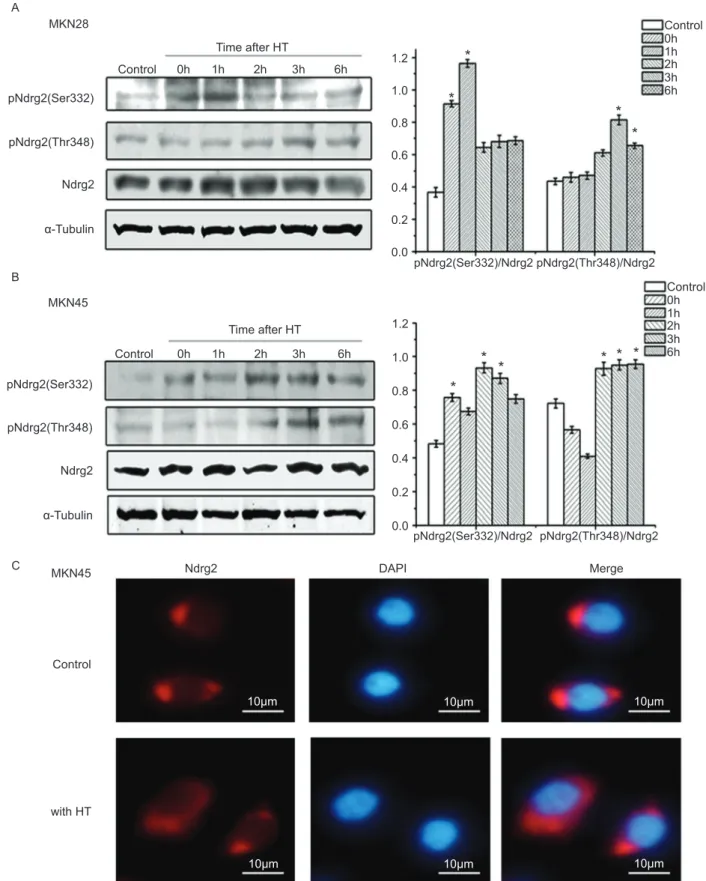
Hyperthermia increased Ndrg2 phosphorylation in gastric cancer cells
Ndrg2 in gastric cancer cells.
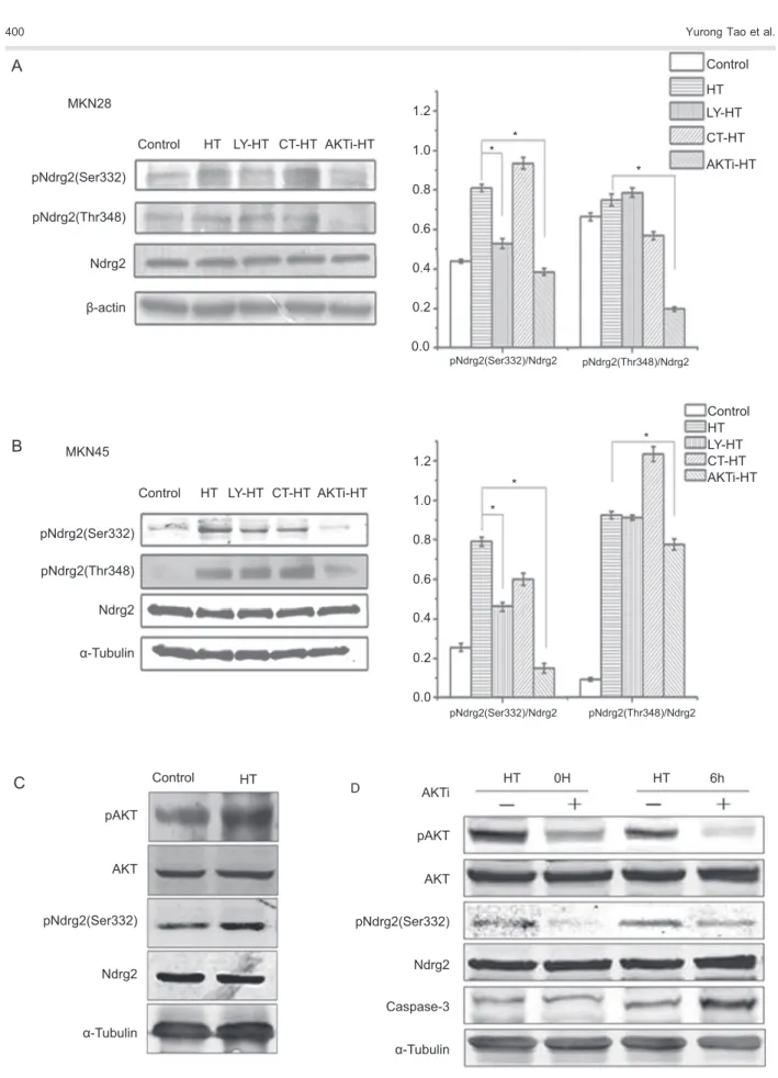
AKT inhibitor attenuated hyperthermia-induced Ndrg2 phosphorylation in gastric cancer cells
Recent studies have shown that Ndrg2 might be a substrate for AKT (14) or other protein kinases (15) under distinct conditions. To identify which kinase was related to Ndrg2 phosphorylation during hyperthermia, MKN28 and MKN45 cells were pretreated for 1 h with the GSK-3 inhibitor CT99021 or AKT inhibitor VIII and then subjected
to heat treatment for 1 h at 426C. After recovery for 3 h at 376C, total and phosphorylated Ndrg2 was detected. We found that Ndrg2 phosphorylation was increased at 3 h after hyperthermia at both Ser332 and Thr348. Phosphorylation of Ndrg2 at Ser332 was decreased by 52.4 and 81.0% by the AKT inhibitor in MKN28 and MKN45 cells, respectively, compared to the hyperthermia group (Figure 4A and B). A similar trend was found for the phosphorylation of Thr348 in MKN28 and MKN45 cells, which was reduced by 73.9 and 15.9% with AKT inhibitor
0.8
A
B
Control MKN28
MKN28 Con
HT
HT Control
MKN45 HT
0.6
0.4
*
*
*
*
*
* *
*
0.2
100 *
*
*
*
EdU-positive cells (%)
50 150
0 Control
With HT
12
0h 12h 24h 48h 72h
24
Incubation time after HT (h) Incubation time after HT (h) 48
0 72 0 12 24 48 72
12 24
Incubation time after HT (h) 48
0 72
Absorbance at 490 nm
1.0
0.0
0.8
0.6
0.4
0.2
Absorbance at 490 nm
1.0
0.0
A B MKN28 MKN45 PI MKN28 * * *
Apoptotic cells (%)
T
rypan blue-positive cells (%)
25 20 15 10 5 0
Con HT-0 HT-12 HT-24 HT-48 HT-72
MKN28 * * * 5 4 3 2 1 0
Con HT-0 HT-12 HT-24 HT-48 HT-72
MKN45
* * *
Apoptotic cells (%)
T
rypan blue-positive cells (%)
25
20
15
10
5
0 Con HT-0 HT-12 HT-24 HT-48 HT-72
MKN45 * * * 6 4 2 0
Con HT-0 HT-12 HT-24 HT-48 HT-72 Control 100 100 101 101 102 102 103 103 HT-0 100 100 101 101 102 102 103 103 HT-12 100 100 101 101 102 102 103 103 HT-24 100 100 101 101 102 102 103 103 HT-48 100 100 101 101 102 102 103 103 HT-72 100 100 101 101 102 102 103 103 Control 100 100 101 101 102 102 103 103 HT-0 103 100 101 101 102 102 103 103 HT-12 Annexin V-FITC 100 100 101 101 102 102 103 103 HT-24 100 100 101 101 102 102 103 103 HT-48 100 100 101 101 102 102 103 103 HT-72 100 100 101 101 102 102 103 103
Figure 2.Hyperthermia (HT) induced apoptosis in gastric cancer cells. MKN28 and MKN45 cells were treated with HT (426C for 1 h) and gastric cancer cells cultured at 376C were used as control (Con).A, Apoptosis rate in MKN28 and MKN45 cells at 0, 12, 24, 48, and 72 h after 1 h of HT. The apoptotic cells were stained with Annexin V/PI and analyzed by flow cytometry. The data are shown in histograms.B,Cell necrosis rates of MKN28 and MKN45 cells were determined using the trypan blue exclusion test for up to 72 h after 1 h of HT. Results are reported as means ± SD for 3 independent experiments. *P,0.05 compared to control (Dunnett test). PI = propidium iodide; FITC = fluorescein isothiocyanate.
MKN28 A
B
C
Time after HT
1.0
Control 0h 1h 2h 3h 6h
Control 0h 1h 2h 3h 6h 0.8
0.6
0.4
0.2 1.2
0.0
1.0
0.8
0.6
0.4
0.2 1.2
0.0 Time after HT
Control
pNdrg2(Ser332)
pNdrg2(Ser332)/Ndrg2 pNdrg2(Thr348)/Ndrg2
pNdrg2(Ser332)/Ndrg2 pNdrg2(Thr348)/Ndrg2 pNdrg2(Thr348)
Ndrg2
Ndrg2
10μm 10μm 10μm
10μm 10μm
10μm
DAPI Merge
α-Tubulin
MKN45
MKN45 pNdrg2(Ser332)
pNdrg2(Thr348)
Ndrg2
Control
with HT α-Tubulin
0h 1h 2h 3h
*
*
*
*
*
*
*
* * *
6h
Control 0h 1h 2h 3h 6h
A
B
C
MKN28
MKN45
1.0
0.8
0.6
0.4
0.2 1.2
0.0
1.0
0.8
0.6
0.4
0.2 1.2
0.0
Control
HT
LY-HT
CT-HT
AKTi-HT
Control HT LY-HT CT-HT AKTi-HT pNdrg2(Ser332)
pNdrg2(Ser332)/Ndrg2 pNdrg2(Thr348)
pNdrg2(Thr348)/Ndrg2
pNdrg2(Ser332)/Ndrg2 pNdrg2(Thr348)/Ndrg2 Ndrg2
β-actin
pNdrg2(Ser332)
pNdrg2(Ser332) pNdrg2(Ser332) pNdrg2(Thr348)
Ndrg2
Ndrg2 Ndrg2
Caspase-3 α-Tubulin
α-Tubulin
α-Tubulin Control HT LY-HT CT-HT AKTi-HT
Control HT
Control
pAKT AKTi D
AKT pAKT
AKT
HT HT 0H HT 6h
LY-HT CT-HT AKTi-HT
* * *
*
*
*
treatment, respectively (Figure 4A and B). However, the GSK-3 inhibitor CT99021 probably did not significantly impact Ndrg2 phosphorylation in MKN28 or MKN45 cells. Following treatment with LY294002, which inhibited the activation of phosphoinositide 3-kinase and hence the activation of AKT, the phosphorylation of Ser332 was decreased dramatically to 34.8 and 41.5% in MKN28 and MKN45 cells, while the phosphorylation of Thr348 remained unchanged in both cell lines (Figure 4A and B). We further detected phosphorylation of AKT in hyperthermia-treated MKN28 cells. As show in Figure 4C, compared to the untreated cells, heat treatment significantly increased AKT phosphorylation. In addition, phosphorylation of Ndrg2 increased correspondingly. These data demonstrate that Ndrg2 phosphorylation could be induced by hyperthermia in an AKT-dependent manner in gastric cancer cells. Ndrg2 Ser332 could be phosphorylated by activation of the classical PI3K/AKT pathway, whereas the ability of AKT to stimulate phosphorylation of Thr348 was partially independent of phosphoinositide 3-kinase activation.
AKT inhibitor promoted hyperthermia-induced apoptosis in gastric cancer cells
We have previously shown that activated AKT enhanced Ndrg2 phosphorylation and abolished the apoptosis induced by free fatty acids in b-TC3 cells (10). In the current study, our data demonstrated that Ndrg2 is a potential substrate of the protein kinase AKT in hyperthermia-treated gastric cancer cells. We further examined the effect of AKT inhibitor on protein expression of the apoptosis marker caspase-3 in heat-treated gastric cells. As shown in Figure 4D, after treatment with the AKT inhibitor, the phosphorylation of AKT and Ndrg2 was suppressed. Meanwhile, AKT inhibitions resulted in an increase in caspase-3 expression levels (Figure 4D).
We further detected the effect of the AKT inhibitor on hyperthermia-induced apoptosis in MKN28 and MKN45 cells. In the hyperthermia group, the percentage of apoptotic cells was 4.4% at 12 h and 14.5% at 24 h, while the apoptosis rate steadily increased from 16.4% at 12 h to 23.1% at 24 h in the AKT inhibitor-hyperthermia group (Figure 5A and B), while it increased slightly to 8.1% in the LY294002-hyperthermia group, and remained
unchanged in the control-hyperthermia group. These data demonstrate that the AKT inhibitor, which suppressed phosphorylation of Ndrg2 at both Ser332 and Thr348, enhanced cell apoptosis induced by hyperthermia in MKN28 cells; while the cell apoptosis was only partly increased by pretreatment of cells with PI3K inhibitors, which suppressed phosphorylation of Ndrg2 only at the Ser332 site. In contrast, the GSK inhibitor, which had no effects on Ndrg2 phosphorylation, did not impact the apoptosis rate of MKN28 cells. Similar results were observed in MKN45 cells (Figure 5A). These results suggest that AKT inhibitor VIII suppressed phosphoryla-tion of Ndrg2 and enhanced hyperthermia-induced apop-tosis in gastric cancer cells. Moreover, phosphorylation of Ndrg2 is negatively related to hyperthermia-induced apoptosis.
Discussion
In this study, we showed that hyperthermia promoted apoptotic cell death in gastric cancer cells 24 h after treatment (Figure 1). Although the expression level of Ndrg2 did not change significantly, phosphorylation of Ndrg2 increased during this process (Figure 3A and B). Further investigation showed that AKT inhibitor VIII attenuated Ndrg2 phosphorylation and enhanced hyperthermia-induced apoptosis (Figures 4 and 5).
Ndrg2 has been reported to be a substrate for several serine-threonine protein kinases including AKT, PKC, SGK1, RSK1, S6K1, and GSK-3 under different experi-mental conditions (14,15). In the present study, our data showed that hyperthermia increased phosphorylation of AKT and Ndrg2 in gastric cancer cells (Figures 3A,B and 4C). In addition, AKT inhibitor VIII resulted in the attenuation of Ndrg2 phosphorylation at both Ser332 and Thr348. Only phosphorylation of Ser332 was reduced after the cell was pretreated with LY294002, a PI3K inhibitor. Furthermore, the GSK-3 inhibitor had no effect on Ndrg2 phosphorylation (Figure 4A). These data suggest that AKT, but not GSK, mediated the phosphor-ylation of Ndrg2 induced by hyperthermia. Furthermore, we found that AKT-regulated phosphorylation of Ndrg2 was partially independent of PI3K activation. This out-come, however, is not rare, as a growing number of
studies have reported mechanisms of PI3K-independent induction of AKT activity (16-18). Moreover, it has been demonstrated that AKT was responsible for the phos-phorylation of GST-Ndrg2 at both the Ser332 and Thr348
sitesin vitro(15). However, another study suggested that Ndrg2 could be phosphorylated by AKT mainly in Thr348 in C2C12 skeletal muscle cells. In the present study, our results showed that the AKT inhibitor reduced Ndrg2
A PI PI MKN28 MKN28 20 10 30 0 0h 24h MKN28 MKN45 MKN45 Control 100 100 101 101 102 102 103 103 100 100 101 101 102 102 103 103 100 100 101 101 102 102 103 103 100 100 101 101 102 102 Annexin V 103 103 100 100 101 101 102 102 103 103 HT 100 100 101 101 102 102 103 103 LY-HT
100 101 102 103
100
101
102
103
CT-HT
HT LY-HT CT-HT
AKTi-HT Control HT-0h HT HT-24h B HT-AKTi-0h HT+AKTi HT+AKTi-24h HT LY-HT CT-HT AKTi-HT
AKTi-HT 100 100 101 101 102 102 103 103 100 100 101 101 102 102 103 103 Control 100 18 16 14 12 10 8 6 4 2 20 0 100 101 101 102 102 103 103 103 100 101 101 102 102 103 103 100 100 101 101 102 102 103 103 100 100 101 101 102 102 103 103 100 100 101 101 102 102 103 103 Annexin V-FITC
Apoptotic cells (%)
Control HT LY-HT CT-HT AKTi-HT 18 16 14 12 10 8 6 4 2 20 0
Apoptotic cells (%)
Apoptotic cells (%)
*
*
*
* *
phosphorylation induced by hyperthermia at both Ser332 and Thr348 (Figure 4A). These differences in AKT-mediated site-specific changes of phosphorylation in Ndrg2 may be due to different experimental conditions or different cell types.
Numerous studies have implied that Ndrg2 plays a tumor suppressor role in many types of cancer cells (19-21). NDRG2 has also been identified as a stress response gene in several studies (13,22). Based on these reports, we had predicted that Ndrg2 might be induced by hyperthermia and might mediate hyperthermia-induced apoptosis. Contrary to our hypothesis, the expression level of Ndrg2 did not change obviously at 24 h when hyperthermia-induced apoptosis occurred in MKN28 cells (Figures 1, 2 and 3). Further investigation revealed that Ndrg2 phosphorylation increased within 6 h after hyperthermia. Intriguingly, we noticed that, compared to the control group, the percentage of apoptotic cells did not change in either cell line during this process (Figure 1). Using a set of kinase inhibitors, we found that AKT-mediated hyperthermia induced Ndrg2 phosphorylation, while the AKT inhibitor suppressed Ndrg2 phosphoryla-tion and enhanced heat stress-induced apoptosis during the early stage of hyperthermia (Figures 3A,B and 4). Thus, these data indicated that AKT-mediated Ndrg2 phosphorylation might be involved in the tolerance of gastric cancer cells to heat-induced stress during the early stage of hyperthermia. Previous studies have demon-strated that the inhibition of the PKB/AKT-dependent survival pathway could promote apoptosis and thermo-sensitization in breast cancer cells (23-25). Although we were unable to gain insights into the function of phosphorylated Ndrg2 in hyperthermia-induced apopto-sis, we did find evidence that AKT inhibitor VIII rendered gastric cancer cells susceptible to hyperthermia-induced apoptosis, partially related to the inhibition of Ndrg2 phosphorylation.
Recent findings also showed that Ndrg2 phosphoryla-tion is involved in the AKT-mediated protecphosphoryla-tion ofb cells against lipotoxicity (10). In addition, our previous studies on
HeLa cells showed that overexpression of Ndrg2 decreased radiation sensitivity, whereas silencing of Ndrg2 caused radiosensitization (12). These results indicated that Ndrg2 may play a protective role in stress-induced apoptosis. In this regard, the biological function of Ndrg2 in cell apoptosis induced by specific stress condi-tions is similar to that of Ndrg1 (26). Like Ndrg2, Ndrg1 has been reported to be a tumor suppressor since its expression is reduced in cancer and metastatic tissues (27). Some studies have shown that hypoxia increased the expression of Ndrg1, which is responsible for reduced apoptosis in drug resistance (28,29). Moreover, the decreased level of Ndrg1 facilitated cell apoptosis that occurred under stress conditions (30). Regarding the role of Ndrg2 in cell apoptosis, we hypothesized that the expression level and post-translational modification of Ndrg2 might contribute to the different cellular response under stress conditions. Another possibility is that Ndrg2 exerts functions as an anti- or pro-apoptosis factor in a context- and cell type-specific manner. Further investiga-tions are required to validate the role of Ndrg2 phosphor-ylation in hyperthermia-induced apoptosis.
Taken together, our data demonstrated that AKT mediated hyperthermia-induced Ndrg2 phosphorylation. Phosphorylation of Ndrg2 was negatively correlated with hyperthermia-induced apoptosis in gastric cancer cells. Further investigation revealed that the AKT inhibitor suppressed Ndrg2 phosphorylation and enhanced hyperthermia-induced apoptosis in gastric cancer cells. Therefore, pre-treatment with the AKT inhibitor might offer a novel strategy to improve the efficacy of hyperthermia treatment in gastric cancer.
Acknowledgments
Research supported by grants from the National Natural Science Foundation of China (#30973437,
#30700918, #30800492, and #801170748) and the Chinese National Key Basic Research and Development Program (#2009CB521704).
References
1. Issels RD, Lindner LH, Verweij J, Wust P, Reichardt P, Schem BC, et al. Neo-adjuvant chemotherapy alone or with regional hyperthermia for localised high-risk soft-tissue sarcoma: a randomised phase 3 multicentre study.Lancet Oncol2010; 11: 561-570, doi: 10.1016/S1470-2045(10)70071-1. 2. Qu X, Zhai Y, Wei H, Zhang C, Xing G, Yu Y, et al.
Characterization and expression of three novel differentiation-related genes belong to the human NDRG gene family.Mol Cell Biochem2002; 229: 35-44, doi: 10.1023/A:1017934810825. 3. Tschan MP, Shan D, Laedrach J, Eyholzer M, Leibundgut
EO, Baerlocher GM, et al. NDRG1/2 expression is inhibited in primary acute myeloid leukemia.Leuk Res2010; 34: 393-398, doi: 10.1016/j.leukres.2009.08.037.
4. Zhou RH, Kokame K, Tsukamoto Y, Yutani C, Kato H, Miyata T. Characterization of the human NDRG gene family: a newly identified member, NDRG4, is specifically expressed in brain and heart.Genomics2001; 73: 86-97, doi: 10.1006/geno.2000.6496.
5. Tepel M, Roerig P, Wolter M, Gutmann DH, Perry A, Reifenberger G, et al. Frequent promoter hypermethylation and transcriptional downregulation of the NDRG2 gene at 14q11.2 in primary glioblastoma.Int J Cancer2008; 123: 2080-2086, doi: 10.1002/ijc.23705.
cell death.Exp Mol Med2007; 39: 705-714, doi: 10.1038/ emm.2007.77.
7. Takahashi K, Yamada M, Ohata H, Honda K, Yamada M. Ndrg2 promotes neurite outgrowth of NGF-differentiated PC12 cells.Neurosci Lett2005; 388: 157-162, doi: 10.1016/ j.neulet.2005.06.055.
8. Lorentzen A, Lewinsky RH, Bornholdt J, Vogel LK, Mitchelmore C. Expression profile of the N-myc Downstream Regulated Gene 2 (NDRG2) in human cancers with focus on breast cancer.BMC Cancer2011; 11: 14, doi: 10.1186/1471-2407-11-14.
9. Yu C, Wu G, Dang N, Zhang W, Zhang R, Yan W, et al. Inhibition of N-myc downstream-regulated gene 2 in prostatic carcinoma.Cancer Biol Ther2011; 12: 304-313, doi: 10.4161/ cbt.12.4.16382.
10. Shen L, Liu X, Hou W, Yang G, Wu Y, Zhang R, et al. NDRG2 is highly expressed in pancreatic beta cells and involved in protection against lipotoxicity.Cell Mol Life Sci 2010; 67: 1371-1381, doi: 10.1007/s00018-010-0258-1. 11. Foletta VC, Prior MJ, Stupka N, Carey K, Segal DH, Jones
S, et al. NDRG2, a novel regulator of myoblast proliferation, is regulated by anabolic and catabolic factors. J Physiol 2009; 587: 1619-1634, doi: 10.1113/jphysiol.2008.167882. 12. Liu J, Zhang J, Wang X, Li Y, Chen Y, Li K, et al. HIF-1 and
NDRG2 contribute to hypoxia-induced radioresistance of cervical cancer Hela cells.Exp Cell Res2010; 316: 1985-1993, doi: 10.1016/j.yexcr.2010.02.028.
13. Wang L, Liu N, Yao L, Li F, Zhang J, Deng Y, et al. NDRG2 is a new HIF-1 target gene necessary for hypoxia-induced apoptosis in A549 cells. Cell Physiol Biochem2008; 21: 239-250, doi: 10.1159/000113765.
14. Burchfield JG, Lennard AJ, Narasimhan S, Hughes WE, Wasinger VC, Corthals GL, et al. Akt mediates insulin-stimulated phosphorylation of Ndrg2: evidence for cross-talk with protein kinase C theta.J Biol Chem2004; 279: 18623-18632, doi: 10.1074/jbc.M401504200.
15. Murray JT, Campbell DG, Morrice N, Auld GC, Shpiro N, Marquez R, et al. Exploitation of KESTREL to identify NDRG family members as physiological substrates for SGK1 and GSK3. Biochem J 2004; 384: 477-488, doi: 10.1042/BJ20041057.
16. Yano S, Tokumitsu H, Soderling TR. Calcium promotes cell survival through CaM-K kinase activation of the protein-kinase-B pathway. Nature 1998; 396: 584-587, doi: 10.1038/25147.
17. Martinez-Lopez N, Varela-Rey M, Fernandez-Ramos D, Woodhoo A, Vazquez-Chantada M, Embade N, et al. Activation of LKB1-Akt pathway independent of phosphoi-nositide 3-kinase plays a critical role in the proliferation of hepatocellular carcinoma from nonalcoholic steatohepatitis. Hepatology2010; 52: 1621-1631, doi: 10.1002/hep.23860. 18. Guo JP, Coppola D, Cheng JQ. IKBKE protein activates Akt independent of phosphatidylinositol 3-kinase/PDK1/ mTORC2 and the pleckstrin homology domain to sustain
malignant transformation.J Biol Chem2011; 286: 37389-37398, doi: 10.1074/jbc.M111.287433.
19. Lusis EA, Watson MA, Chicoine MR, Lyman M, Roerig P, Reifenberger G, et al. Integrative genomic analysis identifies NDRG2 as a candidate tumor suppressor gene frequently inactivated in clinically aggressive meningioma.Cancer Res 2005; 65: 7121-7126, doi: 10.1158/0008-5472.CAN-05-0043. 20. Assamaki R, Sarlomo-Rikala M, Lopez-Guerrero JA, Lasota J, Andersson LC, Llombart-Bosch A, et al. Array compara-tive genomic hybridization analysis of chromosomal imbal-ances and their target genes in gastrointestinal stromal tumors.Genes Chromosomes Cancer2007; 46: 564-576, doi: 10.1002/gcc.20439.
21. Lee DC, Kang YK, Kim WH, Jang YJ, Kim DJ, Park IY, et al. Functional and clinical evidence for NDRG2 as a candidate suppressor of liver cancer metastasis.Cancer Res2008; 68: 4210-4220, doi: 10.1158/0008-5472.CAN-07-5040. 22. Liu N, Wang L, Li X, Yang Q, Liu X, Zhang J, et al. N-Myc
downstream-regulated gene 2 is involved in p53-mediated apoptosis. Nucleic Acids Res 2008; 36: 5335-5349, doi: 10.1093/nar/gkn504.
23. Ma N, Szmitko P, Brade A, Chu I, Lo A, Woodgett J, et al. Kinase-dead PKB gene therapy combined with hyperther-mia for human breast cancer.Cancer Gene Ther2004; 11: 52-60, doi: 10.1038/sj.cgt.7700655.
24. Lee CS, Kim YJ, Jang ER, Myung SC, Kim W. Akt inhibitor enhances apoptotic effect of carboplatin on human epithelial ovarian carcinoma cell lines.Eur J Pharmacol2010; 632: 7-13, doi: 10.1016/j.ejphar.2010.01.004.
25. Cheng Y, Ren X, Zhang Y, Patel R, Sharma A, Wu H, et al. eEF-2 kinase dictates cross-talk between autophagy and apoptosis induced by Akt inhibition, thereby modulating cytotoxicity of novel Akt inhibitor MK-2206. Cancer Res 2011; 71: 2654-2663, doi: 10.1158/0008-5472.CAN-10-2889. 26. Ellen TP, Ke Q, Zhang P, Costa M. NDRG1, a growth and cancer related gene: regulation of gene expression and function in normal and disease states. Carcinogenesis 2008; 29: 2-8, doi: 10.1093/carcin/bgm200.
27. Kovacevic Z, Richardson DR. The metastasis suppressor, Ndrg-1: a new ally in the fight against cancer.Carcinogenesis 2006; 27: 2355-2366, doi: 10.1093/carcin/bgl146.
28. Cangul H. Hypoxia upregulates the expression of the NDRG1 gene leading to its overexpression in various human cancers. BMC Genet 2004; 5: 27, doi: 10.1186/ 1471-2156-5-27.
29. Jung EU, Yoon JH, Lee YJ, Lee JH, Kim BH, Yu SJ, et al. Hypoxia and retinoic acid-inducible NDRG1 expression is responsible for doxorubicin and retinoic acid resistance in hepatocellular carcinoma cells.Cancer Lett2010; 298: 9-15, doi: 10.1016/j.canlet.2010.05.020.