MARÍLIA VIEIRA FEBRÔNIO
Citologia cérvico-vaginal inflamatória associada com
atividade da doença no lúpus eritematoso sistêmico juvenil
Tese apresentada à Faculdade de Medicina da
Universidade de São Paulo para obtenção do
título de Doutor em Ciências
Área de Concentração: Reumatologia
Orientador: Dr. Clovis Artur Almeida da Silva
A
Deus,
AGRADECIMENTOS
À minha mãe Marisa, pelo seu exemplo de integridade, humildade e perseverança, possibilitando que eu pudesse estudar e me formar médica.
A meu irmão Rodrigo, a sua existência fez com que eu aprendesse a “dividir”.
A Randy...sua presença na minha vida fez eu não desistir....
Ao Dr.Clovis....mais do que um orientador, um grande amigo e um “pai”.
Às doutoras Adriana Maluf, Lúcia Campos, Bernadete Liphaus e Paola Lotito, pelo aprendizado, amizade e convivência no serviço de Reumatologia Pediátrica do Instituto da Criança.
Aos meus grandes amigos, Ricardo e Georgiana, pela presença amiga em vários momentos dessa tese, com palavras de estímulo e carinho.
Aos amigos da Reumatologia Pediátrica: Ana Júlia, Mércia, Beth, Rosa, Daniela Lipai, Nadyesda, Aline Islabão, Adriana Jesus, Carlos, Nádia, Kátia, Renata e Aline Miranda, por compartilharmos conhecimento, ajuda e mais que tudo, amizade.
À Dra. Albertina Duarte, pela oportunidade dada em acompanhar o trabalho no ambulatório da Ginecologia do Adolescente.
À Dra. Elsa Gay, pela ajuda na interpretação dos laudos do testes de Papanicolaou.
À Dra. Eloísa Bonfá, agradeço pelas orientações e revisão do artigo para publicação.
À Dra. Rosa Pereira pela ajuda na seleção das pacientes no ambulatório da Raumatologia.
Ao Dr.Ulisses e ao Dr.Crésio pela realização da análise estatística.
A Marisa, bibliotecária do Instituto da Criança, pela ajuda no levantamento bibliográfico.
SUMÁRIO
Resumo Summary
1. Introdução ... 1
2. Objetivos ... 4
3. Métodos ... 6
4. Resultados ... 11
5. Discussão ... 18
6. Conclusões ... 23
7. Anexos ... 25
RESUMO
FEBRÔNIO MV. Citologia cérvico-vaginal inflamatória associada com
atividade da doença no lúpus eritematoso sistêmico juvenil (LESJ) [tese]. São Paulo: Faculdade de Medicina, Universidade de São Paulo; 2006, 57p.
Objetivo: Avaliar a citologia cérvico-vaginal em adolescentes com lúpus eritematoso sistêmico juvenil (LESJ) e comparar com controles. Material e métodos: Cinqüenta e duas adolescentes com LESJ (critérios do American
SUMMARY
FEBRÔNIO MV. Inflammatory cervicovaginal cytology is associated with
disease activity in juvenile systemic lupus erythematosus (LESJ) [thesis]. São Paulo: Faculdade de Medicina, Universidade de São Paulo; 2006, 57p.
Introdução 2
O lúpus eritematoso sistêmico juvenil (LESJ) é uma doença auto-imune multisistêmica de etiologia desconhecida acometendo especialmente mulheres jovens 1. Os recentes avanços no entendimento da genética, mecanismos imunopatológicos e novas terapias do LESJ têm resultado em melhora das taxas de sobrevida de 10 anos em mais de 80% desses pacientes 2,3.
Como as pacientes com LESJ apresentam uma maior sobrevida, uma preocupação com a melhora da qualidade de vida tem sido evidenciada. Neste aspecto, tornam-se adolescentes e iniciam a vida sexual com um maior risco de doenças sexualmente transmissíveis (DST), vaginite e displasia cervical.
Vaginite é uma das infecções mais freqüentes em mulheres de todas as idades4. Além disto, evidências sugerem que vaginite, particularmente candidíase vulvovaginal, é um importante problema para pacientes com doença crônica e para aquelas que utilizam imunosupressores4-6.
Introdução 3
Objetivos 5
1. Avaliar a citologia cérvico-vaginal de adolescentes com LESJ e grupo controle.
Métodos 7
Pacientes
Métodos 8
Avaliação clínica, laboratorial e tratamento LESJ
A avaliação clínica foi realizada pela mesma reumatologista (MVF) e consistiu na revisão de dados sobre manifestações clínicas, exames laboratoriais e terapêuticas utilizadas. Os dados foram obtidos dos prontuários e registrados em um protocolo específico (Anexo 3). As manifestações clínicas do LES foram definidas como: envolvimento cutâneo e de mucosas (eritema malar, lúpus discóide, úlceras orais, vasculite ou fotossensibilidade), comprometimento articular (artralgia ou artrite não-erosiva), doença neuropsiquiátrica (convulsão, psicose, depressão ou neuropatia periférica), envolvimento renal (proteinúria ≥ 0,5g/24h, presença de cilindros celulares, hematúria persistente com ≥ 10 hemácias/campo ou insuficiência renal), doença cárdio-pulmonar (serosite, miocardite, doença pulmonar restritiva e hipertensão pulmonar) e alterações hematológicas (anemia hemolítica, leucopenia com contagem de leucócitos < 4.000/mm3, linfopenia < 1.500/mm3 e trombocitopenia com contagem de plaquetas < 100.000/mm3, na ausência de drogas ou infecção).
No início do estudo, foram determinados atividade de doença e dano cumulativo em todos os pacientes utilizando-se os escores: Systemic Lupus
Erythematosus Disease Activity Index (SLEDAI) 17 e Systemic Lupus
Métodos 9
Avaliação Ginecológica
Um exame clínico sistemático da genitália foi realizado por uma mesma ginecologista e incluiu avaliação da vulva, hímen, vagina e colo uterino, características sexuais secundárias, de acordo com os padrões puberais de Marshall e Tanner19, e coletado o esfregaço para o teste de Papanicolaou na fase folicular do ciclo menstrual (Anexo 3).
As pacientes foram avaliadas de acordo com: idade da menarca; idade da primeira relação sexual; freqüência de relações sexuais (no último mês), número de parceiros sexuais (no último ano); vaginite (candidíase, vaginose bacteriana, Trichomonas vaginallis, HPV, etc) e contracepção (Anexo 3).
Citologia cérvico-vaginal
Os esfregaços para o Papanicolaou foram coletados nas adolescentes virgens com escova Cytobrush ®, que foi inserida na borda do orifício vaginal e gentilmente rodada entre 90° e 180°, e imediatamente rolada sobre o terço externo da lâmina. Nas adolescentes sexualmente ativas, após introdução do espéculo, a citologia foi coletada com escova
Métodos 10
foi espalhado em fina camada sobre o terço médio da lâmina. Após a fixação por imersão em álcool a 95%, os esfregaços foram imediatamente transportados ao laboratório20.
Todos os esfregaços foram avaliados por uma mesma citopatologista do nosso hospital universitário que desconhecia o exame ginecológico. Os esfregaços foram avaliados de acordo com o Sistema de Bethesda 2001 em cinco padrões, com presença ou ausência de infecção pelo HPV: normal (alterações celulares benignas), alterações inflamatórias, células escamosas atípicas de significado indeterminado (CEASI), lesões intra-epiteliais escamosas de baixo ou alto grau (LIE-BG ou LIE-AG) e carcinoma in situ. Foram também avaliadas as identificações dos organismos (Trichomonas
vaginalis, Candida spp, vaginose bacteriana, Actinomyces spp, vírus herpes simples)21.
Análise estatística
Resultados 12
Características demográficas
A distribuição das características demográficas revelou que as pacientes com LESJ e controles foram semelhantes com relação à idade (16,17 ± 1,94 versus 16,13 ± 2,16 anos, p=0,92), à classe sócio-econômica (classe média alta: 21% versus 21%, classe média: 64% versus 52%, classe média baixa: 15% versus 23%, p=0,33) e anos de educação (9,51 ± 1,78 versus 9,7 ± 1,62 anos, p=0,57).
Avaliação Ginecológica
Resultados 13
(1-10), p=1,0] nas pacientes sexualmente ativas com LESJ e controles. Assim como, foram encontradas freqüências similares de contraceptivos (83% versus 90%, p=0,61) e uso de preservativo masculino nas pacientes sexualmente ativas com LESJ e controles (92% versus 70%, p=0,65).
Citologia cérvico-vaginal
Esfregaços normais foram observados em 50% nos dois grupos (p=1,15), alterações inflamatórias em 48% das pacientes com LESJ e em 46% do grupo controle (p=1,0). LIE-BG com presença de infecção pelo HPV foram observadas em apenas uma paciente com LESJ (2%) versus duas controles (4%) (p=1,0) (Tabela 1), apesar que 27 (52%) pacientes com LESJ estavam em uso de um ou mais drogas imunossupressoras (azatioprina em 17, PCE em 15, metotrexate em 11, ciclosporina em 2 e micofenolato mofetil em 1). Por outro lado, entre as 12 pacientes com LESJ e 31 controles sexualmente ativas não foram observadas diferenças em relação à freqüência de citologia normal, inflamatória e LIE-BG (25% versus 29%, p=1,0; 67% versus 65%, p=1,0; 8% versus 6%, p=1,0; respectivamente).
Tabela 1 – Citologia cérvico-vaginal das pacientes com LESJ e controles Citologia cérvico-vaginal, n (%) LESJ (n=52) Controles (n=52) P Citopatologia Normal Inflamatória 26 (50) 25 (48) 26 (50) 24 (46) 1,15 1,0
LIE-BG 1 (2) 2 (4) 1,0
Organismos
Vaginose bacteriana 0 (0) 4 (8) 0,11
Candida spp 7 (14) 0 (0) 0,01
Resultados 14
Na citologia cérvico-vaginal, achados inflamatórios foram significativamente associados com atividade da doença nas pacientes com LESJ com e sem atividade sexual. De fato, citologia com alterações inflamatórias foi observada em 60% das pacientes com SLEDAI ≥ 4 versus 23,5% das pacientes com SLEDAI < 4 (p=0,001). Reforçando esse achado, apenas 37% das pacientes com SLEDAI ≥ 4 tinham citologia normal em comparação com 77% daqueles com SLEDAI < 4 (p=0,001) (Tabela 2).
Tabela 2 - Características da citologia cérvico-vaginal de acordo com atividade da doença
Citologia cérvico-vaginal, n (%)
SLEDAI < 4 (n=17)
SLEDAI ≥ 4 (n=35)
P
Normal 13 (77) 13 (37) 0,001
Inflamatória 4 (23,5) 21 (60) 0,001
LIE-BG 0 (0) 1 (3) 1,0
LESJ - Lúpus Eritematoso Sistêmico Juvenil, LIE-BG – lesão intra-epitelial de baixo grau, SLEDAI - Systemic Lupus Erythematosus Disease Activity Index.
A análise específica das pacientes virgens com LESJ revelou que achados inflamatórios foram também observados mais freqüentemente em pacientes com SLEDAI ≥ 4 em relação as pacientes com SLEDAI < 4 (40% versus 8%, respectivamente, p=0,005) (Tabela 3).
Tabela 3 - Características da citologia cérvico-vaginal em adolescentes virgens com LESJ de acordo com atividade da doença
Citologia cérvico-vaginal, n (%)
SLEDAI < 4 (n=12)
SLEDAI ≥ 4 (n=28)
p
Normal 11 (92) 12 (53) 0,005
Inflamatória 1 (8) 16 (57) 0,005
LESJ - Lúpus Eritematoso Sistêmico Juvenil, SLEDAI - Systemic Lupus Erythematosus
Resultados 15
Em contraste, nenhuma associação estatística foi observada em relação ao SLICC/ACR-DI ≥ 1 ou < 1 e achados inflamatórios (48% versus 50%, p=1,0).
Organismos
Resultados 16
Tabela 4 – Dados clínicos, atividade, dano e tratamento das pacientes com LESJ com e sem infecção vaginal por Candida spp
Variáveis Com
Candida spp (n=7) Sem Candida spp (n=45) p Características clínicas
Cutâneo, n (%) 7 (100) 43 (96) 1,0
Articular, n (%) 6 (86) 40 (89) 1,0
Renal, n (%) 7 (100) 37 (82) 0,57
Cardiopulmonar, n (%) 4 (57) 19 (42) 0,68 Neuropsiquiátrico, n (%) 2 (29) 10 (22) 0,65 Hematológico, n (%) 6 (86) 42 (93) 0,44
Atividade e dano do LESJ
SLEDAI, média ± DP 5,4 ± 4,2 5,5 ± 4,4 0,95 SLICC/ACR-DI ≥ 1, n (%) 3 (4) 7 (16) 0,3
Tratamento
Dose atual de Pd (mg), média ± DP 20,5 ± 5 10,2 ± 8 0,002 Uso atual de IS, n (%) 7 (100) 20 (44) 0,01 LESJ - Lúpus Eritematoso Sistêmico Juvenil, IS – imunossupressor, Pd – prednisona, SLEDAI - Systemic Lupus Erythemathosus Disease Activity Index, SLICC/ACR-DI -
Systemic Lupus International Collaborating Clinics/American College of Rheumatology-Damage Index, DP – desvio padrão.
Resultados 17
de SLICC-ACR/DI ≥ 1 (43% versus 16%, p=0,3) foram comparáveis nas pacientes com LESJ com presença ou ausência de Candida spp na citologia cérvico-vaginal (Tabela 4).
Condiloma acuminado por infecção pelo HPV foi identificado na vulva, vagina e/ou colo pela colposcopia em duas controles sexualmente ativas (6,4%) e em nenhuma paciente com LESJ (p=1,0). Estas pacientes tinham LIE-BG e foram tratadas com excisão eletrocirúrgica por alça.
Vaginoses bacterianas por Gardnerella vaginallis foram observadas em três (10%) das pacientes controles sexualmente ativas e em nenhuma das pacientes com LESJ sexualmente ativas (p=0,54). Estas pacientes apresentaram uma secreção vaginal fétida que era tipicamente esparsa com uma cor acinzentada. O tratamento da vaginose bacteriana foi realizado com metronidazol tópico.
Discussão 19
Este é o primeiro estudo que especialmente estudou a citologia cérvico-vaginal em adolescentes com LESJ e claramente demonstrou uma alta freqüência de achados inflamatórios e infecção fúngica no teste de Papanicolaou.
Vaginite é um dos maiores problemas em pacientes imunocomprometidas, tais como: diabetes mellitus, HIV e doenças auto-imunes6. De fato, situações patológicas em que o pH vaginal aumenta, pode resultar em supressão do crescimento dos lactobacilos e um hiper-crescimento de bactérias ou fungos potencialmente patogênicos, determinando vaginite4.
Em mulheres saudáveis, as infecções vaginais mais comuns são:
Candida spp, vaginose bacteriana e Trichomonas vaginalis4. Por sua vez, nenhum relato tem sido descrito no lúpus em relação à vaginite, apesar de vários estudos demonstrarem que infecção é a maior causa de morbidade e mortalidade nesses pacientes22.
Discussão 20
defeito ou disfunção dos linfócitos CD4+ T helper tipo 1. Além disso, os linfócitos do sangue periférico das pacientes que estão recebendo altas doses de prednisona estão diminuídos in vivo quando há estimulação antigênica pela Candida spp23. Neste aspecto, as pacientes com LESJ que utilizam drogas imunossupressoras devem ser consideradas candidatas às infecções fúngicas. De fato, todas as pacientes com essa infecção oportunista estavam utilizando terapias com imunossupressor e com altas doses de prednisona.
O trato genital feminino deve ser considerado como um local primário de candidíase, com probabilidade de disseminação sistêmica, pois estudos prévios demonstram que Candida albicans foi o mais freqüente microorganismo oportunista identificado em infecção fatal em adultos com LES24.
No presente estudo, não foi realizada cultura para identificar as espécies de fungos, pois a literatura reforça que habitualmente não é necessário, já que a Candida spp é sensível ao fluconazol,25 clotrimazol ou miconazol4 tópicos, como também foi observado neste estudo.
Discussão 21
evidenciado previamente que adolescentes com LESJ apresentaram um atraso na menarca (13,5 ± 1,4 anos) em comparação com 2578 adolescentes normais Brasileiras (12,5 ± 1,3 anos)31.
Por sua vez, estudos prévios em mulheres adultas com LES demonstraram uma alta prevalência de atipia cervical com LIE-BG variando de 12% a 36%8-10,12 e foi associada com uso de drogas imunossupressoras, particularmente PCE e azatioprina8,12-14. A baixa freqüência de displasia cervical em nossas pacientes, apesar da alta freqüência do uso de imunossupressor, sugere que a atividade sexual é provavelmente o principal fator contribuinte para as alterações no teste de Papanicolaou.
Atualmente, a incidência de anormalidades cervicais entre adolescentes está relacionada primariamente às altas taxas de atividade sexual, promiscuidade e infecção pelo HPV 28,32-34. Uma possível explicação para essa associação é a maior vulnerabilidade do colo uterino “imaturo”, que é coberto predominantemente por um epitélio colunar e metaplásico, com intensa replicação e diferenciação, e esse tipo de epitélio é um hospedeiro ideal para a replicação do HPV33. O uso freqüente de preservativo masculino (camisinha) pelos parceiros das nossas pacientes com LESJ e controles no presente estudo, provavelmente, contribuiu para a baixa incidência de displasia cervical e infecção vulvovaginal pelo HPV, como demonstrado recentemente em mulheres jovens sexualmente ativas35.
Discussão 22
Conclusões 24
1. Displasia cervical e infecção por HPV foram raramente observadas em adolescentes com LESJ e controles. Por sua vez, candidíase vaginal foi evidenciada exclusivamente em pacientes com LESJ. Candidíase vaginal nas adolescentes com lúpus foi associada com medicamentos imunossupressores.
Anexos 26
ANEXO 1
HOSPITAL DAS CLÍNICAS
DA
FACULDADE DE MEDICINA DA UNIVERSIDADE DE SÃO PAULO
TERMO DE CONSENTIMENTO LIVRE E ESCLARECIDO
(Instruções para preenchimento no verso)
_______________________________________________________________
I - DADOS DE IDENTIFICAÇÃO DO SUJEITO DA PESQUISA OU RESPONSÁVEL LEGAL
1.NOME DO PACIENTE .:... .. DOCUMENTO DE IDENTIDADE Nº : ... SEXO : M F DATA NASCIMENTO: .../.../...
ENDEREÇO ... Nº ... APTO: ... BAIRRO: ... CIDADE ... CEP:...EFONE: DDD (...) ... 2.RESPONSÁVEL LEGAL ...
NATUREZA (grau de parentesco, tutor, curado etc.)... DOCUMENTO DE IDENTIDADE :...SEXO: M F
DATA NASCIMENTO.: .../.../...
ENDEREÇO:...Nº...APTO:...BAIRRO:... CIDADE:...CEP:...TELEFONE:DDD(...)... ____________________________________________________________________________
II - DADOS SOBRE A PESQUISA CIENTÍFICA
1. TÍTULO DO PROTOCOLO DE PESQUISA : Citologia cérvico-vaginal em adolescentes do sexo feminino com Lúpus Eritematoso Sistêmico Juvenil
2. PESQUISADOR: Clovis Artur Almeida da Silva
CARGO/FUNÇÃO: Médico Responsável - Unidade de Reumatologia INSCRIÇÃO CONSELHO REGIONAL Nº 64724
UNIDADE DO HCFMUSP: Unidade de Reumatologia Pediátrica do ICr-HC-FMUSP 3. AVALIAÇÃO DO RISCO DA PESQUISA:
SEM RISCO
x
RISCO MÍNIMO RISCO MÉDIORISCO BAIXO RISCO MAIOR
(probabilidade de que o indivíduo sofra algum dano como consequência imediata ou tardia do estudo)
Anexos 27
III - REGISTRO DAS EXPLICAÇÕES DO PESQUISADOR AO PACIENTE OU SEU REPRESENTANTE LEGAL SOBRE A PESQUISA, CONSIGNANDO:
1. justificativa e os objetivos da pesquisa: Atualmente, com a melhora do prognóstico do LESJ, os reumatologistas pediátricos encontram-se tendo que lidar com questões próprias da adolescência vividas por seus pacientes, entre elas o início da sexualidade e necessidade da visita com ginecologista. A citologia cérvico-vaginal ou citologia oncótica (teste de Papanicolau) é coletada rotineiramente no exame ginecológico de adolescentes, mesmo sem o início da atividade sexual, com a finalidade de identificar alterações iniciais de infecções e tumores. Alguns estudos evidenciaram aumento do risco de alterações do teste de Papanicolau em mulheres adultas com lúpus eritematoso sistêmico. A ausência de estudos na faixa etária pediátrica estimulou a realização deste projeto.
2. procedimentos que serão utilizados e propósitos, incluindo a identificação dos procedimentos que são experimentais: Os pacientes serão examinados por ginecologistas especializados em adolescentes e será coletado o teste de Papanicolaou na borda vaginal pelo ginecologista, com escova especial, mesmo em pacientes sem início da atividade sexual. Nos pacientes com atividade sexual será coletado no colo do útero.
3. desconfortos e riscos esperados: A coleta do teste de Papanicolaou na borda vaginal pelo ginecologista, com escova especial, poderá causar um leve desconforto, com dor local. Raramente leva sangramento no local e não ocasiona ruptura do hímen (perda da virgindade).
4. benefícios que poderão ser obtidos: Avaliação completa do ginecologista e identificação de possíveis infecções e tumores vaginais.
5. procedimentos alternativos que possam ser vantajosos para o indivíduo:? Seguimento e tratamento ginecológico nos pacientes que apresentarem alterações.
____________________________________________________________________________________
IV - ESCLARECIMENTOS DADOS PELO PESQUISADOR SOBRE GARANTIAS DO SUJEITO DA PESQUISA: ?
1. acesso, a qualquer tempo, às informações sobre procedimentos, riscos e benefícios relacionados à pesquisa, inclusive para dirimir eventuais dúvidas. O paciente terá acesso a informação e discussão do procedimento do estudo (avaliação do ginecologista e teste de Papanicolaou).
2. liberdade de retirar seu consentimento a qualquer momento e de deixar de participar do estudo, sem que isto traga prejuízo à continuidade da assistência. O paciente poderá retirar seu consentimento a qualquer tempo, sem prejuízo do seu seguimento no nosso Serviço.
Anexos 28
4. disponibilidade de assistência no HCFMUSP, por eventuais danos à saúde, decorrentes da pesquisa. Em casos de eventuais danos à saúde relacionados a pesquisa, o paciente terá acesso ao Ambulatório de Ginecologia Infanto-Puberal do HC-FMUSP.
5. viabilidade de indenização por eventuais danos à saúde decorrentes da pesquisa. Nos casos de eventuais danos à saúde relacionados a pesquisa, o paciente terá acesso ao tratamento no Ambulatório de Ginecologia Infanto-Puberal e Unidade de Reumatologia Pediátrica do ICr do HC-FMUSP.
_______________________________________________________________ V. INFORMAÇÕES DE NOMES, ENDEREÇOS E TELEFONES DOS RESPONSÁVEIS PELO ACOMPANHAMENTO DA PESQUISA, PARA CONTATO EM CASO DE INTERCORRÊNCIAS CLÍNICAS E REAÇÕES
ADVERSAS.
O estudo é retrospectivo, com análise dos prontuários.
____________________________________________________________________________________
VI. OBSERVAÇÕES COMPLEMENTARES:
--
____________________________________________________________________________________
VII - CONSENTIMENTO PÓS-ESCLARECIDO
Declaro que, após convenientemente esclarecido pelo pesquisador e ter entendido o que me foi explicado, consinto em participar do presente Protocolo de Pesquisa
São Paulo, 17 de Outubro de 2003.
____________________________________________ _____________________________________ assinatura do sujeito da pesquisa ou responsável legal assinatura do pesquisador
Anexos 29
ANEXO 2
CLASSE SÓCIO-ECONÔMICA ABA-ABIPEMI
(ALMEIDA, WICKERHAUSER, 1991).
A. Quem é o chefe da família na sua casa? ( ) o próprio entrevistado ( ) outros B. Qual foi o último ano da escola que o chefe da família cursou?
Grau de instrução máximo Pontos Não estudou ou primário incompleto 0 Primário completo ou ginásio incompleto 05 Ginásio completo ou colegial incompleto 10 Colegial incompleto ou universitário incompleto 15 Universitário completo 21 C. Na sua casa tem?
Aparelho de vídeo cassete ( ) não ( ) sim (10 pontos) Máquina de lavar roupas ( ) não ( ) sim (08 pontos) Geladeira ( ) não ( ) sim (07 pontos) Aspirador de pó ( ) não ( ) sim (06 pontos) D. Quantos (cada item abaixo) existem em sua casa?
Item Nenhum item
1 2 3 4 5 6 ou mais
itens Carros 0 ponto 4 pontos 9 pontos 13 pontos 18 pontos 22 pontos 26 pontos TV em
cores
0 ponto 4 pontos 7 pontos 11 pontos 14 pontos 18 pontos 22 pontos Banheiros 0 ponto 2 pontos 5 pontos 7 pontos 10 pontos 12 pontos 15 pontos Empregada
mensalista
0 ponto 5 pontos 11 pontos 16 pontos 21 pontos 26 pontos 32 pontos Rádio 0 ponto 2 pontos 3 pontos 5 pontos 6 pontos 8 pontos 9 pontos
Classes sócio - econômicas ABA-ABIPEMI Total de pontos
A 89 ou mais
B 59 a 88
C 35 a 58
D 20 a 34
Anexos 30
ANEXO 3
PROTOCOLO DE CITOLOGIA CÉRVICO-VAGINAL EM ADOLESCENTES COM LESJ.
A- IDENTIFICAÇÃO
Nome:_____________________________________ Registro:_____________________ Pai:_______________________________________ Mãe:_________________________ Data de nascimento:___________ Idade de início do LES (meses):____________________ Idade atual (meses):____________ Tempo de duração do LES:______________________ Data da 1ª consulta:_____________ Data avaliação:_______________________________ Raça:__________ Sexo:_____________ Procedência atual:_________________________ Naturalidade:_____________________________ T elefone:_________________________ Endereço: _________________________________________________________________ CEP:___________ Bairro_____________ Cidade:______________ Estado____________ Classe sócio-econômica da ABIPEME: __________________________________________ Escolaridade:____________________________ Profissão:__________________________
B- ANTECEDENTES
Doenças familiares: _________________________________________________________ Doenças anteriores: _________________________________________________________ Vacinação: ________________________________________________________________ Transfusão sangüínea: _______________________________________________________ Uso de cafeína: ( ) Sim ( ) Não (quantidade/tempo): _________________________ Uso de cigarro: ( ) Sim ( ) Não (quantidade/tempo): _________________________ Uso de álcool: ( ) Sim ( ) Não (quantidade/tempo): _________________________
Uso de drogas: ( ) Sim ( ) Não (quantidade/tempo): _________________________
C – MANIFESTAÇÕES CLÍNICAS DO LES
1. Sintomas constitucionais sim ( ) não ( ) 1.a Febre sim ( ) não ( ) 1.b Perda de peso sim ( ) não ( ) Sistema retículo-endotelial sim ( ) não ( ) 2.a Adenomegalia sim ( ) não ( ) 2.b Hepatomegalia sim ( ) não ( ) 2.c Esplenomegalia sim ( ) não ( ) 3. Cutâneo-mucoso sim ( ) não ( ) 3.a Eritema malar sim ( ) não ( ) 3.b Vasculite cutânea sim ( ) não ( )
3.c Fotossensibilidade sim ( ) não ( ) 3.d Livedo reticular sim ( ) não ( )
Anexos 31
6.b Intersticial sim ( ) não ( ) Sistema nervoso central sim ( ) não ( ) 7.a.Cefaléia importante sim ( ) não ( ) 7.b Convulsão sim ( ) não ( ) 7.c Distúrbio do comportamento sim ( ) não ( ) 7.d Acidente vascular cerebral sim ( ) não ( ) 7.e Psicose sim ( ) não ( ) 7.f Coréia sim ( ) não ( ) 7.g Amaurose fugaz sim ( ) não ( ) 7.h Coma sim ( ) não ( ) 7.i Mielite transversa sim ( ) não ( ) 8. Rim sim ( ) não ( ) 8.1 Hipertensão arterial sim ( ) não ( )
Peso e altura (curva de Marcondes - classe IV Santo André)
D – MANIFESTAÇÕES LABORATORIAIS DO LES
1. Renal sim ( ) não ( ) 1.a Hematúria sim ( ) não ( ) 1.b Leucocitúria sim ( ) não ( ) 1.c Cilindrúria sim ( ) não ( ) 1.d Proteinúria sim ( ) não ( ) 1.e Insuficiência renal sim ( ) não ( )
2.f Biópsia Classe histológica OMS I ( ) IIa ( ) IIb ( ) III ( ) IV ( ) V ( ) VI ( ) 2. Hematológico sim ( ) não ( )
2.a Anemia hemolítica sim ( ) não ( ) 3.b Leucopenia sim ( ) não ( )
3.c Linfopenia sim ( ) não ( ) 3.d Plaquetopenia sim ( ) não ( )
3.e Alterações de coagulação sim ( ) não ( ) 3.f ↓ C3, ↓ C4, ↓ CH50 sim ( ) não ( ) 4. Autoanticorpos sim ( ) não ( ) 4.a FAN sim ( ) não ( ) 4.b anti-DNA sim ( ) não ( ) 4.c anti-Sm sim ( ) não ( ) 4.d anti-RNP sim ( ) não ( ) 4.e anti-RO sim ( ) não ( ) 4.f anti-La sim ( ) não ( ) 4.g anti P sim ( ) não ( ) 4.h anti Clp IgM sim ( ) não ( ) 4.i anti Clp IgG sim ( ) não ( ) 4.j anticoagulante lúpico sim ( ) não ( )
E. ATIVIDADE, CURSO E GRAVIDADE:
• SLEDAI atual___________________________________________________________
• Curso do LES: policíclico-contínuo ( ) e remissivo ( )
• Gravidade do LES: leve ( ) e moderado-grave ( )
F. TRATAMENTO UTILIZADO
• corticosteróides (prednisona/metilprednisolona/hidrocortisona)
Anexos 32
• pulsoterapia com ciclofosfamida
n° pulsos (dose/indução) _____________________________________________________ n° pulsos (dose/manutenção) __________________________________________________ dose total_______________ tempo de uso_______________________________________ indicação _________________________________________________________________ efeitos colaterais ___________________________________________________________
• metotrexate
dose semanal:______________ dose total:______________________________________ via de administração (VO,IM,SC):___________ tempo de uso:_______________________ efeitos colaterais: ___________________________________________________________
• azatioprina
dose diária:______________ dose total:_________________________________________ tempo de uso: ______________________________________________________________ efeitos colaterais: ___________________________________________________________
• ciclosporina
dose diária:______________ dose total: _________________________________________ tempo de uso: ______________________________________________________________ efeitos colaterais: ___________________________________________________________
• micofenolato mofetil
dose diária:______________ dose total:_________________________________________ tempo de uso: ______________________________________________________________ efeitos colaterais: ___________________________________________________________
• cloroquina/ hidroxicloroquina
dose diária:______________ dose total:_________________________________________ tempo de uso: ______________________________________________________________ efeitos colaterais: ___________________________________________________________
• Outras drogas:
doses diária: _______________________________________________________________ tempo de uso:____________________ efeitos colaterais: __________________________
G- HISTÓRIA GINECOLÓGICA:
Menarca __________________________________________________________________ Ciclos menstruais (intervalo/quantidade) _________________________________________ Ciclos menstruais (duração) ___________________________________________________ Dismenorréia (freqüência/ intensidade/ duração) __________________________________ Infecções genitais prévias ____________________________________________________ Atividade sexual (primeira vez/ freqüência/ no de parceiros) __________________________ Masturbação (freqüência ) ____________________________________________________ Presença de lubrificação vaginal _______________________________________________ Presença de libido/orgasmo ___________________________________________________ Contracepção ( tipo de método) ________________________________________________ EXAME FÍSICO GINECOLÓGICO:
Tanner ___________________________________________________________________ Grandes e pequenos lábios ___________________________________________________ Clitóris ___________________________________________________________________ Hímen ____________________________________________________________________ Vagina ___________________________________________________________________ Colo _____________________________________________________________________ Muco cervical ______________________________________________________________ Toque vaginal ______________________________________________________________ RESULTADO DO TESTE DE PAPANICOLAOU:
Anexos 33
ANEXO 4
TRABALHO ENVIADO PARA PUBLICAÇÃO NA REVISTA LUPUS
INFLAMMATORY CERVICOVAGINAL CYTOLOGY IS ASSOCIATED WITH DISEASE ACTIVITY IN JUVENILE SYSTEMIC LUPUS ERYTHEMATOSUS
Marilia V Febrônio1, Rosa M R Perreiral1, Eloísa Bonfá1, Albertina D Takiuti2, Elsa A.G. Pereyra2, Clovis A. A. Silva3.
From Rheumatology Division1, Gynecology Division2, Pediatric Rheumatology Unit of the Department of Pediatrics3, University of São Paulo, São Paulo, Brazil.
This study was supported by Conselho Nacional de Desenvolvimento Científico e Tecnológico – CNPQ (grants 304756/2003-2 to EB and 302469/2005-2 to CAAS).
Address reprint requests to: Marilia V Febrônio
Disciplina de Reumatologia, Faculdade de Medicina da Universidade de São Paulo
Av. Dr. Arnaldo 455, 3º andar, São Paulo - SP - Brazil, CEP - 01246-903 FAX: 00 55 11 3061-7490
E-mail – [email protected]
Anexos 34
SUMMARY
To evaluate cervicovaginal cytology in adolescents with juvenile systemic lupus erythematosus (JSLE) and to compare them to controls. Fifty-two female adolescents with JSLE (ACR criteria) were compared to 52 age-matched healthy controls. All Pap smears were evaluated by the same cytopathologist blinded to gynecology examination (Bethesda 2001). The mean age of JSLE patients and controls were similar (16.17±1.94 vs. 16.13±2.16 years, p=0.92). The cervicovaginal cytology was found to be similar in both groups, although sexual intercourses in the last month were less frequent in JSLE than controls (23% vs. 59.6%, p=0.0003). Only one patient (2%) with JSLE versus two controls (4%) had cervical dysplasia (LGSIL) and human papilomavirus (p=1.0). Remarkably, inflammatory cervicovaginal cytology was observed in 21 (60%) of patients with SLEDAI ≥ 4 and only 4(23%) of those with SLEDAI<4 (p=0.001). Likewise, a higher frequency of inflammatory changes were also observed in virgin JSLE (57% vs. 8%, p=0.005). Of interest, Candida spp vaginitis was observed in 7 JSLE (14%) versus none in controls (p=0.012) and was associated to immunosuppressive drugs (p=0.01) and high dose of prednisone (p=0.002). Our findings supports the notion that female genital tract is a target organ in SLE since cervical inflammation is associated to disease activity independently of sexual activity.
Keywords: Juvenile systemic lupus erythematosus, cervicovaginal cytology, pap smears, vaginal infection, adolescent
Anexos 35
INTRODUCTION
Juvenile systemic lupus erythematosus (JSLE) is a multisystem autoimmune disease of unknown etiology affecting especially young females.1 The recently expanded understanding of the genetic, immunopathologic mechanisms and novel therapies of JSLE has resulted in improved 10-year survival rates that are now found to be over 80% in these patients.2,3
As JSLE patients are living longer, clinical attention has begun to shift away to improve their quality of life. In this regard, upon becoming adolescents, female JSLE became sexually active with a consequent higher risk of sexually transmitted disease (STD), vaginitis and cervical dysplasia.
Of note, vaginitis is one of the most common infections in women in all age groups.4 Furthermore evidence suggests that vaginitis, particularly vulvovaginal candidiasis, is an even greater problem for patients that suffer from chronic illness and immunosuppression.4-6
Likewise, lupus itself may be associated with an enhanced risk of cervical dysplasia, since the described frequency in adult SLE ranging of 24% to 36% is much higher than that observed for the control group.7-11 In addition, human papillomatovirus (HPV) infection10 and immunosuppressive drugs particularly intravenous cyclophosphamide (IVCYC) and azathioprin 12-14 may also induce the development of abnormal Papanicolaou (Pap) smears in SLE patients.
Anexos 36
METHODS
Patients and controls: Fifty-five consecutive female JSLE patients with age between 10-19 years old regularly followed at the Pediatric Rheumatology Unit or the Rheumatology Division, University of São Paulo, were selected for this study from 2004 to 2005. All patients fulfilled the American College of Rheumatology (ACR) SLE classification criteria.15 Inclusion criteria was presence of menarche and exclusion criteria were current pregnancy and diabetes mellitus. Three patients had parental refusal. After exclusion, the final group consisted of 52 JSLE patients. The control group consisted of 52 healthy female adolescents age-matched followed at the Adolescent Gynecology Unit of our University Hospital. The Local Ethical Committee approved this study and an informed consent was obtained from all participants and when necessary from their respective parents. The Brazilian socio-economic classes were classified according the Associação
Brasileira dos Institutos de Pesquisa de Mercados.16
Anexos 37
SLE disease activity and cumulative damage at the time of study entry were measured in all patients, using the SLE Disease Activity Index (SLEDAI)17 and the Systemic Lupus International Collaborating Clinics/ACR (SLICC/ACR) Damage Index.18 Data concerning the current dosage of prednisone, methotrexate, azathioprine, IVCYC, cyclosporine and mycofenolate mofetil were determined.
Gynecologic evaluation: A systematic clinical examination of the genitalia was performed by the same expert gynecologist at study entry and includes evaluation of vulva, hymen, vagina and cervix, secondary sexual characteristics according to Marshall and Tanner's pattern of pubertal changes19 and collection of specimens for a Pap test in the follicular phase of menstrual cycle.
Patients were evaluated according to: age at menarche; age of the first sexual relation; frequency of sexual intercourse (in the last month), number of sexual partners (in the last year); vaginitis (candidiasis, bacterial vaginosis, Trichomonas vaginallis, HPV, etc) and contraception.
Cervicovaginal cytology: The Pap smears were collected in virgin adolescents with Cytobrush ®, the brush was inserted in the band of vaginal ostium and gently rotated 90º to 180º, and immediately rolled over the outer third of the slide. In sexually active adolescents, the cervicovaginal cytology was collected with Cytobrush ® and spatula Ayre, after insertion of the speculum. The cervix was visualized and the spatula Ayre was inserted in the ostium and rotated 360º inder gently pressure, after inserted the Cytobrush ® for two thirds in the endocervical canal and rotated 90º to 180º. The material of the Cytobrush ® was rolled over the outer third of the slide and the material on the spatula Ayre was spread in a thin layer in one movement over the middle third of the slide. After the fixation by immersion in 95% ethanol, the specimens were immediately transported to the laboratory.20
Anexos 38
according to the Bethesda Classification System in 5 patterns with presence or absence of HPV infection: normal (benign cellular changes), inflammatory changes, atypical squamous cells of uncertain significance (ASCUS), high- or low-grade squamous intraepithelial lesions (HGSIL or LGSIL), and carcinoma
in situ. The identification of the organisms (Trichomonas vaginalis, Candida
spp, bacterial vaginosis, Actinomyces spp, herpes simplex virus) was also evaluated.21
Statistical analysis: Results were presented as the mean ± standard deviation (SD) or median for continuous and number (%) for categorical variables. Data were compared by t test in continuous variables to evaluate differences among JSLE and control group, and among JSLE subgroups. For categorical variables differences were assessed by chi-square test or Fisher’s exact test. In all the statistical tests the level of significance was set at 5% (p <0.05).
Anexos 39
RESULTS
Demographic features: The distribution of demographic features revealed that JSLE patients and controls were comparable regarding mean current age (16.17±1.94 vs. 16.13±2.16 years, p=0.92), Brazilian socio-economic class (p=0.33) and number of school years (9.51 ± 1.78 vs. 9.7 ± 1.62 years, p=0.57).
Gynecologic evaluation: The secondary sexual characteristics according to Marshall and Tanner's pattern of pubertal changes,19 were similar in both groups with regard to the predominance of M4P4 (48% vs. 46%, p=1.0). Of note, the mean age of menarche of JSLE patients (12.82 ± 1.62 years, range 10 to 17) was higher than control group (11.54 ± 1.45 years, range 9 to 14, p=0.00004). On the other hand, the frequency of sexually active patients was lower in JSLE patients than controls (23% vs. 60%, p=0.0003). In contrast, no difference was observed concerning the mean age of the first sexual intercourse (15.33 ± 1.72 vs. 14.87 ± 1.99 years, p=0.48), the mean number of sexual intercourses in the last month (4.33 ± 4.73 vs. 5.45 ± 4.02 days, p=0.44) and the median number of the partners in the last year [1 (1-5) vs. 1 (1-10), p=1.0] in JSLE and controls sexually active. Furthermore, a similar frequency of contraception (83% vs. 90%, p=0.61) and condom use in partners of those sexually active (92% vs. 70%, p=0.65) was found in JSLE and control group.
Anexos 40
observed regarding frequency of normal, inflammatory and LGSIL comparing sexually actives JSLE patients and sexually actives controls (25% vs. 29%, p=1.0; 67% vs. 65%, p=1.0; 8% vs. 6%, p=1.0; respectively).
Interestingly, inflammatory changes in cervicovaginal cytology were significantly associated with disease activity in JSLE with and without sexual activity. In fact, Pap smears with inflammatory changes was observed in 60% of patients with SLEDAI ≥ 4 versus 23.5% of patients with SLEDAI < 4 (p=0.001). Reinforcing this finding, only 37% of patients with SLEDAI ≥ 4 had normal cervicovaginal cytology whereas 77% of those with SLEDAI < 4 had normal pattern (p=0.001) (Table 2). Likewise, the specific analysis of the virgin JSLE patients revealed that inflammatory changes was also observed more frequently in patients with SLEDAI>4 than in those with SLEDAI<4 (57% vs.8%, respectively, p=0.005) (Table 3). In contrast, no statistical association was observed in relation to SLICC/ACR-DI ≥ 1 or < 1 and inflammatory changes (48% vs. 50%, p=1.0).
Organisms: Remarkably, 7 (14%) of JSLE patients evidenced
Candida spp in Pap smears compared to 0% of controls (p=0.01) (Table 1). All of them had inflammatory changes in Pap smears and five were virgin. Likewise, in sexually active individuals, a higher frequency of Candida spp was observed in lupus compared to controls (17% vs. 0%, respectively), although this difference did not reach statistical significance (p=0.07). Only 2 patients had candidiasis with vaginal discomfort and leucorrhea, and 5 had only leucorrhea. All patients with Candida spp were treated with oral and topic fluconazole therapy for 7 days and showed symptomatic improvement. Of note, JSLE patients with Candida spp were under higher mean current dose of prednisone compared to those without this vaginal organism (20.5 ± 5 vs. 10.2 ± 8 mg, p=0.002). Likewise, the percentage of the immunosuppressive drugs use (azathioprine and/or IVCYC) was higher in patients with Candida spp (100% vs. 44%, p=0.01) (Table 4). In addition, frequencies of clinical findings were evenly observed in JSLE with or without
Anexos 41
(86% vs. 93%, p=0.44), cutaneous (100% vs. 96%, p=1.0), renal (100% vs. 82%, p=0.57), cardiopulmonary (57% vs. 42%, p=0.68), neuropsychiatry (29% vs. 22%, p=0.65). The mean of SLEDAI score (5.4 ± 4.2 vs. 5.5 ± 4.4, p=0.95) and percentage of SLICC-ACR/DI score ≥ 1 (43% vs. 16%, p=0.3) were comparable in JSLE patients with the presence or absence of Candida
spp in Pap smears (Table 4).
Condylomata acuminata by HPV infection was identified in vulva, vagina and/or cervix by colposcopy in two sexually actives controls (6.4%) and none in JSLE patients (p=1.0). These patients had LGSIL and were treated with loop electrosurgical excisional procedure.
Bacterial vaginosis by Gardnerella vaginallis were observed in 3 (10%) of sexually actives controls compared to none sexually actives JSLE patients (p=0.54). These patients presented a malodorous vaginal discharge that was tipically thin and dull gray in color. The treatment of bacterial vaginosis consisted of metronidazol topical.
Anexos 42
DISCUSSION
This is the first study that specifically addressed cervicovaginal cytology in adolescents JSLE and clearly demonstrates a high frequency of inflammatory changes in Pap smears and fungal vaginitis.
Vaginitis is a major problem for immunocompromised states such as diabetes mellitus, HIV and autoimmune diseases.6 In fact, pathological situations in which the vaginal pH rises may result in suppression of the growth of lactobacilli and overgrowth of potentially pathogenic bacteria or yeasts, and leads to vaginitis.4
In health women, the most common vaginal infections are Candidal vaginitis, bacterial vaginosis and Trichomonas vaginalis.4 In contrast, no data is available in lupus regard vaginitis, in spite of several studies demonstrating that infection is a major cause of morbidity and mortality in lupus patients.22
In the present study, Candida spp was identified as the major pathogenic organism of vaginitis in JSLE. Excessive epithelial adhesion and colonization of the Candida spp are related to the adherence capability of the fungus as well as the poor innate immunity in the compromised host. Furthermore, vaginal candidiasis is caused by defective or dysfunctional CD4+ T helper 1-type cell-mediated immune reactivity. Moreover, peripheral blood lymphocytes from patients who are receiving high doses of prednisone have been shown to have markedly depressed in vivo lymphocyte transformation to antigenic stimulation by Candida spp.23 In this regard, JSLE patients receiving immunosupressive drugs should be considered candidates for fungal infections. In fact, all patients with this opportunistic infection evaluated herein were under immunosupressive therapy and a higher dose of prednisone.
Anexos 43
In our study we have not performed culture to identify the yeast species because it is largely accepted that it is not necessary in view of the fact that most Candida spp will be sensitive to topical fluconazole,25 clotrimazole or miconazole4 as also observed herein.
Of interest, cervical dysplasia, particularly low-grade squamous intraepithelial lesions by the Bethesda Classification System, was rarely observed in JSLE and controls. This finding is consistent with others studies with healthy adolescents that showed cervical dysplasia in 1.9% to 37% and LGSIL in 3% to 24.5%.26-30 The most likely explanation for our finding is the lower frequency of sexual activity possibly related to delayed menarche in JSLE compared to controls. Indeed, we have previously showed that adolescents with JSLE presented a late occurrence of their first period (13.5 ± 1.4 years) in comparison with 2578normal Brazilian adolescents (12.5 ± 1.3).31
Of note, previous studies in women with SLE have demonstrated an increased prevalence of atypical cervical smears with LGSIL ranging of 12% to 36%8-10,12 and were associated to immunosuppressive drugs use, particularly IVCYC and azathioprine.8,12-14 The low incidence of cervical dysplasia in our patients in spite of high frequency and long-term immunosuppressive use, suggest that sexual activity is probably the main contributing factor for this Pap smears abnormality.
Actually, the incidence of cervical abnormalities among adolescents is related primarily to increasing rates of sexual activity, promiscuity and HPV infection.28,32-34 A possible explanation for this association is inherent vulnerability of the “immature” cervix that is covered predominantly columnar and metaplastic epithelium and reflects rapid replication and differentiation, this epithelial type is perfect host for HPV replication.33 The frequent use of condom use by JSLE and controls partners in the present study probably have contributed to the low incidence of cervical and vulvovaginal HPV infection as recently demonstrated for young sexually active women.35
Anexos 44
are not exposed to sexually transmited genital infection, reinforcing the role of disease activity in cervical inflammatory alterations. Moreover, since the later were not secondary to local infection in the majority of the patients studied herein we hypothesize that female genital tract is indeed a target organ in lupus.
REFERENCES
1. Benseler SM, Silverman ED. Systemic lupus erythematosus. Pediatr Clin
N Am 2005; 52: 443-467.
2. Arkachaisri T, Lehman TJ. Systemic lupus erythematosus and related disorders of childhood. Curr Opin Rheumatol 1999; 11: 384-392.
3. Stichweh D, Arce E, Pascual V. Update on pediatric systemic lupus erythematosus. Curr Opin Rheumatol 2004; 16: 577-587.
4. Aagaard-Tillery KM, Holmgren CM, Scott JR. Gynecologic Problems in Women with Autoimmune Diseases. In: Lockshin M, Branch DW.
Handbook of Systemic Autoimune Diseases, Volume 4. Reproductive and Hormonal Aspects of Systemic Autoimmune Diseases. 1st edition. Elsevier, 2006, pp 141-160.
5. Fidel PL Jr. Immunity in vaginal candidiasis. Curr Opin Infect Dis2005; 18: 107-111.
6. Fidel PL Jr, Sobel JD. Immunopathogenesis of recurrent vulvovaginal candidiasis. Clin Microbiol Rev1996; 9: 335-348.
7. Blumenfeld Z, Lorber M, Yoffe N, Scharf Y. Systemic lupus erythematosus: predisposition for uterine cervical dysplasia. Lupus 1994; 3: 59-61.
8. Nyberg G, Eriksson O, Westberg NG. Increased incidence of cervical atypia in women with systemic lupus erythematosus treated with chemotherapy. Arthritis Rheum 1981; 24: 648-650.
9. Dhar JP, Kmak D, Bhan R, Pishorodi L, Ager J, Sokol RJ. Abnormal cervicovaginal cytology in women with lupus: a retrospective cohort study.
Anexos 45
10. Tam LS, Chan AY, Chan PK, Chang AR, Li EK. Increased prevalence of squamous intraepithelial lesions in systemic lupus erythematosus: association with human papillomavirus infection. Arthritis Rheum 2004;
50: 3619-3625.
11. Dhar JP, Essenmacher L, Ager J, Sokol RJ. Ominous cervical cytopathology in women with lupus. Int J Gynaecol Obstet. 2005; 89:
295-296.
12. Bateman H, Yazici Y, Leff L, Peterson M, Paget SA. Increased cervical dysplasia in intravenous cyclophosphamide treated patients with SLE: a preliminary study. Lupus 2000; 9: 542-4.
13. Ognenovski VM, Marder W, Somers EC et al. Increased incidence of cervical intraepithelial neoplasia in women with Systemic Lupus Erythematosus treated with intravenous cyclophosphamide. J Rheumatol 2004; 31: 1763-1737.
14. Bernatsky S, Ramsey-Goldman R, Gordon C, et al. Factors associated with abnormal Pap results in systemic lupus erythematosus.
Rheumatology 2004; 43: 1386-1389.
15. Hochberg MC. Updating the American College of Rheumatology revised criteria for the classification of systemic lupus erhytematosus. Arthrits
Rheum 1997; 40: 1725.
16. Almeida PM, Wickerrhauser H. Critério de classe econômica da Associação Brasileira de Anunciantes (ABA) e Associação Brasileira dos Institutos de Pesquisa de Mercado (ABIPEME) 1991, pp 1-29.
17. Brunner HI, Silverman ED, To T, Bombardier C, Feldman BM. Sensitivity of the Systemic Lupus Erhytematosus Disease Activity Index, British Isles Lupus Assessement Group Index, and Systemic Lupus Activity Measure in the evaluation of clinical change in childhood-onset systemic lupus erhytematosus. Arthrits Rheum 1999; 42: 1354-1360.
18. Brunner HI, Silverman ED, To T, Bombardier C, Feldman BM. Risk factors for damage in childhood-onset systemic lupus erythematosus: cumulative disease activity and medication use predict disease damage.
Anexos 46
19. Marshall JC, Tanner JM. Variations in patterns of pubertal changes in boys and girls. Arch Dis Child 1970; 45: 13-23.
20. Fokke HE, Salvatore CM, Schipper MEI, Bleker OP. A randomized trial of three methods of obtaining Papanicolaou semars. Eu J Obstet Gynecol
Reprod Bio 1993; 48: 103-106.
21. Solomon D, Davey D, Kurman R et al. The 2001 Bethesda System: terminology for reporting results of cervical cytology. JAMA 2002; 287:
2114-9.
22. Greenberg SB. Infections in the immunocompromised rheumatologic patient. Crit Care Clin 2002; 18: 931-956.
23. Folb PI, Trounce JR. Immunological aspects of candida infection complicating steroid and immunosuppressive drug therapy. Lancet 1970;
2: 1112-1114.
24. Hellmann DB, Petri M, Whiting-O'Keefe Q. Fatal infections in systemic lupus erythematosus: the role of opportunistic organisms. Medicine
(Baltimore) 1987; 66: 341-348.
25. Goswami D, Goswami R, Banerjee U et al. Pattern of Candida species isolated from patients with diabetes mellitus and vulvovaginal candidiasis and their response to single dose oral fluconazole therapy. J Infect2006;
52: 111-117.
26. Tarkowski TA, Koumans EH, Sawyer M et al. Epidemiology of human papillomavirus infection and abnormal cytologic test results in an urban adolescent population. J Infect Dis 2004; 189: 46-50.
27. Wright JD, Davila RM, Pinto KR et al. Cervical dysplasia in adolescents.
Obstet Gynecol 2005; 106: 115-120.
28. Utagawa ML, Pereira SM, Cavaliere MJ, Maeda MY, Shih LW, Shirata NK. Cervical intraepithelial neoplasia in adolescents: study of cytological findings between 1987 and 1995 in Sao Paulo State-Brazil. Arch Gynecol
Obstet 1998; 262: 59-64.
Anexos 47
30. Simsir A, Brooks S, Cochran L, Bourquin P, Ioffe OB. Cervicovaginal smear abnormalities in sexually active adolescents. Implications for management. Acta Cytol 2002; 46: 271-276.
31. Silva CA, Leal MM, Leone C et al. Gonadal function in adolescents and young women with juvenile systemic lupus erythematosus. Lupus 2002;
11: 419-425.
32. Mount SL, Papillo JL. A study of 10,296 pediatric and adolescent Papanicolaou smear diagnoses in northern New England. Pediatrics 1999; 103: 539-545.
33. Moscicki AB. Human Papilloma Virus, Papanicolaou Smears, and the College Female. Pediatr Clin N Am 2005; 52: 167-177.
34. Hillard PJA. Pediatric and adolescent gynecology. In: Scott JR, Saia PJ, Hammond CB, Spellacy WN. Danforth’s Obstetrics & Gynecology, 9th edition. Lippincott Williams & Wilkins, 1999, pp 529-540.
35. Winer RL, Hughes JP, Feng Q, et al. Condom use and the risk of genital human papillomavirus infection in young women. N Engl J Med 2006;
Anexos 48
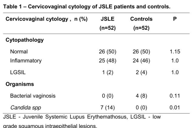
Table 1 – Cervicovaginal cytology of JSLE patients and controls. Cervicovaginal cytology , n (%) JSLE
(n=52)
Controls (n=52)
P
Cytopathology
Normal Inflammatory
26 (50) 25 (48)
26 (50) 24 (46)
1.15 1.0
LGSIL 1 (2) 2 (4) 1.0
Organisms
Bacterial vaginosis 0 (0) 4 (8) 0.11
Candida spp 7 (14) 0 (0) 0.01
Anexos 49
Table 2 - Characteristics of JSLE cervicovaginal cytology according to disease activity.
Cervicovaginal cytology, n (%) SLEDAI < 4 (n=17)
SLEDAI ≥ 4 (n=35)
p
Normal 13 (77) 13 (37) 0.001
Inflammatory 4 (23) 21 (60) 0.001
LGSIL 0 (0) 1 (3) 1.0
JSLE - Juvenile Systemic Lupus Erythemathosus, LGSIL - low grade squamous intraepithelial lesions, SLEDAI - Systemic Lupus Erythematosus Disease Activity Index.
Anexos 50
Table 3 - Characteristics of JSLE cervicovaginal cytology in virgin adolescents according to disease activity.
Cervicovaginal cytology, n (%) SLEDAI < 4 (n=12)
SLEDAI ≥ 4 (n=28)
p
Normal 11 (92) 12 (53) 0.005
Inflammatory 1 (8) 16 (57) 0.005
Anexos 51
Table 4 – Clinical features, activity, damage and treatment of JSLE patients with and without vaginal infection by Candida spp.
Variables With Candida spp
(n=7)
Without Candida spp
(n=45)
p
Clinical features
Cutaneous n (%) 7 (100) 43 (96) 1.0
Articular, n (%) 6 (86) 40 (89) 1.0
Renal, n (%) 7 (100) 37 (82) 0.57
Cardiopulmonary, n (%) 4 (57) 19 (42) 0.68 Neuropsychiatry, n (%) 2 (29) 10 (22) 0.65 Hematological abnormalities, n (%) 6 (86) 42 (93) 0.44
JSLE activity and damage
SLEDAI, mean ± SD 5.4 ± 4.2 5.5 ± 4.4 0.95 SLICC/ACR-DI ≥ 1, n (%) 3 (4) 7 (16) 0.3
Treatment
Current dose of Pd (mg), mean ± SD 20.5 ± 5 10.2 ± 8 0.002 Current use of IS, n (%) 7 (100) 20 (44) 0.01 JSLE - Juvenile Systemic Lupus Erythemathosus, IS – immunosuppressive drugs, Pd – prednisone, SLEDAI - Systemic Lupus Erythemathosus Disease Activity Index, SLICC/ACR-DI - Systemic Lupus International Collaborating Clinics/American College of Rheumatology-Damage Index, SD - standard deviation.
Referências 53
1. Benseler SM, Silverman ED. Systemic lupus erythematosus. Pediatr Clin N
Am 2005; 52: 443-467.
2. Arkachaisri T, Lehman TJ. Systemic lupus erythematosus and related disorders of childhood. Curr Opin Rheumatol 1999; 11: 384-392.
3. Stichweh D, Arce E, Pascual V. Update on pediatric systemic lupus erythematosus. Curr Opin Rheumatol 2004; 16: 577-587.
4. Aagaard-Tillery KM, Holmgren CM, Scott JR. Gynecologic Problems in Women with Autoimmune Diseases. In: Lockshin M, Branch DW. Handbook
of Systemic Autoimune Diseases, Volume 4. Reproductive and Hormonal Aspects of Systemic Autoimmune Diseases. 1st edition. Elsevier, 2006, pp 141-160.
5. Fidel PL Jr. Immunity in vaginal candidiasis. Curr Opin Infect Dis 2005; 18: 107-111.
6. Fidel PL Jr, Sobel JD. Immunopathogenesis of recurrent vulvovaginal candidiasis. Clin Microbiol Rev 1996; 9: 335-348.
7. Blumenfeld Z, Lorber M, Yoffe N, Scharf Y. Systemic lupus erythematosus: predisposition for uterine cervical dysplasia. Lupus 1994; 3: 59-61.
8. Nyberg G, Eriksson O, Westberg NG. Increased incidence of cervical atypia in women with systemic lupus erythematosus treated with chemotherapy.
Referências 54
9. Dhar JP, Kmak D, Bhan R, Pishorodi L, Ager J, Sokol RJ. Abnormal cervicovaginal cytology in women with lupus: a retrospective cohort study.
Gynecol Oncol 2001; 82: 4-6.
10. Tam LS, Chan AY, Chan PK, Chang AR, Li EK. Increased prevalence of squamous intraepithelial lesions in systemic lupus erythematosus: association with human papillomavirus infection. Arthritis Rheum 2004; 50: 3619-3625.
11. Dhar JP, Essenmacher L, Ager J, Sokol RJ. Ominous cervical cytopathology in women with lupus. Int J Gynaecol Obstet. 2005; 89: 295-296.
12. Bateman H, Yazici Y, Leff L, Peterson M, Paget SA. Increased cervical dysplasia in intravenous cyclophosphamide treated patients with SLE: a preliminary study. Lupus 2000; 9:542-4.
13. Ognenovski VM, Marder W, Somers EC, Johnston CM, Farrehi JG, Selvaggi SM, McCune WJ. Increased incidence of cervical intraepithelial neoplasia in women with Systemic Lupus Erythematosus treated with intravenous cyclophosphamide. J Rheumatol 2004; 31: 1763-1737.
14. Bernatsky S, Ramsey-Goldman R, Gordon C, Joseph L, Boivin JF, Rajan R, Allen A, Moore AD, Leung MH, Clarke A. Factors associated with abnormal Pap results in systemic lupus erythematosus. Rheumatology 2004; 43: 1386-1389.
15. Hochberg MC. Updating the American College of Rheumatology revised criteria for the classification of systemic lupus erhytematosus. Arthrits
Rheum 1997; 40: 1725.
Referências 55
17. Brunner HI, Silverman ED, To T, Bombardier C, Feldman BM. Sensitivity of the Systemic Lupus Erhytematosus Disease Activity Index, British Isles Lupus Assessement Group Index, and Systemic Lupus Activity Measure in the evaluation of clinical change in childhood-onset systemic lupus erhytematosus. Arthrits Rheum 1999; 42: 1354-1360.
18. Brunner HI, Silverman ED, To T, Bombardier C, Feldman BM. Risk factors for damage in childhood-onset systemic lupus erythematosus: cumulative disease activity and medication use predict disease damage. Arthritis
Rheum 2002; 46: 436-444.
19. Marshall JC, Tanner JM. Variations in patterns of pubertal changes in boys and girls. Arch Dis Child 1970; 45: 13-23.
20. Fokke HE, Salvatore CM, Schipper MEI, Bleker OP. A randomized trial of three methods of obtaining Papanicolaou semars. Eu J Obstet Gynecol
Reprod Bio 1993; 48: 103-106.
21. Solomon D, Davey D, Kurman R, Moriarty A, O’Connor D, Prey M, Raab S, Sherman M, Wilbur D, Wright T, Young N. The 2001 Bethesda System: terminology for reporting results of cervical cytology. JAMA 2002; 287:2114-9.
22. Greenberg SB. Infections in the immunocompromised rheumatologic patient. Crit Care Clin 2002; 18: 931-956.
23. Folb PI, Trounce JR. Immunological aspects of candida infection complicating steroid and immunosuppressive drug therapy. Lancet 1970; 2: 1112-1114.
24. Hellmann DB, Petri M, Whiting-O'Keefe Q. Fatal infections in systemic lupus erythematosus: the role of opportunistic organisms. Medicine
Referências 56
25. Goswami D, Goswami R, Banerjee U, Dadhwal V, Miglani S, Lattif AA, Kochupillai N. Pattern of Candida species isolated from patients with diabetes mellitus and vulvovaginal candidiasis and their response to single dose oral fluconazole therapy. J Infect 2006; 52: 111-117.
26. Tarkowski TA, Koumans EH, Sawyer M, Pierce A, Black CM, Papp JR, Markowitz L, Unger ER. Epidemiology of human papillomavirus infection and abnormal cytologic test results in an urban adolescent population. J
Infect Dis 2004; 189: 46-50.
27. Wright JD, Davila RM, Pinto KR, Merritt DF, Gibb RK, Rader JS, Mutch DG, Gao F, Powell MA. Cervical dysplasia in adolescents. Obstet Gynecol 2005; 106: 115-120.
28. Utagawa ML, Pereira SM, Cavaliere MJ, Maeda MY, Shih LW, Shirata NK. Cervical intraepithelial neoplasia in adolescents: study of cytological findings between 1987 and 1995 in Sao Paulo State-Brazil. Arch Gynecol
Obstet 1998; 262: 59-64.
29. Prussia PR, Gay GH, Bruce A. Analysis of cervico-vaginal (Papanicolaou) smears, in girls 18 years and under. West Indian Med J 2002; 51: 37-39.
30. Simsir A, Brooks S, Cochran L, Bourquin P, Ioffe OB. Cervicovaginal smear abnormalities in sexually active adolescents. Implications for management. Acta Cytol 2002; 46: 271-276.
31. Silva CA, Leal MM, Leone C, Simone VP, Takiuti AD, Saito MI, Kiss MHB. Gonadal function in adolescents and young women with juvenile systemic lupus erythematosus. Lupus 2002; 11: 419-425.
Referências 57
33. Moscicki AB. Human Papilloma Virus, Papanicolaou Smears, and the College Female. Pediatr Clin N Am 2005; 52: 167-177.
34. Hillard PJA. Pediatric and adolescent gynecology. In: Scott JR, Saia PJ, Hammond CB, Spellacy WN. Danforth’s Obstetrics & Gynecology, 9th edition. Lippincott Williams & Wilkins, 1999, pp 529-540.