ÓRGÃO OFICIAL dA SOCIEdAdE PORTUGUESA dE REUMATOLOGIA
45 ARTIGO ORIGINAL
1- Faculdade de Medicina da Universidade do Porto 2. Reumatologia, Centro Hospitalar S. João
3. Unidade Reumatologia Pediátrica, Centro Hospitalar S. João
Adverse events occurred in 2 patients, including de-layed second dose after the diagnosis of cryptococco-sis and respiratory tract infection with concomitant hy-pogammaglobulinemia needing of immunoglobulin re-placement and antibiotic therapy.
Conclusions: Rituximab might have a role in the treat-ment of JSLE and JIA. However controlled studies and long term follow-up are needed to evaluate its safety and efficacy.
Keywords: JSLE; JIA; Rituximab, Pediatric Rheuma-tology
IntroductIon
Juvenile idiopathic arthritis (JIA) and juvenile systemic lupus erythematosus (JSLE) are probably the most cli -nical relevant chronic rheumatic disorders of pediatric age where autoimmunity and inflammation are crucial for the development of the disease1, 2.
JIA represents a heterogeneous group of arthritis of unknown etiology presenting before 16 years and las -ting at least 6 weeks and it is subdivided into 7 distinct subtypes3. There is evident heterogeneity among JIA subtypes with respect to clinical, demographic, gene -tic and pathophysiological features, translating into dis-tinct responses to therapies currently available4,5. The discovery of new immunological markers is expected to improve diagnosis and treatment6.
The incidence of JIA ranges between 1 per 100,000 in Japan to more than 20 per 100,000 children/year in northern Europe1. Prevalence rates varies between 10 and 150 per 100000 children7, 8, but is probably un-derdiagnosed9.
Unlike JIA, JSLE is substantially the same disease as in adults10. JSLE represents about 10% to 20% of the to-tal cases of systemic lupus erythematosus (SLE)2. Most studies use either 16 years or 18 years as the upper cutoff age for JSLE2. The incidence is between 0,36 and
Anti CD20 (Rituximab) therapy
in refractory pediatric rheumatic diseases
Reis J1, Aguiar F2, Brito I3
ACTA REUMATOL PORT. 2016;41:45-55
AbstrAct
Objectives: We aim to report the efficacy and safety of rituximab (RTX) in patients diagnosed with juvenile systemic lupus erythematosus (JSLE) or juvenile idio-pathic arthritis (JIA) refractory to conventional treat-ment.
ÓRGÃO OFICIAL dA SOCIEdAdE PORTUGUESA dE REUMATOLOGIA
46
Anti CD20 (RituximAb) theRApy in RefRACtoRy peDiAtRiC RheumAtiC DiseAses
0,9 per 100000 children/year in North American and European studies11and the prevalence ranges from 3,3 to 8,8 per 100000 children2.
Despite the similarities, JSLE patients have more acti ve disease at presentation and over time12. JSLE car-ries a higher risk of developing lupus nephritis, malar rash, anti-DsDNA antibody positivity and hemolytic anemia13. Usually JSLE patients also receive more in-tensive drug therapy and accumulate more related damage12.
Recent discoveries regarding the inflammation pro-cess and autoimmunity allowed the specific management of the aberrant responses present in both disea -ses. These discoveries resulted mainly in major im-provements in short and medium-term outcomes and better control of acute-life threatening events14.
These patients are usually treated with a combina-tion of nonsteroidal anti-inflammatory drugs (NSAID), corticosteroids, disease-modifying anti-rheumatic drugs (DMARDs) such as methotrexate (MTX), sulfasalazine or leflunomide (LEF), and immunosuppres -sive agents such as azathioprine (AZT), cyclophos-phamide (CYC) or mycophenolate mofetil (MMF).
However, not all patients respond to these treat-ments and they are sometimes associated with toxici-ties that limit long-term use or diminish compliance15. Such patients are therefore candidates for treatment with biologic agents.
In recent years, several non-controlled studies have demonstrated that rituximab (RTX) might be effective in JSLE and JIA16-23in accordance with the important role that B cells might have in autoimmune diseases through three different ways: producing antibodies, presenting antigens to T-cells and producing cy-tokines24, 25. However large clinical trials in adults eva -luating RTX for renal26and non-renal lupus27did not prove its efficacy, contrary to rheumatoid arthritis28, but the failure might be more related with the study de-sign than with RTX itself29.
RTX is a chimeric monoclonal antibody (mAb) ini-tially approved for the treatment of B cell malignan-cies30. Currently it is also approved for rheumatoid arthritis, granulomatosis with polyangiitis and micros -copic polyangiitis in adults31, but its use in JIA and JSLE is still off-label. RTX binds with high affinity to cells that express CD20 antigen in their surface. This antigen is only present on B cells (malignant or not)32. CD20 antigen is not expressed in lymphoid progeni-tors in bone marrow allowing repopulation after stop-ping the treatment24. Plasma cells do not express CD20
antigen either and they are not directly depleted by RTX. Consequently not all antibodies decrease after treatment24.
RTX causes peripheral B cell depletion by two dif-ferent mechanisms: antibody-dependent cellular cy-totoxicity (ADCC) and complement-dependent cyto-toxicity (CDC)33. For ADCC occur it is necessary that mAb Fc interacts with Fc receptors for IgG (Fc Rs) on the surface of effector cells, localized in the reticu-loendothelial system (RES), especially in the liver34. Fc RIIA and Fc RIIIA are two of the most important Fc R in human, and RTX s efficacy increases if mAb Fc affinity to Fc RIIIA is improved 24. Circulating B cells are rapidly cleared at RES while B cells residing in lym-phoid tissues are slowly eliminated by this mechanism as they need to have access to the circulation. The time needed to eliminate cells that does not circulate is even longer as they depend solely on the CDC34.
Binding of RTX to podocytes membranes has also been reported, inducing remodeling of glomeruli that might explain the early decrease on proteinuria in lu-pus nephritis even before immune changes occur35.
Therefore, we aim to report our single center expe-rience with the use of RTX in patients diagnosed with JSLE and JIA resistant to other treatments in terms of safety and efficacy.
MAterIAls And Methods
We made a retrospective review of all medical records of patients with JSLE or JIA treated with RTX between January 2009 and January 2015 in the Pediatric Rheumatology Unit of a central hospital. This study was approved by the ethics committee along with all the treatments made.
The patients with JSLE included fulfilled the Ameri -can College of Rheumatology criteria for the diagnosis of SLE36. The patients with JIA were classified accor -ding to the criteria of the International League of As-sociations for Rheumatology3.
The medical records were reviewed for patients’ de-mographic characteristics, age at diagnosis, baseline rheumatic disease, previous and current medical treat-ments. Indication for RTX, age at the time of the first treatment with RTX, protocol used and follow-up time were also collected.
Ju-ÓRGÃO OFICIAL dA SOCIEdAdE PORTUGUESA dE REUMATOLOGIA
47 Reis J et Al
RTX was indicated for refractory class IV lupus nephritis (in three patients), JSLE with refractory multisystem involvement (in one patient), and severe poly -arthritis (in a patient with JIA who had anti-TNFa -in-duced lupus-like nephritis).
Ten cycles of RTX were performed, in a total of 23 infusions (Table II). The most common dosage used
was 750 mg/m2(maximum of 1 g) administered 2
weeks apart. One patient had also administration of CYC during the first RTX cycle. Three patients needed more than one cycle of RTX. The median time for a new cycle of RTX was 8 months (6–10 months).
The median follow-up time after receiving the first cycle of RTX was 24 months (12–70 months).
It was possible to reduce the dose of oral corticos-teroids (CS) in all patients, including discontinuation of oral CS in the patient with JIA. The mean daily oral CS dose before RTX was 23 mg/day (20-25mg/day). At the last evaluation it was 11 mg/day (0–20 mg/day). During and/or after RTX patients with JSLE continued therapy with MMF.
The scores of disease activity also improved. The SLEDAI scores decreased from a median of 15, 5 (11-18) before RTX, to 3 (0 – 6) at the last evaluation. The JADAS-27 score also diminished from 40.4 before RTX to 3.5 at the last evaluation.
It was recorded one case of respiratory tract infection 3 months after RTX with concomitant hypogamma-globulinemia (IgG of 423 mg/dL) and one case of in-venile Arthritis Disease Activity Score (JADAS-27)38,
respectively. We also collected laboratory measures for JSLE (hemoglobin, platelets, erythrocyte sedimentation rate, complement C3 and C4, antidoublestran -ded DNA antibodies, serum immunoglobulins, plas-ma creatinine, urine protein, and urinary sediment). Brain natriuretic peptide was also collected for one pa-tient with JSLE. For the papa-tient with JIA we collected the laboratory measures as in JSLE, but included the number of active and restrictive joints affected, and ex-cluded the complement levels and auto-antibodies.
All serious adverse events, medically important in-fections and infusion reactions were also evaluated.
results
Five patients were included (Table I). Four had JSLE (all female, 3 caucasian and 1 black) and 1 had extended oligoarticular JIA (caucasian male). The median age at diagnosis was 10 years (16 months – 17years) and the median evolution time until receiving RTX was 6 years (5 months – 15 years). All of them have received pre-vious treatments that included high-dose prednisolone (n=5), methylprednisolone pulses (n=5), cyclophos-phamide pulses (n=3), methotrexate (n=2), hydroxy-chloroquine (n=3), mycophenolate mofetil (n=3), aza-thioprine (n=2), leflunomide (n=1) and etanercept (n=1).
tAble I. deMogrAphIc, therApeutIc And clInIcAl chArActerIstIcs of pAtIents
Patients 1 2 3 4 5
Gender F F F F M
Race Caucasian Caucasian Caucasian Black Caucasian Disease JSLE JSLE JSLE JSLE Extended oJIA Age at diagnosis 10y 10y 10y 17y 1y
Disease duration before RTX 15y 6y 7y 5m 5y Age at first administration of RTX 25y 16y 17y 17y 6y
Medication before RTX MP, CYC, MP, CYC, MP, CYC, MP, PDN, MP, PDN, MTX, PDN, AZA, PDN, MMF PDN, MMF, MTX, HCQ LEF, Etanercept MMF, HCQ AZA, HCQ
RTX indication Anemia of Refractory Refractory Refractory Severe polyarthritis CD+ refractory class IV class IV lupus multisystem (previous anti-TNFa
class IV lupus lupus nephritis involvement induced lupus-like nephritis nephritis nephritis)
AZA=azathioprine; CYC = cyclophosphamide; F= Female; M= Male; y= years; m = months; oJIA = oligoarticular Juvenile idiopathic arthritis; JSLE = juvenile systemic lupus erythematosus; HCQ = hydroxychloroquine; LEF = leflunomide; MMF = mycophenolate mofetil;
ÓR
GÃO
OFI
CI
AL
d
A
S
OCI
Ed
Ad
E
P
ORT
U
GU
ES
A
d
E
REU
MA
T
OL
OGI
A
4
8
A
nt
i
CD20
(Ri
t
u
x
imA
b)
t
he
RA
p
y
in
Re
f
RA
Ct
oR
y
pe
Di
A
t
Ri
C
Rhe
u
mA
t
iC
Di
se
A
se
s
tAble II. treAtMent chArActerIstIcs And outcoMe of the pAtIents
Patients 1 2 3 4 5
Follow-up time after 12 months 19 months 70 months 24 months 28 months
first RTX treatment
No. of doses 4 4 3 2 8
Regimen of RTX used* 1st 750 (0, 15 d) 1st 750 (0, 15 d) + 750 (0, 15, 30d) 750 (0, 50 d) 1st375 (0, 15d)
(days of administration) 2nd750 (0, 15 d) CYC 375 (1d) 2nd750 (0, 15 d)
2nd 750 (0,15 d) 3rd750 (0, 15 d)
4th750 (0, 15 d)
Time to retreatment 10 months 8 months None None 7 months, 9 months
and 8 months
Adverse events None None Respiratory tract Cryptococcosis None
infection and transient hypogammaglobulinemia
Outcome Hematologic response. Initial response. Global improvement, but Improvement. Initial complete response Renal improvement, but Retreated later with persistent proteinuria Deterioration after with multiple flare. persistent proteinuria renal improvement stopping MMF In Complete remission
another hospital nowadays Drug dosage previous MMF: 2,5 g/d MMF: 2g/d MMF: 2g/d MMF: 2g/d PDN: 20 mg/d
to RTX PDN: 25 mg/d PDN: 20 mg/d PDN:25 mg/d PDN:20mg/d
Drug Dosage at last MMF: = MMF: = MMF:= MMF:↓0,5g/d PDN: ↓20 mg/d
evaluation PDN:↓5mg/d PDN:↓10mg/d PDN:↓15mg/d PDN:↓5mg/d (stopped)
ÓRGÃO OFICIAL dA SOCIEdAdE PORTUGUESA dE REUMATOLOGIA
49 Reis J et Al
fection by Cryptococcus diagnosed a month after the first RTX infusion.
Next we present the clinical particularities for each patient (Table III):
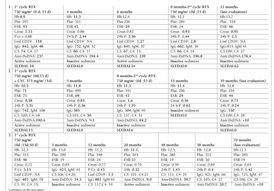
Patient 1: Caucasian female diagnosed with SLE at 10 years old (1999) initially with mucocutaneous, ar-ticular, hematologic involvement and positivity to anti-nuclear antibodies (ANAs) and anti-DsDNA antibodies. At 14 years old (2003) developed nephrotic proteinuria and cylindruria with complement consumption. Kid-ney biopsy revealed class IV lupus nephritis. She was treated with methylprednisolone pulses (MP) and CYC and later with AZT and prednisolone (PDN). Due to leucopenia AZT was switched to MMF. She kept stable with PDN and MMF until March 2013. She developed asthenia, peripheral edema, increasing blood pressure, anemia of chronic disease, complement consumption, and worsening of renal function. MMF was increased to 2,5 g/day, but maintained disease activity during the next months. The kidney biopsy showed class IV lupus nephritis with great activity and mild chronicity. There was no significant response to the treatment with MP pulses. For these reason she received the first RTX cy-cle. There was hematological response and renal im-provement but maintained persistent proteinuria. The dose of MMF and PDN was reduced. Six months later she developed again edema and nephrotic proteinuria with complement consumption and active sediment. MMF was increased to 2,5 g/day, along with losartan and furosemide. The sediment became inactive and proteinuria turned subnephrotic. Another cycle of RTX was made and at the last evaluation the patient pre-sented with no edema, inactive sediment, normal blood pressure, but maintaining proteinuria.
Patient 2: Caucasian female diagnosed with SLE at 10 years old (2006) presenting with renal involvement. The biopsy showed class IV lupus nephritis. Clinical-ly stable after induction treatment with CYC and main-tenance treatment with MMF. She stopped hydroxy-chloroquine due to dermatologic toxicity. In 2012 de-veloped progressive proteinuria with active sediment that did not respond to treatment optimization. The kidney biopsy revealed class IV lupus nephritis. She was treated with RTX plus CYC. Five months later the proteinuria had significantly decreased together with inactive sediment. The platelet values and hemoglobin values also improved. Because proteinuria worsened again it was decided to do another cycle of RTX, eight months after the first one. At the last evaluation the la -b oratory results showed improvement in all parameters
evaluated.
Patient 3: Caucasian female diagnosed with SLE at 10 years old (2002) with hemolytic anemia and severe thrombocytopenia together with lupus nephritis class IV. Patient achieved hematologic response but kept lu-pus nephritis refractory to induction treatment with CYC and maintenance with PDN and MMF. Since 2009 she developed severe nephrotic proteinuria and hy-pertension without complement consumption. A new biopsy confirmed the previous result. She made pul ses of MP, followed by PDN with unsatisfactory response and was decided to start RTX. There was a significant decrease in proteinuria and normalization of anti--DsDNA antibodies. Three months later she was hos-pitalized due to lower respiratory tract infection that was treated successfully with antibiotic therapy. The laboratory results showed hypogammaglobulinemia [IgG 423 mg/dL (N 650 – 1500]) and a B cell count of 1,46 cel/mm3. Intravenous immunoglobulin (IvIg) was administered. Since then she is clinically stable with-out hypertension and reduced proteinuria.
Patient 4: Black female hospitalized at another hos-pital in 2012 with 17 years old due to prolonged febrile syndrome, lymphadenopathies, fatigue, and weight loss. She was then transferred to our hospital already with oral ulcers, arthritis and malar rash. Laboratory exams showed positive ANAs, anti-DsDNA, anti-ri-bonucleoproteins antibodies and normochromic, nor-mocytic anemia with positive Coombs. She was diagno sed with JSLE and was treated with PDN and hydroxychloroquine (HCQ). After discharge she start-ed MTX and rstart-educstart-ed PDN (20 mg/day).
ÓR
GÃO
OFI
CI
AL
d
A
S
OCI
Ed
Ad
E
P
ORT
U
GU
ES
A
d
E
REU
MA
T
OL
OGI
A
5
0
A
nt
i
CD20
(Ri
t
u
x
imA
b)
t
he
RA
p
y
in
Re
f
RA
Ct
oR
y
pe
Di
A
t
Ri
C
Rhe
u
mA
t
iC
Di
se
A
se
s
tAble III. evolutIon After rtX treAtMent
1 1stcycle RTX 8 months-2ndcycle RTX 12 months
750 mg/m2(0 d; 15 d) 4 months 6 months 750 mg/m2(0d ;15 d) (last evaluation)
Hb:8,9 Hb: 11,3 Hb:12,4 Hb: 12,1 Hb:13,7
Plat: 297 Plat: 311 Plat:236 Plat: 284 Plat.: 318
ESR: 93 ESR:45 ESR: 29 ESR: 24 ESR: 46
Creat: 1.13 Creat: 0.86 Creat 0.83 Creat : 0,93 Creat: 0,91
P:cr = 0.88 24 h P: 2,44 24h P: 3.36 24h P: 1,44 24h P: 2,5
Linf CD19+: 118 Linf CD19+: NA Linf CD19+: 1,27 Linf CD19+: 2,8 Linf CD19+: NA
IgG: 842; IgM: 48 IgG: 752; IgM: 32 IgG 641; IgM: 37 IgG 662; IgM: 56 IgG:814; IgM 41 C3: 74; C4: 17 C3: 80; C4: 17 C3: 67; C4: 13; C3: 66; C4: 14 C3:109; C4: 27 Anti-DsDNA: 237 Anti-DsDNA: 194.4 Anti-DsDNA: 239 Anti-DsDNA: 196.8 Anti-DsDNA:178,4
Activesediment Inactivesediment Activesediment Inactivesediment Inactivesediment
SLEDAI: 18 SLEDAI:8 SLEDAI:16 SLEDAI:8 SLEDAI:62
2 1stcycle RTX
750 mg/m2(0d;15 d) 8 months-2ndcycle RTX
+ CYC 375 mg/m2(1d) 5 months 750 mg/m2(0d ;15 d) 15 months 19 months (last evaluation)
Hb: 10,5 Hb: 11,8 Hb: 11,3 Hb: 11,4 Hb: 12,8
Plat: 71 Plat: 499 Plat: 341 Plat: 296 Plat: 254
ESR: 71 ESR: 69 ESR: 43 ESR: 29 ESR: 44
Creat: 0,58 Creat: 0.83 Creat: 1,6 Creat: 0.56 Creat: 0.75
24h P: 3.31 24h P: 0,36 24h P: 1,55 24 h P :0.62 24h P: 0.24
IgG: 742; IgM: 108 IgG:385 IgG: 504; IgM: 95 Inactivesediment IgG: 550 C3: 100; C4: 18 C3:103; C4: 50 C3: 137; C4: 31 SLEDAI:0 C3:109; C4: 28
Anti-DsDNA:190,4 Anti-DsDNA: NA Anti-DsDNA: 84,2 Anti-DsDNA: <10
Active sediment Inactivesediment Inactive sediment Inactivesediment
SLEDAI:15 SLEDAI:0 SLEDAI:6 SLEDAI:0
3 1stcycle RTX
750 mg/m2 70 months
(0d ;15d;30 d) 3 months 12 months 20 months 48 months 58 months (last evaluation)
Hb: 12,3 Hb: 13,8 Hb: 11,2; Hb: 11.1; Hb: 12,3; Hb: 12,8 Hb:12.5
Plat: 313 Plat: 290 Plat: 348 Plat: 265 Plat: 280 Plat: 311 Plat: 283
ESR: 46 ESR: 19 ESR: 24 ESR:10 ESR: 19 ESR: 20 ESR:19
Creat: 0,72 Creat: 0,83 Creat: 0,77 Creat: 0.70 Creat: 0.59 Creat: 0,69 Creat: 0,65 P:Cr: 5,15 IgG :423; IgM: 61 P:Cr: 0.43 24h :0.32 24h P: 1,43 24h P: 0.4 24h P: 1.42 Linf CD19+: NA C3:106; C4:29; Linf CD19+: 1,5 C3: 101; C4:21 C3: 111; C4: 24 IgG: 921; IgM: 66 IgG: 723; IgM: 47
tAble III. contInuAtIon
1stcycle RTX
750 mg/m2 70 months
(0d ;15d;30 d) 3 months 12 months 20 months 48 months 58 months (last evaluation)
Anti-DsDNA: 25.8 SLEDAI:0 Anti-DsDNA: 18,9 SLEDAI:8 SLEDAI:4 Inactivesediment Inactivesediment
Activesediment Inactivesediment SLEDAI:0 SLEDAI:4
SLEDAI:16 SLEDAI:0
4 1stcycle RTX Emigrated to France
750 mg/m2(0d;50 d) 7 months Follow-up on local hospital 12 months 24 months (last evaluation)
Constitutional symptoms No constitutional symptoms. Suspended mycophenolate Constitutional symptoms No constitutional symptoms
Polyarthritis Oligoarthritis mofetil and augmented Polyarthritis No polyarthritis
Hb: 6.1; WBC: 3.20 ; Plat: 64 Hb: 13.4; WBC: 3.0 ;Plat: 308 prednisolone Hb: 12.4; WBC 3.55 ; Plat:305 Hb 11,2; WBC: 3,45 ; Plat 352
ESR: 103 ESR: 31 ESR: 36 ESR: 49
Creat:0,42 Creat:0.56 Creat: 0.54 Creat: 0,52
24h P: 0.4 C3: 132; C4: 44 C3: 132 ; C4: 44 C3:102; C4: 24
C3: 98; C4: 19 BNP: 57.4 BNP: 70.5 BNP: 59.1
BNP: 2100 Anti-DsDNA: >800 Anti-DsDNA: >800 Anti-DsDNA: >800
Anti-DsDNA: >800 No proteinuria and inactive No proteinuria and inactive No proteinuria and inactive
Inactivesediment sediment sediment sediment
Heart failure with myocarditis NYHA I SLEDAI:6 SLEDAI:2
Systolic dysfunction of left SLEDAI:2 ventricle(EF 24%)
SLEDAI:11
5 1stcycle RTX 7 months-2nd 16 months-3rd 24 months-4th
375 mg/m2 cycle RTX cycle RTX cycle RTX 28 months
(0d;15 d) 750 mg/m2 (0d ;15 d) 12 months 750 mg/m2(0d;15 d) 22 months 750mg/m2 (0d ;15) (last evaluation)
A. joints: 12 A. joints: 4 A. joints: 0 A. joints: 11 A. joints: 1 Worsening of polyarthritis A. joints:2 R. joints: 12 R. joints: 3 R. joints:2 R. joints: 12 R. joints: 3 Hb:12.2 R. joints:0
Hb: 12,0 Hb:11.8 Hb: 13.0 Hb:12.2 Hb: 13.4 Plat: 445 Hb:13.1
Plat: 558 Plat: 484 Plat: 447 Plat: 482 Plat: 519 ESR: 24 Plat: 322
ESR: 24 ESR: 13 ESR: 2 ESR: 47 ESR: 7 Creat: 0.29 ESR: 8
Creat: 0,30 Creat: 0.29 Creat: 0.34 Creat: 0.29 Creat:0.30 P:Cr:NA Creat: 0.28 P:Cr: 0,087 P:Cr: 0.098 P:Cr: 0,094 P:Cr:NA P:Cr:NA Linf CD19+: 358 P:Cr: 0,095
IgG:1220; IgM: 106 Linf CD19+: 298 Linf CD19+:1.0 Linf CD19+: 260 Linf CD19+:4.7 Inactivesediment Linf CD19+: 0,0
Inactivesediment IgG: 1030;IgM: 32 IgG: 879; IgM: 19 IgG: 914;IgM: 30 IgG: 659; IgM:7 JADAS 27: 39.4 since last RTX cycle
JADAS 27: 40.4 Inactivesediment Inactivesediment Inactivesediment Inactivesediment Inactivesediment
JADAS 27: 14 JADAS 27: 3 JADAS 27: 39.4 JADAS 27:8 JADAS 27: 5
ÓR
GÃO
OFI
CI
AL
d
A
S
OCI
Ed
Ad
E
P
ORT
U
GU
ES
A
d
E
REU
MA
T
OL
OGI
A
5
1
Re
is
J
e
t
Al
Hb = haemoglobin concentration (g/dL); Plat = platelet count (x 109/L) ; ESR= Erythrocyte sedimentation rate (mm/h) ; Creat= Creatinine serum concentration (mg/dL) ; P:cr= protein –creatinine ratio
on urine sample; 24P= 24-hour urine protein (grams) ; Ig = Immunoglobulin (mg/dL); Anti-DsDNA= Anti-double-stranded DNA antibody (UI/ml); WBC=White blood cell count (x 109/L);
Linf CD19+= lymphocytes CD19+ (per mm3); NYHA= New York Heart association functional classification of heart failure; C3 andC4= complement (mg/dL); EF= systolic ejection fraction; RCP=
reactive C-protein (mg/mL); BNP = B-type natriuretic peptide (ng/mL); A.joints= active joints; R.joints= restrictive joints; RTX= Rituximab; CYC= Cyclophosphamide; NA = Not available; SLEDAI=
ÓRGÃO OFICIAL dA SOCIEdAdE PORTUGUESA dE REUMATOLOGIA
52
Anti CD20 (RituximAb) theRApy in RefRACtoRy peDiAtRiC RheumAtiC DiseAses
pulse, Cryptococcus was detected in blood cultures. She was treated with amphotericin B and fluconazole. Fifty days later, after an improvement of systolic func-tion and laboratory analyses she made a second pulse of RTX and reduced PDN to 30 mg/day. After discharge she initiated again HCQ (400 mg/day) and reduced PDN to 15 mg/day, while maintaining 1,5g/day of MMF. She maintained clinically stable with no signs of heart failure. She immigrated to France. When she returned MMF had been suspended, the PDN augmented to 20 mg/day and she referred febrile spikes and polyarthri-tis. She resumed therapy with MMF (1,5 g/day) and PDN (15 mg/day) and at the last evaluation she was asymptomatic but slightly anemic.
Patient 5: Caucasian male, 8 years old, diagnosed with extended oligoarticular JIA since 16 months of age, with uveitis and clinical evolution dependent of CS and refractory to treatment with MTX and LEF. He started etanercept (0,4 mg/Kg/dose) in September of 2010 resulting in complete clinical response and suspension of CS. In December of 2011 he was hospita -lized due to a nephrotic syndrome together with acute kidney lesion, hypertension with complement con-sumption and positive ANAs. The biopsy revealed membranoproliferative glomerulonephritis, interpre -ted as possible glomerulonephritis lupus–like induced by anti-TNFa. It was decided to stop etanercept and administrate PDN and MTX, with clinical and renal improvement. However arthritis recurred even with opti -mized dosage of CS and MTX. Because Abatacept was not available, he made one cycle of RTX, and a second cycle eight months later, with clinical and analytical improvement. PDN was reduced to 2,5 mg/day. In Ja -nuary 2014 he presented again with polyarthritis. PDN was augmented to 5 mg/day and made one more cycle of RTX obtaining clinical remission again. In October of 2014 the polyarthritis worsened again and another cycle was administrated. Since that time B cells have been completely depleted and achieved clinical remis-sion. No signs of renal lesion were observed after stop-ping etanercept.
dIscussIon
The American College of Rheumatology published re-cently recommendations for the Treatment of JIA39with an update on systemic JIA released in 201340. There are no approved drugs for treatment of juvenile lupus nephritis and the treatment relies on off-label use of
medications approved for any other reason, as is the case of RTX41. Frequently the therapeutic strategies used are similar to what it is done in adult age. How-ever, various adverse events limit their use compared to adults10,12,42and the optimal dosage and safety is not determined41.
Besides the side effects, the conventional therapies also have limitations in their efficacy. The induction treatment of proliferative lupus nephritis with conven-tional first-line treatments as CYC or MMF has report-ed failure rates ranging from 10% to 43%43. In JIA rates of non-response to therapy with NSAIDs, CS or DMARDs vary from 5% to 30 %44.
As mentioned before, an increasing number of open-label, retrospective studies and case reports have demonstrated that RTX might be effective in JSLE and
JIA16-23. However assessing the efficacy after RTX is
complicated by the fact that it takes a variable amount of time to respond and in the meanwhile patients might need adjustments in conventional therapy to control the clinical situation before obtain a response45, as in our case.
Three of the patients received RTX due to refracto-ry lupus nephritis. All of them had lupus nephritis clas-sified as class IV according to World Health Organization. Class IV represents 40 to 60% of all cases as des -cribed in literature and has the worst prognosis 10. Kidn ey involvement has a great impact in morbidity and mortality, and it has a major impact in the choice of immunosuppressive therapy14, being usually the main indication for RTX treatment20, as in our case.
ÓRGÃO OFICIAL dA SOCIEdAdE PORTUGUESA dE REUMATOLOGIA
53 Reis J et Al
seen in patient 3 that maintained stable remission du ring 70 months. However, in patient 1 and 2, the res -pon se was not permanent as shown by the increase of SLEDAI scores by more than 3 points, suggesting a moderate flare46, and the increase of proteinuria and serum creatinine, requiring an adjustment of the an-giotensin-converting-enzyme inhibitor and conven-tional therapy followed by another cycle of RTX. This is in accordance with literature where is reported that 30% needed more than one cycle and account for 58% of the cycles20. Although the degree of B cell depletion usually correlates with better responses29, relapses may occur even with a low B cell level as observed in patient 1, probably reflecting RTX’s mechanism of action.
In the case of patient 4, after RTX treatment used to control refractory multisystem involvement, SLEDAI score fell significantly; there was an improvement in hemoglobin concentration, platelets and leukocytes, along with decrease in liver enzymes. With respect to the heart failure, the findings from magnetic resonance were indicative of myocarditis, which was treated as possible autoimmune manifestation. Clinically signi -ficant myocarditis in JSLE is quite uncommon, however it might be the initial manifestation14. The improve-ment might also be attributed to the previous use of RTX. In the only case report founded, RTX was used with success to treat myocarditis in childhood lupus with improvement after a week47. However, it was not possible to rule out cryptococcosis as a cause.
Overall, patients showed a good hematological res -ponse, even if that was not the reason to administrate RTX. There was an improvement in hemoglobin con-centration and platelet’s count, as seen in other larger cohorts20. Evidence also suggests that depletion of B cell can be efficacious and safe for treatment of au-toimmune thrombocytopenia and/or auau-toimmune hemolytic anemia in JSLE refractory to first line treat-ment47.
Patient 5, diagnosed with AIJ, required a total of four cycles. The worsening of polyarthritis, with concomi-tant increase of JADAS-27 score, was the main reason for repeating RTX 18and was always preceded by an in-crease of CD19+ B cell count48. Nevertheless, some stud-ies find no correlation between B cell count and symp-toms in JIA18. At least for JIA the evidence suggests that 75% of patients required repeat cycles of RTX to sup-press disease activity, with remission rates improving over time, as shown by improvement in JADAS-27 score. This suggests that patients with highly refracto-ry JIA can achieve high levels of improvement by
con-tinued treatment with RTX18. Patient 5 also has a past history of “lupus-like” nephritis after using etanercept. A growing body of evidence suggests that TNFa an-tagonists can induce lupus like nephritis especially with etanercept49,50.
The dosage of RTX typically used in our study was 750 mg/m2, with exception of the first cycle in patient 5 where we used 375 mg/m2, taking into account his age. Whenever possible the second dose was adminis-tered 14 days apart. We only included CYC (375mg/m2) in patient 2 because the other patients have reached the maximum cumulative dose. The dosage of RTX we used was in accordance with the published literature16,19,20,22, although there are reports of lower doses (375 mg/m2) used weekly during 4 weeks18,21,23or with two weeks apart17. These schemes usually combine RTX with CYC in the treatment of SLE in the majority of the patients, but not in the case of JIA18.
In our opinion it was important to spare the use of CYC in JSLE patients since it is known for its capacity to induce irreversible infertility and increase the risk of malignancy42.
Furthermore, we managed to decrease the mean dose of CS between the evaluation previously to RTX and the last evaluation, consistent with the published literature16-23. All the patients with JSLE maintained the therapy with MMF. Data from other larger cohorts indicate that in the majority of the patients at least ano -ther immunosuppressive drug is maintained along with
PDN21,22. This reduction in CS is especially important
since children have more damages related to their use, including cataracts and avascular necrosis. 50.9% of the damage observed in JSLE appears to be related to the use of CS compared to 29.3% in adults12. The ab-normal growth, delayed puberty and alterations in body image can have a major impact in adolescents’ identity and relationships contributing to a worst com-pliance with treatments10.
In the case of lupus nephritis it was demonstrated to be possible to induce remission with rituximab while keeping maintenance therapy with MMF, allowing re-duction or total withdraw of corticosteroids51,52.
However several studies showed that JSLE is more severe, frequently needing higher doses of oral CS and immunosuppressive therapies and having more admis -sions in intensive care units compared to adults53-55. So the complete withdrawal of CS might not be possible.
-ÓRGÃO OFICIAL dA SOCIEdAdE PORTUGUESA dE REUMATOLOGIA
54
Anti CD20 (RituximAb) theRApy in RefRACtoRy peDiAtRiC RheumAtiC DiseAses
ted to the infusion. As mentioned before, we report one respiratory tract infection with concomitant hy-pogammaglobulinemia and one Cryptococcus’ infec-tion. In literature concerning pediatric age, the most common adverse events reported are mild infusion re-actions and infections 16-23,which is similar to what is re-ported in adults26,27. The reported incidence of infec-tions requiring hospital admission is of 86,9/1000 per-son-year22,and alarge cohort showed that only 2% of JSLE treated with RTX needed IvIg replacement20. Al-though in adults IgG levels are usually unchanged af-ter RTX, because CD20 antigen is not present in plas-ma cells45, prolonged deficiency of immunoglobulin or CD20+B cell was documented in some pediatric cases even after 12 months21. Although very rare the European Medicines Agency recently actualized RTX pro -duct information to include the occurrence of pro-gressive multifocal leukoencephalopathy and reactiva-tion of hepatitis B virus31.
conclusIon
The overall response to RTX was favorable in our case series, since it was possible to reduce the use of corti-costeroids without compromising disease control. It was also possible to induce remission of lupus nephri-tis with RTX without CYC while maintaining therapy with MMF and PDN. Some patients needed more than one cycle with a mean time of 8.4 months. This is pro -bably related to the RTX’s mechanism of action and with the degree of depletion of B cells, especially in the youngest patients. The drug was globally safe. Limita-tions to this study include its retrospective nature and the small sample size.
Controlled studies are needed to demonstrate the efficacy and the safety at long term. Rituximab might be able to spare children from deleterious effects of cor-ticosteroids and other immunosuppressive drugs. However, we cannot rule out RTX interference with the immune system and future consequences upon chro -nic usage.
correspondence to
Brito I
Al. Prof. Hernâni Monteiro, 4200-319 Porto, PORTUGAL E-mail: [email protected]
references
1. Cassidy JT, Petty RE. Chronic Arthritis in Childhood. In: Cassidy JT, Petty RE, Laxer RM and Lindsley CB. Textbook of Pediatric Rheumatology. Philadelphia:Elsevier Saunders 2010:211-235.
2. Kamphuis S, Silverman ED. Prevalence and burden of pediatric-onset systemic lupus erythematosus. Nat Rev Rheumatol 2010; 6: 538-546.
3. Petty RE, Southwood TR, Manners P, et al. International League of Associations for Rheumatology classification of juvenile idio-pathic arthritis: second revision, Edmonton, 2001. J Rheuma-tol 2004; 31: 390-392.
4. Stoll ML, Cron RQ. Treatment of juvenile idiopathic arthritis in the biologic age. Rheum Dis Clin North Am 2013; 39: 751-766. 5. Stoll ML, Cron RQ. Treatment of juvenile idiopathic arthritis: a revolution in care. Pediatr Rheumatol Online J 2014; 12: 13. 6. Borchers AT, Selmi C, Cheema G, Keen CL, Shoenfeld Y, Gersh
-win ME. Juvenile idiopathic arthritis. Autoimmun Rev 2006; 5: 279-298.
7. Prieur AM, Le Gall E, Karman F, Edan C, Lasserre O, Goujard J. Epidemiologic survey of juvenile chronic arthritis in France. Comparison of data obtained from two different regions. Clin Exp Rheumatol 1987; 5: 217-223.
8. Ravelli A, Martini A. Juvenile idiopathic arthritis. Lancet 2007; 369: 767-778.
9. Manners PJ, Diepeveen DA. Prevalence of juvenile chronic arth-ritis in a population of 12-year-old children in urban Australia. Pediatrics 1996; 98: 84-90.
10. Malattia C, Martini A. Paediatric-onset systemic lupus erythe-matosus. Best Pract Res Clin Rheumatol 2013; 27: 351-362. 11. Pons-Estel GJ, Alarcon GS, Scofield L, Reinlib L, Cooper GS.
Un-derstanding the epidemiology and progression of systemic lupus erythematosus. Semin Arthritis Rheum 2010; 39: 257-268. 12. Brunner HI, Gladman DD, Ibanez D, Urowitz MD, Silverman
ED. Difference in disease features between childhood-onset and adult-onset systemic lupus erythematosus. Arthritis Rheum 2008; 58: 556-562.
13. Mina R, Brunner HI. Update on differences between childhood-onset and adult-childhood-onset systemic lupus erythematosus. Arthritis Res Ther 2013; 15: 218.
14. Silverman E, Eddy A. Systemic Lupus Erythematosus. In: Cas-sidy JT, Petty RE, Laxer RM and Lindsley CB. Textbook of Pe-diatric Rheumatology. Philadelphia:Elsevier Saunders 2010:315 -343
15. Al-Mayouf SM, Alenazi A, Aljasser H. Biologic agents therapy for Saudi children with rheumatic diseases: indications and safety. Int J Rheum Dis 2014.
16. Lehman TJ, Singh C, Ramanathan A, et al. Prolonged improve-ment of childhood onset systemic lupus erythematosus follo-wing systematic administration of rituximab and cyclophos-phamide. Pediatr Rheumatol Online J 2014; 12: 3.
17. Ale’ed A, Alsonbul A, Al-Mayouf SM. Safety and efficacy of com-bined cyclophosphamide and rituximab treatment in recalci-trant childhood lupus. Rheumatol Int 2014; 34: 529-533. 18. Alexeeva EI, Valieva SI, Bzarova TM, et al. Efficacy and safety of
repeat courses of rituximab treatment in patients with severe refractory juvenile idiopathic arthritis. Clin Rheumatol 2011; 30: 1163-1172.
19. Podolskaya A, Stadermann M, Pilkington C, Marks SD, Tullus K. B cell depletion therapy for 19 patients with refractory sys-temic lupus erythematosus. Arch Dis Child 2008; 93: 401-406. 20. Watson L, Beresford MW, Maynes C, et al. The indications, ef-ficacy and adverse events of rituximab in a large cohort of pa-tients with juvenile-onset SLE. Lupus 2015; 24: 10-17. 21. Jansson AF, Sengler C, Kuemmerle-Deschner J, et al. B cell
ÓRGÃO OFICIAL dA SOCIEdAdE PORTUGUESA dE REUMATOLOGIA
55 Reis J et Al
Rheumatol 2011; 30: 87-97.
22. Tambralli A, Beukelman T, Cron RQ, Stoll ML. Safety and Effi-cacy of Rituximab in Childhood-onset Systemic Lupus Erythe-matosus and Other Rheumatic Diseases. J Rheumatol 2015. 23. Nwobi O, Abitbol CL, Chandar J, Seeherunvong W, Zilleruelo
G. Rituximab therapy for juvenile-onset systemic lupus erythe-matosus. Pediatr Nephrol 2008; 23: 413-419.
24. Bluml S, Mckeever K, Ettinger R, Smolen J, Herbst R. B-cell tar-geted therapeutics in clinical development. Arthritis Res Ther 2013; 15 Suppl 1: S4.
25. Morbach H, Wiegering V, Richl P, et al. Activated memory B cells may function as antigen-presenting cells in the joints of children with juvenile idiopathic arthritis. Arthritis Rheum 2011; 63: 3458-3466.
26. Rovin BH, Furie R, Latinis K, et al. Efficacy and safety of rituxi-mab in patients with active proliferative lupus nephritis: the Lu-pus Nephritis Assessment with Rituximab study. Arthritis Rheum 2012; 64: 1215-1226.
27. Merrill JT, Neuwelt CM, Wallace DJ, et al. Efficacy and safety of rituximab in moderatelytoseverely active systemic lupus ery -thematosus: the randomized, double-blind, phase II/III syste-mic lupus erythematosus evaluation of rituximab trial. Arthri-tis Rheum 2010; 62: 222-233.
28. Lopez-Olivo MA, Amezaga Urruela M, Mcgahan L, Pollono EN, Suarez-Almazor ME. Rituximab for rheumatoid arthritis. Coch-rane Database Syst Rev 2015; 1: Cd007356.
29. Favas C, Isenberg DA. B-cell-depletion therapy in SLE—what are the current prospects for its acceptance? Nat Rev Rheuma-tol 2009; 5: 711-716.
30. Grillo-Lopez AJ, White CA, Varns C, et al. Overview of the cli-nical development of rituximab: first monoclonal antibody ap-proved for the treatment of lymphoma. Semin Oncol 1999; 26: 66-73.
31. MabThera : EPAR - Product Information. http://www.ema.eu-ropa.eu/docs/en_GB/document_library/EPAR_-_Summary_f or_the_public/human/000165/WC500025815.pdf.Accessed in 10/12/2014
32. Grillo-Lopez AJ. Rituximab: an insider’s historical perspective. Semin Oncol 2000; 27: 9-16.
33. Eisenberg R. Update on rituximab. Ann Rheum Dis 2005; 64 Suppl 4: iv55-57.
34. Gong Q, Ou Q, Ye S, et al. Importance of cellular microenvi-ronment and circulatory dynamics in B cell immunotherapy. J Immunol 2005; 174: 817-826.
35. Fornoni A, Sageshima J, Wei C, et al. Rituximab targets podo-cytes in recurrent focal segmental glomerulosclerosis. Sci Transl Med 2011; 3: 85ra46.
36. Tan EM, Cohen AS, Fries JF, et al. The 1982 revised criteria for the classification of systemic lupus erythematosus. Arthritis Rheum 1982; 25: 1271-1277.
37. Bombardier C, Gladman DD, Urowitz MB, Caron D, Chang CH. Derivation of the SLEDAI. A disease activity index for lupus pa-tients. The Committee on Prognosis Studies in SLE. Arthritis Rheum 1992; 35: 630-640.
38. Consolaro A, Ruperto N, Bazso A, et al. Development and vali-dation of a composite disease activity score for juvenile idio-pathic arthritis. Arthritis Rheum 2009; 61: 658-666. 39. Beukelman T, Patkar NM, Saag KG, et al. 2011 American
Col-lege of Rheumatology recommendations for the treatment of ju-venile idiopathic arthritis: initiation and safety monitoring of therapeutic agents for the treatment of arthritis and systemic
features. Arthritis Care Res (Hoboken) 2011; 63: 465-482. 40. Ringold S, Weiss PF, Beukelman T, et al. 2013 update of the
2011 American College of Rheumatology recommendations for the treatment of juvenile idiopathic arthritis: recommendations for the medical therapy of children with systemic juvenile idio-pathic arthritis and tuberculosis screening among children re-ceiving biologic medications. Arthritis Rheum 2013; 65: 2499--2512.
41. Mina R, Von Scheven E, Ardoin SP, et al. Consensus treatment plans for induction therapy of newly diagnosed proliferative lu-pus nephritis in juvenile systemic lulu-pus erythematosus. Arth-ritis Care Res (Hoboken) 2012; 64: 375-383.
42. Morgan TA, Watson L, Mccann LJ, Beresford MW. Children and adolescents with SLE: not just little adults. Lupus 2013; 22: 1309-1319.
43. Boneparth A, Ilowite N. Comparison of renal response para-meters for juvenile membranous plus proliferative lupus neph-ritis versus isolated proliferative lupus nephneph-ritis: a cross-sectio-nal across-sectio-nalysis of the CARRA Registry. Lupus 2014; 23: 898-904. 44. Ungar WJ, Costa V, Hancock-Howard R, Feldman BM, Laxer RM. Cost-effectiveness of biologics in polyarticular-course ju-venile idiopathic arthritis patients unresponsive to disease-mo-difying antirheumatic drugs. Arthritis Care Res (Hoboken) 2011; 63: 111-119.
45. Jayne D. Role of rituximab therapy in glomerulonephritis. J Am Soc Nephrol 2010; 21: 14-17.
46. Mosca M, Bombardieri S. Assessing remission in systemic lupus erythematosus. Clin Exp Rheumatol 2006; 24: S-99-104. 47. Aggarwal P, Singh S, Suri D, Rawat A, Narula N, Manojkumar
R. Rituximab in childhood lupus myocarditis. Rheumatol Int 2012; 32: 1843-1844.
48. Trouvin AP, Jacquot S, Grigioni S, et al. Usefulness of monito-ring of B cell depletion in rituximab-treated rheumatoid arthri-tis patients in order to predict clinical relapse: a prospective ob-servational study. Clin Exp Immunol 2015; 180: 11-18. 49. Piga M, Chessa E, Ibba V, et al. Biologics-induced autoimmune
renal disorders in chronic inflammatory rheumatic diseases: sys-tematic literature review and analysis of a monocentric cohort. Autoimmun Rev 2014; 13: 873-879.
50. Ramos-Casals M, Brito-Zeron P, Munoz S, et al. Autoimmune di-seases induced by TNF-targeted therapies: analysis of 233 ca-ses. Medicine (Baltimore) 2007; 86: 242-251.
51. Pepper R, Griffith M, Kirwan C, et al. Rituximab is an effective treatment for lupus nephritis and allows a reduction in main-tenance steroids. Nephrol Dial Transplant 2009; 24: 3717-3723. 52. Condon MB, Ashby D, Pepper RJ, et al. Prospective observatio-nal single-centre cohort study to evaluate the effectiveness of treating lupus nephritis with rituximab and mycophenolate mo-fetil but no oral steroids. Ann Rheum Dis 2013; 72: 1280-1286. 53. Hersh AO, Von Scheven E, Yazdany J, et al. Differences in long-term disease activity and treatment of adult patients with child-hood- and adult-onset systemic lupus erythematosus. Arthritis Rheum 2009; 61: 13-20.
54. Hui-Yuen JS, Imundo LF, Avitabile C, Kahn PJ, Eichenfield AH-Levy DM. Early versus later onset childhood-onset systemic lu-pus erythematosus: Clinical features, treatment and outcome. Lupus 2011; 20: 952-959.