CLINICAL SCIENCE
Increased IgE serum levels are unrelated to allergic
and parasitic diseases in patients with juvenile
systemic lupus erythematosus
Bernadete L. Liphaus, Adriana A. Jesus, Clovis A. Silva, Antonio Coutinho, Magda Carneiro-Sampaio
Instituto da Crianc¸a (ICr) do Hospital das Clı´nicas da Faculdade de Medicina da Universidade de Sa˜o Paulo, Sa˜o Paulo/SP, Brazil.
OBJECTIVE:The aim of this study was to assess the IgE serum levels in juvenile systemic lupus erythematosus patients and to evaluate possible associations with clinical and laboratory features, disease activity and tissue damage.
METHODS: The IgE serum concentrations in 69 consecutive juvenile systemic lupus erythematosus patients were determined by nephelometry. IgG, IgM and IgA concentrations were measured by immunoturbidimetry. All patients were negative for intestinal parasites. Statistical analysis methods included the Mann-Whitney, chi-square and Fisher’s exact tests, as well as the Spearman rank correlation coefficient.
RESULTS: Increased IgE concentrations above 100 IU/mL were observed in 31/69 (45%) juvenile systemic lupus erythematosus patients. The mean IgE concentration was 442.0¡163.4 IU/ml (range 3.5-9936.0 IU/ml). Fifteen of the 69 patients had atopic disease, nine patients had severe sepsis and 56 patients presented with nephritis. The mean IgE level in 54 juvenile systemic lupus erythematosus patients without atopic manifestations was 271.6¡699.5 IU/ml, and only nine of the 31 (29%) patients with high IgE levels had atopic disease. The IgE levels did not statistically differ with respect to the presence of atopic disease, severe sepsis, nephritis, disease activity, or tissue damage. Interestingly, IgE concentrations were inversely correlated with C4 levels (r = -0.25,p= 0.03) and with the SLICC/ACR-DI score (r = -0.34,p= 0.005). The IgE concentration was also found to be directly correlated with IgA levels (r = 0.52, p= 0.03).
CONCLUSIONS: The present study demonstrated for the first time that juvenile systemic lupus erythematosus patients have increased IgE serum levels. This increase in IgE levels was not related to allergic or parasitic diseases. Our results are in line with the hypothesis that high IgE levels can be considered a marker of immune dysregulation.
KEYWORDS: Juvenile Systemic Lupus Erythematosus; IgE; Nephritis; Intestinal Parasite; Allergic Disease.
Liphaus BL, Jesus AA, Silva CA, Coutinho A, Carneiro-Sampaio M. Increased IgE serum levels are unrelated to allergic and parasitic diseases in patients with juvenile systemic lupus erythematosus. Clinics. 2012;67(11):1275-1280.
Received For Publication onJune 11, 2012;First review completed onJuly 20, 2012;Accepted for publication onJuly 20, 2012 E-mail: [email protected]
Tel.: 55 11 3898-1078
INTRODUCTION
Systemic lupus erythematosus (SLE) is a complex auto-immune disease with respect to its underlying genetics, and it is characterized by the disruption of immune tolerance, leading to a hyperactive Th2 response, polyclonal activation of B lymphocytes, immune-complex deposition, and tissue damage (1-5). In SLE patients, the Th2 response is evidenced by the significant production of interleukins (ILs) 4, 5, and 10, which is similar to the interleukin profile in patients with allergic disorders (4-8).
Of note, the polyclonal activation of B lymphocytes in SLE patients results in the production of autoantibodies, particu-larly of the IgG and IgM classes, and, rarely, antinuclear IgE autoantibodies and IgE immune complexes (1-4,7,9,10). Indeed, IgE plays a central role in host immunity against parasitic infections and in the pathogenesis of atopic diseases (5,11). Recently, IgE has also been considered a biomarker for immune dysregulation, as observed in patients with partial T cell immunodeficiencies (5). To date, few studies have evaluated the IgE levels in SLE patients (4,6,7). High total IgE serum levels have been associated with disease activity and nephritis in adult SLE patients (4,12-15). However, to our knowledge, no studies have evaluated the association of IgE with juvenile SLE (JSLE).
Therefore, the aim of this study was to assess total IgE serum concentrations in JSLE patients and to evaluate possible associations between increased IgE levels and clinical and laboratory lupus features, disease activity and tissue damage.
Copyrightß2012CLINICS– This is an Open Access article distributed under the terms of the Creative Commons Attribution Non-Commercial License (http:// creativecommons.org/licenses/by-nc/3.0/) which permits unrestricted non-commercial use, distribution, and reproduction in any medium, provided the original work is properly cited.
METHODS
Sixty-nine consecutive patients diagnosed with JSLE based on the American College of Rheumatology (ACR) revised classification criteria were enrolled in this study (16,17). At the onset of disease, all patients were younger than 16 years of age. This study was approved by the local ethics committee, and informed consent was obtained from all patients/parents participating in the study. The exclu-sion criteria included bacterial, viral, fungal or parasitic infection at the time of study entry.
Clinical evaluation and treatment
Patients and parents were systematically inquired regard-ing the followregard-ing allergic manifestations: atopic dermatitis, acute or chronic urticaria, allergic rhinitis, asthma, and drug and food reactions. The presence of infection (recurrent pyogenic infections, mycobacteriosis, fungal infections, herpes zoster and/or severe sepsis) was also assessed. Severe sepsis was defined according to the international pediatric sepsis consensus conference definitions (18).
Medical records were evaluated for patients clinical findings, which included cutaneous, hematological, renal, musculoskeletal and neuropsychiatric manifestations. The renal histologic class according to the World Health Organization classification criteria was also registered (19). Disease activity and disease-related tissue damage were determined for each patient upon enrollment based on the SLE Disease Activity Index 2K (SLEDAI 2K) and the Systemic Lupus International Collaborating Clinics/ACR Damage Index (SLICC/ACR-DI) scores, respectively (20-22). Disease activity was arbitrarily defined as equivalent to a SLEDAI 2K score$4.
All patients were prescribed glucocorticoid therapy, and fifty-eight patients were taking one of the following immunosuppressive agents: intravenous cyclophospha-mide, azathioprine, methotrexate, or mycophenolate mofe-til.
Laboratory evaluation
Total serum IgE concentrations were determined by nephelometry (Dade Behring/Siemens, Deerfield, USA). According to recommendations by the manufacturer and several previous studies on IgE levels, the cut-off value for an elevated IgE level was set at 100 IU/ml (23-26). Serum IgG, IgM and IgA levels were determined by immunoturbi-dimetry (Roche Diagnostics, Indianapolis, USA).
The following laboratory parameters were also analyzed: complete blood cell count, urinalysis, and erythrocyte sedimentation rate, determined using the Westergren method; C reactive protein, determined by nephelometry; and serum complement components C3 and C4, determined by nephelometry. Serum levels of C1q were determined by radial immunodiffusion. Antinuclear antibodies (ANAs) were detected by indirect immunofluorescence in HEp-2 cells, and anti-dsDNA antibodies were determined by both indirect immunofluorescence on Crithidia luciliae and by quantitative ELISA.
Three consecutive stool samples were collected from each patient. Stool analyses were performed by a blinded, qualified technician by microscopic examination for the detection of protozoan oocysts, cysts, helminthic eggs, and larvae using techniques published by Faust et al., as well as
modified techniques published by Rugai and Lutz, Hoffman, Pons and Janer.
Statistical analysis
Continuous variables were analyzed using a Mann-Whitney test, and categorical variables were evaluated using a chi-square or Fisher’s exact test, as appropriate (27). Correlation analyses were performed using the Spearman rank correlation coefficient (27). p-values,0.05 were considered statistically significant.
RESULTS
The mean age of the patients upon enrollment was 15.8¡3.7 years, and 58 of the 69 patients were female. The
mean disease duration and age at disease onset were 6.9¡3.6 and 8.8¡3.3 years, respectively. The clinical and
laboratory characteristics of the JSLE patients are presented in Table 1.
Allergic manifestations were reported by 15 (21.7%) JSLE patients; specifically, three had atopic dermatitis, nine presented with allergic rhinitis and/or asthma and three presented with both respiratory and cutaneous allergic features. Severe sepsis was observed in nine (13%) patients. Fifty-six (81.2%) patients presented with nephritis, 39 of whom underwent renal biopsy. Of the patients who under-went renal biopsy, seven (17.9%) had focal proliferative nephritis (class III), ten (25.6%) had diffuse proliferative nephritis (class IV), and 17 (43.6%) had membranous nephritis (class V).
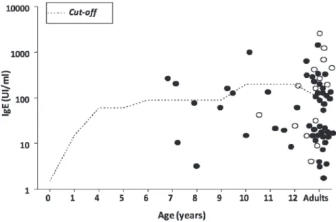
The total IgE concentrations in the JSLE patients ranged from 3.5 to 9936.0 IU/ml. Increased IgE concentrations ($100 IU/ml) were observed in 31 of the 69 (45%) patients, and the mean IgE level was 442.0¡163.4 IU/ml (Figure 1).
The mean IgG, IgM and IgA concentrations were 1387.9¡557.4, 115.8¡59.0 and 207.7¡152.3 mg/dl, respec-tively.
The IgE levels ranged from 6.1 to 9936.0 IU/ml in the JSLE patients with atopic disease, 3.5 to 9936.0 IU/ml in patients with active disease (SLEDAI 2K$4), 3.5 to 2934.0 IU/ml in patients with severe sepsis and 3.5 to 4920.0 IU/ml in patients with nephritis. In JSLE patients without atopic manifestations (n = 54), the mean IgE level
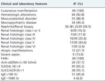
Table 1 -Clinical and laboratory findings in 69 patients with juvenile systemic lupus erythematosus.
Clinical and laboratory features N˚(%)
Cutaneous manifestation 69 (100)
Hematologic alterations 64 (92.8)
Musculoskeletal disorder 55 (80.9)
Neuropsychiatric disease 34 (49.3)
Nephritis/Renal biopsy 56 (81.2)/39 (56.5) Renal histologic class I or II 4/39 (10.3) Renal histologic class III 7/39 (17.9)
Renal histologic class IV 10/39 (25.6)
Renal histologic class V 17/39 (43.6)
Renal histologic class VI 1/39 (2.6)
Atopic manifestations 15 (21.7)
Severe sepsis 9 (13.0)
FAN+ 69 (100)
Anti-dsDNA+($50 IU/ml) 26 (37.7)
SLEDAI 2K$4 45 (65.2)
SLICC/ACR-DI$1 38 (55.1)
IgE$100 IU 31 (45.0)
was 271.6¡699.5 IU/ml. Nine of the 31 patients (29%) with
high IgE levels had atopic disease. The mean IgE level in 18 patients with severe active disease (SLEDAI 2K$10) was 371.7¡691.1, which was more than two-fold higher than
that in the 12 patients with an SLEDAI 2K score of zero (133.6¡171.2, p= 0.4). Thirty-four patients had severe
nephritis (renal histologic class III, IV, or V), with IgE concentrations ranging from 3.5 to 4920.0 IU/ml. Of note, four patients had IgE values.1000 IU/ml, and one of these patients presented with a primary C1q deficiency and atopic manifestations. IgE serum levels did not statistically differ with respect to the presence of atopic manifestations, severe sepsis, nephritis, severe nephritis, disease activity, or tissue damage (p.0.05) (Table 2).
Additionally, IgE serum concentrations were inversely correlated with C4 levels (r = -0.25, p= 0.03) and with the patient SLICC/ACR-DI scores (r = -0.34,p= 0.005). The IgE levels were also directly correlated with patient IgA levels (r = 0.52, p= 0.03) (Figure 2). No correlation was observed between IgE serum levels and C3 levels, IgG levels, IgM levels, anti-dsDNA levels or SLEDAI 2K scores.
DISCUSSION
The present study showed that JSLE patients have an increased serum IgE concentration regardless of the presence of atopic manifestations or parasitic disease.
SLE pathogenesis is complex, and there remain contro-versies concerning the involvement of immunoglobulin E in the pathogenesis of the disease (1-5). The dysregulation of immune tolerance results in aberrant Th2 responses and polyclonal activation of B lymphocytes along with the production of autoantibodies, including those of IgE isotype (1-5,10). The human Th2 immune response, characterized by the significant production of IL-4, IL-5, IL-10, and IgE, is mainly observed in atopic diseases and in some parasitic infections; however, elevated IgE production has also been observed in patients with partial T cell immunodeficiencies and autoimmune diseases (5,7,8).
Although IgE synthesis is tightly controlled by regulatory T cells, B cells, and cytokines, the role of immunoglobulin E in autoimmune diseases has not been fully elucidated (11). The most striking observation reported in the literature is that elevated IgE production and allergic and autoimmune manifestations frequently occur in patients with partial T cell immunodeficiencies even though elevated IgE levels and autoimmune and inflammatory diseases are traditionally associated with hyperactivity of the adaptive immune system (5). Moreover, mouse models have recently highlighted that an increased IgE level frequently accompanies different partial T cell immunodeficiencies that result in autoimmunity (5). Taken together, these observations suggest that IgE levels increase when there is an imbalance between the immuno-genic and toleroimmuno-genic signals in effector T cells; thus, elevated IgE levels can be considered a biomarker of immune dysregulation (5).
Although some authors have postulated that IgE is not related to connective tissue disease pathogenesis, others have claimed that IgE plays an essential role in connective tissue disorders (7,28). The latter authors state that, through the release of vasoactive mediators from basophils and mast
Figure 1 -IgE serum concentrations in 69 patients with juvenile systemic lupus erythematosus. Individual values were plotted in the graph with normal values for IgE obtained from a standard curve previous established21. White circles represent patients WITH atopic manifestations, while black circles represent patients WITHOUT atopic manifestations.
Table 2 -Total IgE serum concentrations (IU/ml) in 69 patients with juvenile systemic lupus erythematosus (JSLE) according to clinical features, disease activity and tissue damage.
Clinical Feature (N˚)
Present Mean¡SD
Absent
Mean¡SD p-value
Atopic disease (15)
cells, IgE can cause increased vasopermeability, which may be important in causing the deposition of circulating immune complexes in glomerulonephritis pathogenesis (28,29). The demonstration of increased IgE serum levels in SLE patients with renal involvement and the detection of IgE immune complex deposition in renal biopsies further implicate IgE in the pathogenesis of lupus nephritis (13,29,30). High IgE levels have also been reported in adult SLE patients without nephritis, suggest-ing that IgE may have a role in SLE disease and not only in nephritis (4,12,14,15,31,32). Furthermore, antinuclear and anti-DNA autoantibodies of the IgE isotype were observed in adult SLE patients, and these IgE autoanti-bodies did not correlate with the serum IgE concentration (4,9,10,33). Anti-IgE IgG autoantibodies have also been observed in SLE patients with lymphoadenopathy, articu-lar involvement and anti-DNA antibodies (9). In addition, increased IgE serum levels were observed in children of mothers with lupus, regardless of the presence of allergic disease in the mothers (34). Another study showed elevated IgE levels in male SLE patients compared with female patients (35). To the best of our knowledge, this was the first study to demonstrate increased IgE levels in JSLE patients.
SLE and allergic disorders share certain immunological abnormalities (6,7) because the prevalence of IgE-mediated and/or IgE-associated disorders, such as allergic reactions to drugs, atopic dermatitis, asthma, allergic rhinitis and allergic conjunctivitis, has been reported to be elevated in patients with lupus (6,12,36,37). In contrast, recent studies observed a similar prevalence of IgE-related disorders in SLE subjects compared with patients without SLE (30,38-41). In our study, 15 (21.7%) JSLE patients had at least one respiratory and/or cutaneous allergic manifestation, which is similar to the rate of atopic disorders found in healthy subjects (40,42).
The current study showed that JSLE patients harbor higher IgE concentrations in a manner independent of the presence of allergic disease. Our observations are in line with recent reports stating that patients with lupus are not at an increased risk of IgE-mediated allergic disorders. However, our observations differ from those of previous studies reporting evidence of an increased incidence of atopic conditions in SLE patients (12,13,38,41).
We also observed increased IgE serum levels in JSLE patients independent of the presence of parasitic infections.
However, the IgE serum concentrations varied widely, which may be due to differences in environment- and patient-specific factors, such as contact with antigens, race, gender and age. Witting et al. (24) showed that an IgE level of 100 IU/ml is the upper-limit cutoff for elevated IgE diagnostic sensitivity and specificity for all patient cohorts. Thus, one possible limitation of this study is the lack of comparison with age- and gender-matched healthy controls. Regarding lupus activity, patients with severe active disease (SLEDAI 2K$10) had IgE levels at least two times higher than the IgE levels of patients with inactive disease, although this difference was not statistically significant. Interestingly, IgE concentrations correlated inversely with C4 levels, which could suggest that the complement cascade was activated and its components were consumed, as shown by the reduction of C4 levels, which is also considered a marker of disease activity. The latter state-ment that IgE levels could be a marker of disease activity requires further investigation because the correlation between IgE and C4 levels was relatively weak. Although serum IgE levels have been reported to vary according to disease activity in adult SLE patients (14,15,31,39), our findings could not confirm this correlation in a definitive manner in patients with JSLE.
Interestingly, IgE serum concentrations were inversely correlated with SLICC/ACR-DI scores, suggesting a pro-tective role for increased IgE levels. This finding is contrary to current knowledge that patients with active disease have high IgE levels and are consequently at a higher risk of organ damage. In addition to the observed direct correlation between IgE and IgA serum levels, the inverse relationship between IgE levels and SLICC/ACR-DI scores supports the hypothesis that increased IgE levels can be considered a marker of immune dysregulation, which may be important in the generation of immune complexes.
Lupus nephritis, which has various histological patterns and variable clinical outcomes, is one of the most important SLE-associated morbidities. Although the pathogenic mechanism in each histological type of nephritis remains unclear, some findings point to a role for both Th1 and Th2 immune responses in renal damage (7,28). Th1 cytokines have been related to diffuse lupus nephritis, while Th2 cytokines have been associated with membranous lupus nephritis (7,13,28-30). In the present study, 81.2% of the JSLE patients had lupus nephritis, and IgE levels $100 UI/ml were observed in 36.2% of these patients. However, the
mean IgE levels were similar in patients with and without nephritis, as well as in patients with severe renal disease. This observation is in contrast with the published works with adult SLE patients, which showed a significant correlation between serum IgE concentration and nephritis activity, and previous reports, which showed that IgE renal deposits in lupus patients correlate with a poor prognosis (13,30,39).
All patients with JSLE were taking glucocorticoids, and the down-regulation of allergic inflammation could have been associated with the use of this medication, as glucocorticoids can increase IL-10 gene transcription and decrease both IL-4 and IL-5 gene transcription (43).
Finally, despite the variety of factors that can influence IgE production, the present study demonstrated for the first time that JSLE patients have higher IgE concentrations, suggesting that increased IgE levels could play a role in lupus pathogenesis. However, the specific mechanisms underlying the elevation of IgE levels in children with lupus remain to be clarified, and further studies are needed.
ACKNOWLEDGMENTS
This work was supported by FAPESP (Fundac¸a˜o de Amparo a` Pesquisa do Estado de Sa˜o Paulo) under grant number 08/58238.
AUTHOR CONTRIBUTIONS
All authors drafted the manuscript, critically reviewed the manuscript for intellectual content, and approved the final version to be published. Liphaus BL contributed to the study conception and design, data analysis and interpretation. Jesus AA contributed to the data acquisition, analysis and interpretation. Silva CA contributed to data interpretation and critically reviewed the manuscript. Coutinho A contributed to the study conception and data interpretation. Carneiro-Sampaio M contributed to data interpretation and manuscript intellectual content.
REFERENCES
1. Mok CC & Lau CS. Pathogenesis of systemic lupus erythematosus. J Clin Pathol. 2003;56(7):481-90.
2. Carneiro-Sampaio M, Liphaus BL, Jesus AA, Silva CA, Oliveira JB, Kiss MHB. Undertanding Systemic Lupus Erythematosus Physiopathology in Light of Primary Immunodeficiencies. J Clin Immunol. 2008;28(Suppl 1):S34-41, http://dx.doi.org/10.1007/s10875-008-9187-2.
3. Liphaus BL, Kiss MHB. The role of apoptosis proteins and complement components in the etiopathogenesis of systemic lupus erythematosus. Clinics. 2010;65(3):327-33, http://dx.doi.org/10.1590/S1807-59322010000 300014.
4. Atta AM, Sousa CP, Carvalho EM, Sousa-Atta MLB. Immunoglobulin E and systemic lupus erythematosus. Braz J Med Biol Res. 2004;37(10):1497-501.
5. Liston A, Enders A, Siggs OM. Unravelling the association of partial T-cell immunodeficiency and immune dysregulation. Nat Rev Immunol. 2008;8(7):545-58, http://dx.doi.org/10.1038/nri2336.
6. Shahar E, Lorber M. Allergy and SLE: common and variable. Isr J Med Sci. 1997;33(2):147–9.
7. Ring GH, Lakkis FG. Breakdown of self-tolerance and the pathogenesis of autoimmunity. Semin Nephrol. 1999;19:25-33.
8. Romagnani S. Lymphokine production by human T cells in disease states. Annu Rev Immunol. 1994;12:227-57, http://dx.doi.org/10.1146/ annurev.iy.12.040194.001303.
9. Gruber BL, Kaufman LD, Marcheses MJ, Roth W, Kaplan AP. Anti-IgE autoantibodies in systemic lupus erythematosus.Prevalence and biologic activity. Arthritis Rheum. 1988;31(8):1000-6, http://dx.doi.org/10.1002/ art.1780310810.
10. Atta AM, Santiago MB, Guerra FG, Pereira MM, Sousa Atta ML. Autoimmune response of IgE antibodies to cellular self-antigens in systemic Lupus Erythematosus. Int Arch Allergy Immunol 2010;152(4):401-6, http://dx.doi.org/10.1159/000288293.
11. Geha RS. Regulation of IgE synthesis in humans. J Allergy Clin Immunol. 1992;90(2):143-50, http://dx.doi.org/10.1016/0091-6749(92)90064-9. 12. Goldman JA, Klimek GA, Ali R. Allergy in systemic lupus
erythematosus.IgE level and reaginic phenomenon. Arthritis Rheum. 1976;19(4):669-76, http://dx.doi.org/10.1002/1529-0131(197607/ 08)19:4,669::AID-ART1780190403.3.0.CO;2-E.
13. Laurent J, Lagrue G, Sobel A. Increased serum IgE levels in patients with lupus nephritis. Am J Nephrol. 1986;6(5):413-4.
14. Rebhun J, Quismorio F, Dubois E, Heiner DC. Systemic lupus erythematosus activity and IgE. Ann Allergy. 1983;50(1):34-6. 15. Mikecz K, Sonkoly I, Meszaros C, Szegedi G. Serum IgE in systemic
lupus erythematosus. Acta Med Hung. 1985;42(1-2):59-65.
16. Tan EM, Cohen AS, Fries JF, et al. The 1982 revised criteria for the classification of SLE. Arthritis Rheum. 1982;25(11):1271-7, http:// dx.doi.org/10.1002/art.1780251101.
17. Hochberg MC. Updating the American College of Rheumatology revised criteria for the classification of systemic lupus erythematosus [letter]. Arthritis Rheum. 1997;40(9):1725, http://dx.doi.org/10.1002/art.17804-00928.
18. Goldstein B, Giroir B, Randolph A. International pediatric sepsis consensus conference: definitions for sepsis and organ dysfunction in Pediatrics; International Consensus Conference on Pediatric Sepsis. Pediatr Crit Care Med. 2005;6(1):2-8, http://dx.doi.org/10.1097/ 01.PCC.0000149131.72248.e6",-1,"xxx/72248.e6.
19. Cameron JS. Lupus nephritis in childhood and adolescence. Pediatr Nephrol. 1994;8(2):230-49, http://dx.doi.org/10.1007/BF00865490. 20. Bombardier C, Gladman DD, Urowitz MB, Caron D, Chang CH.
Committee on Prognosis Studies in SLE. Derivation of the SLEDAI. A disease activity index for lupus patients. Arthritis Rheum. 1992;35(6):630-40, http://dx.doi.org/10.1002/art.1780350606.
21. Gladman DD, Ibanez D, Urowitz MB. Systemic lupus erythematosus disease activity index 2000. J Rheumatol. 2002;29(2):288-91.
22. Gladman DD, Urowitz MB, Goldsmith CH, Fortin P, Ginzler E, Gordon C, et al. The reliability of the Systemic Lupus International Collaborating Clinics/American College of Rheumatology Damage Index in patients with systemic lupus erythematosus. Arthritis Rheum. 1997;40(5):809-13, http://dx.doi.org/10.1002/art.1780400506.
23. Dati F, Ringel KP. Reference values for serum IgE in healthy non-atopic children and adults. Clin Chem. 1982;28:1556.
24. Wittig HJ, Belloit J, De Fillippi I, Royal G. Age-related serum immunoglobulin E levels in healthy subjects and in patients with allergic disease. J Allergy Clin Immunol. 1980;66(4):305-13, http:// dx.doi.org/10.1016/0091-6749(80)90026-3.
25. Mancini I, Sole´ D, Naspitz CK. Nı´veis se´ricos de IgE total em crianc¸as brasileiras normais no primeiro ano de vida. J Pediatr (Rio J). 1996;72(2):98-102, http://dx.doi.org/10.2223/JPED.589.
26. Spalding SM, Wald V, Bernd LAG. IgE se´rica total em ato´picos e na˜o-ato´picos na cidade de Porto Alegre. Ver Ass Med Barsil. 2000;46(2):93-7. 27. Rosner B. Fundamentals of biostatistics. 5thed. Duxburg, CA: Thomsom
Learning; 2000.
28. Robertson MR, Potter EV, Roberts ML, Patterson R. Immunoglobulin E in renal disease. Nephron. 1976;16(4):256-71, http://dx.doi.org/10.1159/ 000180610.
29. McPhaul Jr JJ, Newcomb RW, Mullins JD, Thompson Jr AL, Lordon RE, Rogers PW. Participation of immunoglobulin E (IgE) in immune-mediated glomerulonephritis. Kidney Int. 1974;5(4):292-9, http:// dx.doi.org/10.1038/ki.1974.39.
30. Tuma SN, Llach F, Sostrin S, Dubois EL, Massry G. Glomerular IgE deposits in patients with lupus nephritis. Am J Nephrol. 1981;1(1):31-6. 31. Wozniacka A, Sysa-Jedrzejowska A, Robak E, Samochocki Z, Zak-Prelich M. Allergic diseases, drug adverse reactions and total immunoglobulin E levels in lupus erythematosus patients. Mediators Imflamm. 2003;12(2):95-9, http://dx.doi.org/10.1080/0962935031000097709. 32. Sekigawa I, Tokano Y, Yoshike T, Iida N, Hashimoto H, Ogawa H.
Relationship between serum IgE and autoantibodies levels in SLE patients. Clin Exp Rheumatol. 2003;21(5):683.
33. Egido J, Crespo S, Lahoz C, Garcia R, Lopez-Trascada M, Hernando L. Evidence of an immediate hypersensitivity mechamism in systemic lupus erythenmatyosus. Ann Rheum Dis. 1980;39(4):312-7, http:// dx.doi.org/10.1136/ard.39.4.312.
34. Sasai K, Furukawa S, Hashimoto H, Yabuta K. Increased levels of serum IgE in children of mothers with systemic lupus erythematosus. Allergy. 1995;50(4):370-3, http://dx.doi.org/10.1111/j.1398-9995.1995.tb01163.x. 35. Sekigawa I, Yanada M, Iida N, Hashimoto H, Ogawa H. Comparison of
serum IgE levels between female and male SLE patients, with reference to gender differences in the incidence of SLE. Clin Exp Rheumatol. 2004;22(3):384-5.
36. Petri M & Allbritton J. Antibiotic allergy in systemic lupus erythema-tosus: a case control study. J Rheumatol. 1992;19(2):265-9.
37. Sequeira JF, Cesic D, Keser, Bukelica M, Karanagnostis S, Khamashta MA, Huges GR. Allergic disorders in systemic lupus erythematosus. Lupus. 1993;2(3):187-91, http://dx.doi.org/10.1177/096120339300200311. 38. Sekigawa I, Yoshiike T, Iida N, Hashimoto H, Ogawa H. Allergic
diseases in systemic lupus erythematosus: prevalence and immunologi-cal considerations. Clin Exp Rheumatol. 2003;21(1):117-21.
39. Elkayam O, Tamir R, Pick AI, Wysenbeek A. Serum IgE concentrations, disease activity, and atopic disorders in systemic lupus erythematosus. Allergy. 1995;50(1):94-6.
41. Sekigawa I, Yoshiike T, Iida N, Hashimoto H, Ogawa H. Allergic disorders in systemic lupus erythematosus prevalence and family history. Lupus. 2002;11(7):426-9, http://dx.doi.org/10.1191/ 0961203302lu221oa.
42. Becker LC. Allergy in systemic lupus erythematosus. Hopkins Med J. 1973;133(1):38-44.