Cells Permanently Transduces and Transcribes Human
DNA
David M. Goldenberg1,2*, Robert J. Rooney3, Meiyu Loo2, Donglin Liu2, Chien-Hsing Chang2
1Garden State Cancer Center, Center for Molecular Medicine and Immunology, Morris Plains, New Jersey, United States of America,2Immunomedics, Inc., Morris Plains, New Jersey, United States of America,3Genome Explorations, Inc., Memphis, Tennessee, United States of America
Abstract
After demonstrating, with karyotyping, polymerase chain reaction (PCR) and fluorescencein-situhybridization, the retention of certain human chromosomes and genes following the spontaneous fusion of human tumor and hamster cellsin-vivo, it was postulated that cell fusion causes the horizontal transmission of malignancy and donor genes. Here, we analyzed gene expression profiles of 3 different hybrid tumors first generated in the hamster cheek pouch after human tumor grafting, and then propagated in hamsters and in cell cultures for years: two Hodgkin lymphomas (GW-532, GW-584) and a glioblastoma multiforme (GB-749). Based on the criteria of MAS 5.0 detection P-values #0.065 and at least a 2-fold greater signal expression value than a hamster melanoma control, we identified 3,759 probe sets (ranging from 1,040 to 1,303 in each transplant) from formalin-fixed, paraffin-embedded sections of the 3 hybrid tumors, which unambiguously mapped to 3,107 unique Entrez Gene IDs, representative of all human chromosomes; however, by karyology, one of the hybrid tumors (GB-749) had a total of 15 human chromosomes in its cells. Among the genes mapped, 39 probe sets, representing 33 unique Entrez Gene IDs, complied with the detection criteria in all hybrid tumor samples. Five of these 33 genes encode transcription factors that are known to regulate cell growth and differentiation; five encode cell adhesion- and transmigration-associated proteins that participate in oncogenesis and/or metastasis and invasion; and additional genes encode proteins involved in signaling pathways, regulation of apoptosis, DNA repair, and multidrug resistance. These findings were corroborated by PCR and reverse transcription PCR, showing the presence of human alphoid (a)-satellite DNA and theF11Rtranscripts in additional tumor transplant generations. We posit thatin-vivofusion discloses genes implicated in tumor progression, and gene families coding for the organoid phenotype. Thus, cancer cells can transduce adjacent stromal cells, with the resulting progeny having permanently transcribed genes with malignant and other gene functions of the donor DNA. Using heterospecific in-vivo cell fusion, genes encoding oncogenic and organogenic traits may be identified.
Citation:Goldenberg DM, Rooney RJ, Loo M, Liu D, Chang C-H (2014)In-VivoFusion of Human Cancer and Hamster Stromal Cells Permanently Transduces and Transcribes Human DNA. PLoS ONE 9(9): e107927. doi:10.1371/journal.pone.0107927
Editor:Thomas Dittmar, Witten/Herdecke University, Germany
ReceivedMarch 27, 2014;AcceptedAugust 19, 2014;PublishedSeptember 26, 2014
Copyright:ß2014 Goldenberg et al. This is an open-access article distributed under the terms of the Creative Commons Attribution License, which permits unrestricted use, distribution, and reproduction in any medium, provided the original author and source are credited.
Data Availability:The authors confirm that all data underlying the findings are fully available without restriction. The data files have been deposited in the Gene Expression Omnibus, and can be viewed at http://www.ncbi.nlm.nih.gov/geo/query/acc.cgi?acc = GSE58277.
Funding:The initial transplantation and characterization studies were supported in part by American Cancer Society Grant IN-581, USPHS grants CA11327 and CA12374 from the National Institutes of Health, and the Damon Runyon Memorial Fund for Cancer Research (to DMG). The funders had no role in study design, data collection and analysis, decision to publish, or preparation of the manuscript.
Competing Interests:All authors are employed by Immunomedics, Inc., or Genome Explorations, Inc., but this does not alter their adherence to PLOS ONE policies on sharing data and materials.
* Email: dmg.gscancer@att.net
Introduction
Primary human tumor transplants, particularly to immunosup-pressed rodents, such as nude and NOD/SCID mice, are used as preclinical models for evaluating tumor biology and drug sensitivity [1–7]. These studies are based on the supposition that such xenografts retain the properties and critical genotypes of their donor tumors, thus being predictive for clinical translation. However, we and others have demonstrated that such transplants can induce tumors in their rodent recipients, such as golden hamsters [8–10], nude/SCID mice [11–24], and immunosup-pressed rats [25], although infrequently (either because of low incidence or rare testing). One plausible explanation is the horizontal transfer of oncogenic DNA [25–27]. Indeed, lateral
carcinoma cells, were metastatic and lethal in nude mice or immunocompetent mice of the same genetic background [14]. This induction of stromal tumors in host animals after xenotrans-plantation of human epithelial cancers has been confirmed by others [15–25], thus suggesting that cancer xenografts be carefully evaluated for horizontal oncogenesis [13,24]. How this transfor-mation or induction occurred was not elucidated, but a viral role has been discussed [17].
In some of these experiments involving primary human tumor transplants, transfer of functional human genetic information by
in-vivo cell hybridization of the donor tumor and recipient host cells, showing chromosomal, immunological, or genetic features of both partners [9,29–33], was proposed as the mechanism for induction of these tumors that exhibited highly invasive and metastatic behavior in their animal hosts [34,35]. For example, we reported that after long-term propagation of human-hamster hybrid tumors derived from a glioblastoma multiforme [33] and two Hodgkin lymphomas, human DNA and genes could be confirmed by fluorescence in-situ hybridization (FISH) and polymerase chain reaction (PCR), and their donor organoid features by histology [36,37]. Translation of some of these gene products was found by immunohistochemistry (IHC) in the glioblastoma multiforme transplants, even after propagation for over a year [36].
These results indicate that human genes can remain functional within human-hamster hybrid tumors propagated in the animal host, emphasizing the horizontal transmission of human DNA implicated with malignancy and the organoid features of the original patient donor tumors. However, the scope of human DNA transduced and transcribed in these interspecies hybrid cells has not been investigated. Accordingly, we examined (i) if such formalin-fixed, paraffin-embedded (FFPE) tumor grafts, which were stored for over 40 years since they were made, could be tested globally for the expression of transcribed human genes, (ii) if human genes are retained during long-term serial passage, and (iii) if there are specific human gene families indigenous to these human-hamster hybrid tumors. By using tumors and hosts of different species, we are able to identify each party’s genetic contribution, which is especially problematic when attempting to prove cell-cell fusion in humans, whether involving normal-normal, malignant-normal-normal, or malignant-malignant fusions.
We postulate that these results of heterospecific fusions provide a general mechanism of tumor DNA transfer to stromal cells that results in genetic instability, heterogeneity, and aneuploidy, leading to stable genomic changes associated with cancer progression, while also retaining the tumor’s original organoid phenotype, as well as other genes derived from the donor human tumor. This merging of tumor and normal genomes into a new population of malignant hybrid cells could be a mechanism whereby a cancer escapes host immunity by reducing the immunological disparity between the tumor and its host [34,35]. Various aspects of the role of cell-cell fusion in cancer are now gaining increased attention [35,38–47].
Results
Human mRNA transcripts present in each of four different human-hamster hybrid tumor FFPE samples (Table 1) were identified by analysis of total RNA, in comparison to a control hamster melanoma line (CCL-49), using Affymetrix Human U133 X3P arrays. Probe sets with MAS 5.0 detectionP-values#0.065 in a hybrid sample, a detection P-value.0.065 in the hamster control, and an expression signal value that was at least 2-fold greater in the hybrid sample than in the hamster control, were
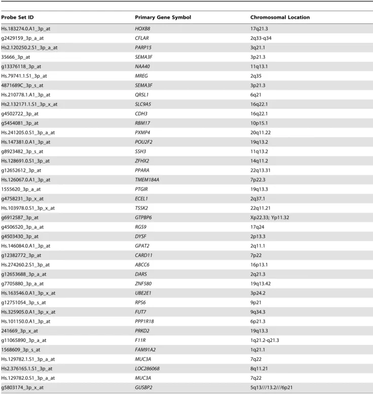
considered to represent expressed human gene transcripts. Using these criteria, we identified a total of 3759 probe sets (ranging from 1040 to 1303 probe sets in at least one hybrid sample), which unambiguously mapped to 3107 unique Entrez Gene IDs (Table S1), representing genes from all human chromosomes. Among these, 39 probe sets passed all of the expression criteria in all four hybrid specimens (Figure 1, Table 2), with 34 probe sets detecting 33 unique Entrez Gene IDs (Table S2), two probe sets detecting either MUC3A or MUC1B, and the remaining probe sets detecting an uncharacterized gene (LOC286068), GUSBP2 or mutlpleGUSBpseudogenes, andFAM91A2or multiple unchar-acterized genes. Thus, at least 33 unique human genes were transcribed in these FFPE tissues from 3 different human tumor xenografts representing different transplant generations, including two for GW-532, propagated serially for months to years as highly metastatic tumors.
As listed in Table S3, transcripts of the genes expressed in all four hybrid samples include five encoding transcription factors that are known to regulate cell growth and differentiation (HOXB8, PPARA, POU2F2, ZFHX2, and ZNF580), and five encoding cell adhesion and transmigration-associated proteins that participate in tumorigenesis and/or invasion/metastasis (CDH3, FUT7, F11R, MUC3A, and SEMA3F). In addition, genes whose products are associated with signaling pathways, regulation of apoptosis, DNA repair, and multidrug resistance, also were identified (namely, PRKD2, ECEL1, CARD11, CFLAR, PARP15, andMRP6).
Recognizing that the degraded nature of the FFPE RNA and the high background of hamster RNA in the FFPE hybrid samples could interfere with the sensitivity of MAS 5.0 detectionP-values, we relaxed the detectionP-value criterion by requiring a detection
P-value#0.065 in only one of the four hybrid samples, instead of all four, and produced a larger list of human genes that potentially were commonly expressed in all of the hybrid samples. This second list contained 1120 probe sets, representing 982 unique Entrez Gene IDs (Table S4). These results indicate the presence of genes for CD20 (MS4A1), CD22, and CD44 (signaling compo-nent of the macrophage migration inhibitor factor (MIF)-CD74-CD44 receptor complex), thus corroborating the previous PCR results for the presence of CD20 and, also, CD74 genes in the GW-532 and GW-584 lymphoma hybrid tumors [36,37]. A number of other human genes, such as those encoding CD24, CD27, CD47, CD52, CD84, CD151, and tenascin XB (TNXB), were found to be transcribed in these hybrid cell lines when the detectionP-value criterion was relaxed (Table S4).
activation, and apoptosis (Table S6). These results, from both the pathway and functional enrichment analyses, indicate that the various sets of human genes expressed in each hybrid tumor sample affect related cellular processes, and thereby likely produce similar effects on cellular function and growth.
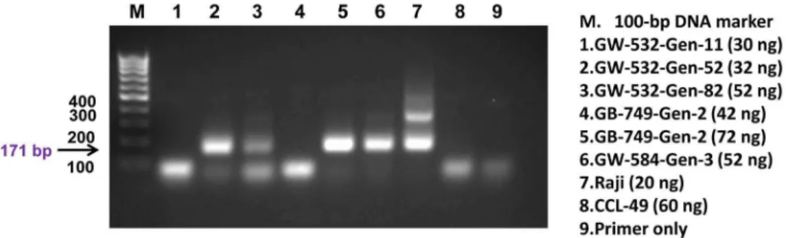
To further corroborate the microarray findings, PCR was performed on six additional FFPE tissue samples: three from GW-532 (generations 11, 52, and 82), one from GW-584 (generation 3), and two from GB-749 (both of generation 2), to assess the presence of human DNA in these tissue blocks, using a pair of primers directed to the 171-bp monomer of human alpha satellite DNA [56]. As shown in Figure 2, four of the 6 samples (GW-532 generations 52 and 82, GW-584 generation 3, and GB-749 generation 2) were positive for the expected PCR product of humanalpha satellite DNA (the 171-bp), which was detected also
in the DNA of human lymphoma Raji cells (positive control), but not in the DNA of CCL-49 hamster melanoma cells (negative control). Moreover, we were able to confirm the expression of the
F11Rgene detected by the cDNA microarray studies in two of the six samples by one-step reverse transcription-PCR, using human hepatic cancer HepG2 cells as the positive control [57]. As shown in Figure 3, the presence of a 141-bp band was prominent in both GW-532 generation 11 and GW-584 generation 3, as well as in human HepG2 cells (positive control), but not in the tissue of a hamster spleen (negative control). These results were confirmed in a repeat experiment (Figure S1), using CCL-49 cells as the negative control.
Discussion
In this study, we utilized human gene expression microarrays to provide further evidence that human genes can remain functional within metastatic human-hamster hybrid tumors propagated in the animal host, and corroborated such findings with additional samples showing the presence of human alphoid (a) satellite DNA
and theF11Rtranscripts by PCR and reverse transcription-PCR, respectively. Our results demonstrate that human tumors trans-planted to rodents can merge their DNA with the genome of the animal host, as an example of the larger program of tumor-stromal crosstalk. Cancer cells depend and are influenced by their ‘‘soil’’ or stromal microenvironment [58–60], but it is also known that there can be genetic interchange [61,62]. The reciprocal horizontal transfer of genetic material between stromal and tumor cells could explain the heterogeneity and genetic diversity and evolution of cancer cell populations, not only between different patient tumors of the same cancer type, but even different tumors of the same patient, as observed in genetic analyses of human tumor specimens
[63–65]. Cell-cell fusion enables the transfer of chromosomes and genetic material from one cell to another, and has been shown to result in viable hybrid progeny capable of replication for different periods, but usually not long-term or as permanent cell lines [66]. By using heterospecific cell-cell fusionin-vivo, genes controlling oncogenesis and organoid traits in the donor cancer cells may be elucidated in the fused progeny.
The fusion of tumor and myeloid cells was proposed at the beginning of the 20th century by various German pathologists, such as Aichel, Dor, Hallion, and Kronthal, as cited with the first experimental results and discussion of spontaneous fusionin-vivo
in 1968 [34]. This was based on the development of highly aggressive and metastatic tumors after grafting four different human cancers, with one of ovarian cancer origin (GW-127)
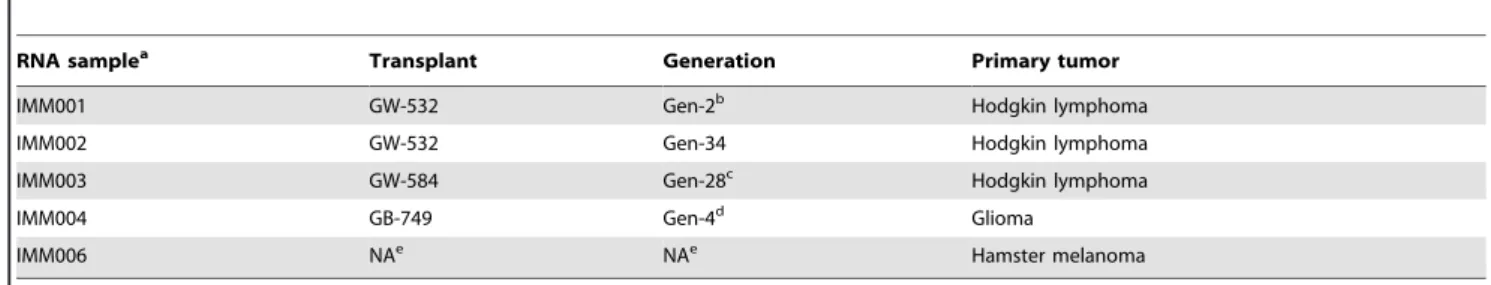
Table 1.Characteristics of test articles used in the microarray study.
RNA samplea Transplant Generation Primary tumor
IMM001 GW-532 Gen-2b Hodgkin lymphoma
IMM002 GW-532 Gen-34 Hodgkin lymphoma
IMM003 GW-584 Gen-28c Hodgkin lymphoma
IMM004 GB-749 Gen-4d Glioma
IMM006 NAe NAe Hamster melanoma
aPrepared from FFPE specimens as indicated, except IMM006, which was prepared from CCL-49, a Syrian golden hamster melanoma cell line acquired from ATCC. bHuman genes ofCD74,CXCR4,CD19,CD79b, andVIMwere detected by PCR (Ref. 37).
cHuman genes ofCD74,CXCR4,CD20, andCD79bwere detected by PCR (Ref. 37). dThe expression of CD74, CXCR4 and PLAGL2 were detected by IHC staining (Ref. 36). eNot applicable.
doi:10.1371/journal.pone.0107927.t001
Figure 1. Clustered heat map of the 39 human probe sets detected in all four hybrid tumor samples.The heat map depicts expression signal values for 39 Affymetrix Human U133_X3P probe sets detected in FFPE sections from all four hybrids tested (IMM001-004) and a hamster control (IMM006). Prior to unsupervised hierarchical clustering, the MAS 5.0 signal values were log2-transformed and row mean centered. Samples were clustered by Complete Linkage based on Pearson correlation; probe sets were clustered by Complete Linkage based on Euclidean distance. Criteria for detectable human gene expression included MAS 5.0 Detection p-values#0.065 in the hybrid sample and .0.065 in the hamster control, and $2-fold increased signal in the hybrid sample vs. the hamster control.
showing hamster chromosomes, but also retention of human antigens [8,9,29,30]. A series of subsequent studies described the transplantation of diverse human cancers to the cheek pouch of unconditioned (non-immunosuppressed) golden hamsters, and also showed metastases in their hamster hosts as early as 3–4 weeks after grafting, and the presence of both human and hamster markers within the cancer cells. The transplants displayed mostly hamster properties while retaining features of their human origin, including human chromosomes, isoenzyme patterns, antigens, and stathmokinetic properties in response to colchicine that was more
compatible with human than hamster cells [30–33]. Over the course of about 15 years, while grafting more than 1200 primary human cancers to hamsters (cheek pouch site) or nude mice (subcutaneous site), 15 (1.25%) highly aggressive and metastatic tumors resulted from the hamster transplants [35]. These were derived from diverse solid and hematopoietic human tumors, and could be propagatedin-vitroorin-vivofor years as permanent cell lines, showing rapid growth and metastatic features typical of a hamster tumor [10,33,35].
Table 2.The 39 probe sets determined to be positive in all hybrid FFPE specimens.
Probe Set ID Primary Gene Symbol Chromosomal Location
Hs.183274.0.A1_3p_at HOXB8 17q21.3
g2429159_3p_a_at CFLAR 2q33-q34
Hs2.120250.2.S1_3p_a_at PARP15 3q21.1
35666_3p_at SEMA3F 3p21.3
g13376118_3p_at NAA40 11q13.1
Hs.79741.1.S1_3p_at MREG 2q35
4871689C_3p_s_at SEMA3F 3p21.3
Hs.210778.1.A1_3p_at QRSL1 6q21
Hs2.132171.1.S1_3p_x_at SLC9A5 16q22.1
g4502722_3p_at CDH3 16q22.1
g5454081_3p_at RBM17 10p15.1
Hs.241205.0.S1_3p_a_at PXMP4 20q11.22
Hs.147381.0.A1_3p_at POU2F2 19q13.2
g8923482_3p_s_at SSH3 11q13.2
Hs.128691.0.S1_3p_at ZFHX2 14q11.2
g12652612_3p_at PPARA 22q13.31
Hs.126067.0.A1_3p_at TMEM184A 7p22.3
1555620_3p_a_at PTGIR 19q13.3
g4758231_3p_x_at ECEL1 2q37.1
Hs.103978.0.S1_3p_x_at TSSK2 22q11.21
g6912587_3p_at GTPBP6 Xp22.33; Yp11.32
g4506520_3p_a_at RGS9 17q24
g4503430_3p_at DYSF 2p13.3
Hs.146084.0.A1_3p_at GPAT2 2q11.1
g12382772_3p_at CARD11 7p22
Hs.274260.2.S1_3p_at ABCC6 16p13.1
g12653688_3p_a_at DARS 2q21.3
g7705880_3p_a_at ZNF580 19q13.42
Hs.163546.0.A1_3p_x_at UBE2E1 3p24.2
g12751054_3p_s_at RPS6 9p21
Hs.325905.0.A1_3p_x_at FUT7 9q34.3
Hs.101150.0.A1_3p_at PPP1R18 6p21.3
241669_3p_x_at PRKD2 19q13.3
g11065890_3p_a_at F11R 1q21.2-q21.3
1568609_3p_s_at FAM91A2 1q21.1
Hs.129782.1.S1_3p_a_at MUC3A 7q22
Hs2.376165.1.S1_3p_at LOC286068 8q11.21
Hs.129782.0.S1_3p_a_at MUC3A 7q22
g5803174_3p_x_at GUSBP2 5q13///13.2///6p21
Since gene probes were not available then, it was only recently that FFPE tissues from these earlier transplants were subjected to FISH, PCR, and IHC methods to demonstrate the presence of both species’ genetic markers and translation of human genes in some of these permanent transplants, even after years in the foreign, animal host [36,37]. For example, the glioblastoma multiforme (GW-749) was reported in 1974 to be a human-hamster hybrid tumor based on retention of up to 15 human and many hamster chromosomes in the same malignant cells, as classified by Giemsa staining, even with definite identification of chromosomes karyotyped from the patient’s lymphocytes, thus being a heterosynkaryon [33]. More recently, the GW-749 xenograft tumor was shown to have retained 7 transcribed human genes(CD74, CXCR4, PLAGL2, GFAP, VIM, TP53, EGFR), of whichCD74, CXCR4, and PLAGL2, continued to be translated to their respective proteins that were visualized by IHC, as well as hamster X chromosome and human pancentromeric DNA in the same nuclei by FISH [36]. Surprisingly, these genes are known to have an association with malignancy and, in particular glial tumors, as well as VIM associated with mesenchymal cells. The transplants continued to express features of the original glioma tumor grafted, even after propagation in hamsters for ,1 year [36].
Similar analyses were reported recently for two lymphomas grafted to hamsters [37], one of which was described in 1970 and shown to resemble its donor human tumor although gaining highly metastatic properties in the hamster [10]. FISH and PCR analyses showed that these two Hodgkin lymphoma-derived hybrid tumors
displayed both hamster and human DNA in the same nuclei by FISH, while also retaining the human genes, CD74, CXCR4, CD19, CD20, CD71, CD79b, and VIM. It is noteworthy that the GB-749 glioblastoma hybrid tumor showed retention of glioma-related genes (PLAGL2, GFAP), whereas the lymphoma-derived hybrid tumor retained several B-cell antigen receptor (BCR)-related genes (CD19, CD20, CD71, CD79b). Three human genes,
CD74,CXCR4, andVIM, were common to both the glioblastoma and lymphoma transplants. Both vimentin and CXCR4 are mesenchymal markers associated with epithelial-mesenchymal transition (EMT) whose genes were transcribed in all 3 hybrid tumors examined. It was also suggested that the heterosynkaryons of Hodgkin lymphoma with their Hodgkin Reed-Sternberg (HRS) cells retained their B-cell origin [37], confirming other evidence for a B-cell origin of this neoplasm [67], and again corroborated herein by gene probe analysis disclosing B-cell genes (CD20, CD22) in these specimens. As described, these tumors were observed within 2 weeks of their first transplantation, and showed evidence of metastasis in the hamster within 3–4 weeks [10,37], suggesting that the hamster host’s early response to the foreign tissue graft may have contributed to this process. Indeed, inflammation and wound healing are known to facilitate cell fusion [68–70].
In the current studies, we were interested in surveying the extent by which human DNA could be transferred and continuously transcribed in the hybrid tumors. Gene expression microarray analysis was performed using total RNA isolated from FFPE sections of these hybrid tumors, including two different transplant
Figure 2. PCR of human alpha satellite DNA.The presence of human DNA was demonstrated by the detection of the 171-bp product in GW-532 generation 52 (lane 2), GW-532 generation 82 (lane 3), GB-749 generation 2 (lane 5), and GW-584 generation 3 (lane 6), but not in the negative control of hamster melanoma, CCL-49 (lane 8). The 171-bp and its higher oligomers were detected in the positive control of human Raji lymphoma cells (lane 7). The experimental conditions and the nominal amount of DNA used for each sample are indicated.
doi:10.1371/journal.pone.0107927.g002
Figure 3. One-step reverse transcription PCR.The mRNA transcripts ofthe F11Rgene were detectable in 532 generation 11 (lane 1), GW-584 generation 3 (lane 2), and the positive control of human HepG2 cells (lane 6), but not in the negative control hamster spleen cells (lane 5). The experimental conditions and the nominal amount of RNA used for each sample are indicated.
generations of GW-532. Unexpectedly, we detected a combined total of .3000 human genes amongst all of the samples, representing genes from all 23 pairs of human chromosomes, and found that 33 human genes were ubiquitously expressed in each of the 4 samples from the 3 tumors. Five of these genes encode transcription factors that are known to regulate cell growth and differentiation (HOXB8, PPARA, POU2F2, ZFH2, ZNF580), while another five encode cell adhesion and transmi-gration-associated proteins that are known to participate in tumorigenesis and/or metastatic invasion (CDH3, FUT7, F11R, MUC3A, and SEMA3F). Additional genes whose products can promote metastatic growth were also identified, including two signaling pathway enzymes (PRKD2andECEL1), two apoptosis regulators (CARD11andCFLAR), the DNA repair and apoptosis regulator (PARP15), and the multidrug resistance gene (ABCC6). A representative publication for each of these 16 genes is provided in References S1. It is particularly noteworthy that published reports show that deregulated expression of either PPARA or
POU2F2 can promote oncogenic growth, the developmental function ofPOU2F2andHOXgenes is to maintain cells in a less-differentiated state [71–80], and high expression ofECEL1gene was reported by Kawamoto et al [81] to associate with favorable prognosis in human neuroblastoma. A limitation of this evalua-tion, however, is the fidelity of the RNA extracted from these FFPE tissues, which were over 40 years old, emphasizing that only positive microarray results can be considered informative. This could explain why some of the genes identified in these specimens by PCR [36] were not identified by microarray analysis. In this study, however, both the DNA arrays and PCR identified the retention of transcribed human F11R, which codes for a junctional adhesion molecule. The other human gene detected by RT-PCR,a-satellite DNA, is present in the centromere of all
human chromosomes, comprising the main structural component of heterochromatin. We should also note that the FFPE sections are of various transplant generations made over many years, and at various times studied in vitro. The populations are very uniform, not reflecting different cell populations morphologically. When the GB-749 glioma transplant was studied after transplan-tation, several generations showed the presence, in single cells, of both human and hamster chromosomes based on chromosome banding, and in fact compared to chromosomes identified in the donor patient’s leukocytes. Since these were in single cells, we referred to these as heterosynkaryons. As such tumors were propagated for long periods, the cell population became very uniform, and there was never evidence of purely human tumor cells being propagated and maintained in serial passage.
Recently, the fusion of human bone marrow stromal cells with two human breast cancer cell lines indicated that the hybrid progeny were more metastatic than the parental breast cancers, and that analysis of coding single-nucleotide polymorphisms by RNA sequencing revealed genetic contributions from both parental partners, with between 1239 and 5345 genes from the parental cells retained in the fused cells [66]. However, these fused cells did not show long-term stability, but did retain breast cancer morphology [66]. In contrast, fusion of human cancer cells with normal stromal cells of murine mammary glands resulted in malignant tumors that had a sarcomatous appearance [82]. Two different human breast cancer cell populations injected into mice resulted in malignant cells that showed evidence of fusion in the mouse bone marrow, and were more extensively metastatic than the parental cell lines [83]. Similarly, two separate sets of genes that promote metastasis to bone and lung were combined via fusion of breast cancer cell lines, resulting in stable hybrids propagated long-term in cell culture and in-vivo [70]. Further,
fusion of hematopoietic cells with human and murine epithelial ovarian cancer cells resulted in aggressive tumors of an epithelial phenotype retaining hematopoietic markers [84]. It is interesting that the chemokine receptor, CXCR4, which is a promigration marker, was expressed in the hybrid tumors, similar to our own experience of this chemokine’s gene being transcribed in the three hybrid tumors studied here.
In our own experiments, the transcribed genes are known to be implicated in tumor progression to invasion and metastasis, including those involving EMT that is postulated to advance tumor cells to more malignant features [85,86]. Recently, in fact, fusions of human lung cancer cells from cell lines and human bone marrow-derived mesenchymal stem cells, when co-cultured in-vitro, showed evidence of cell fusion and the convergence to a mesenchymal-like progeny with EMT and stem cell-like proper-ties, even after injection into NOD/SCID mice [87]. Unfortu-nately, although considered by these authors as ‘spontaneous’ cell fusion, it is hardly spontaneous when 2 cell lines are grown together in culture, in contrast to growth of tumors that fuse in-vivowith unselected cells in their microenvironment. Nevertheless, such observations provide experimental evidence that in circum-stances promoting horizontal gene transfer, whether or not truly spontaneous or the result of experimental conditions, new hybrid daughter tumor cells with new properties are generated, with features of more advanced malignancy [45,46,70,82,83]. Other experiments also have indicated that the progeny hybrid cells after fusion can acquire different properties than the parental cells [38,43,70,82,83,88]. Thus, such fusion experiments may help further define genes and gene families participating in the evolution, change, and progression of human cancers by methods that are difficult to apply to humans or human tumor specimens directly.
In order to reproduce this heterospecific hybridization exper-imentally, a murine melanoma was fused with hamster cheek pouch fibroblastsin- vitro, and the chromosomes of the daughter cells and their behavior in- vivo in hamsters and genetically-compatible mice were studied [94]. It was found that the murine-hamster hybrid tumor cells (confirmed karyologically) were more malignant in the hamster than the original murine melanoma was in mice, and that the hybrid tumor cells could not be propagated in genetically-compatible mice. Since the original murine mela-noma could not grow in adult golden hamsters, the hamster genome came to dominate the genome of the hybrid tumor derived from the murine melanoma, retaining malignancy and metastasizability in hamsters but not in mice, while also losing expression of the melanin present in the original murine melanoma [94]. Evidently, the genetic contribution of the normal (fibroblast) cells governed the biological behavior and genetic features of the hybrid progeny, with the exception of malignancy and metastasizability derived from the murine melanoma. Similar experimental results of melanoma fusions with macrophages in mice have corroborated these findings, but where melanin was retained in the hybrid cells [46,95,96]. Thus, these various studies provide evidence of tumor progression after human-hamster, human-murine, and hamster-murine cell fusions.
The interpretation and relevance of these findings to human cancer are both challenging and stimulating. Does synkaryon formation and the progression of tumors to metastasizability constitute an isolated biological phenomenon without clinical relevance? Tumor heterogeneity has been a focus of interpretation and discussion since the beginnings of cancer histopathology, when diverse cell types and multinucleated giant cells were identified in the tumor and in its microenvironment. These gross cellular observations were then confirmed by genetic studies indicating a heterogeneity between different cells of the same tumor and between different metastases compared among themselves or to the primary tumor cells [63–65].
Cell-cell fusion may in fact be one mechanism of a more general process of intercellular DNA transfer. Supernatant from human tumor cell cultures or even cell-free DNA from human tumors or sera from cancer patients have been shown to induce tumors in recipient mice [27,97,98]. Other studies have suggested lateral transfer of non-cellular gene, RNA, or DNA via membrane-derived vesicles, exosomes, or other shed cell constituents [99,100]. However, many of these experiments demonstrating oncogenicity utilized immortalized embryonic murine fibroblasts (NIH-3T3), which are known to be susceptible to transformation [98]. Nevertheless, human mutated gene sequences (e.g.,KRAS) associated with the primary human cancers were transferred to the transformed murine fibroblasts by plasma DNA taken from human cancer patients, which then proved to be malignant in genetically-compatible mice [97]. Others have reported that circulating breast cancer cells exhibit epithelial and mesenchymal traits, with the latter indicating a more aggressive cell population [101]. The basis of this EMT, which has been discussed in many other models of malignancy [83,85,86], was not elucidated, but does stimulate questioning whether this could be due to DNA transfer, possibly via carcinoma-mesenchymal cell fusion, as already discussed in lung cancer x mesenchymal stem-cell fusion studies [101]. Indeed, it has been hypothesized that circulating cancer cells in humans express myeloid markers as a result of cell fusion [102].
These studies suggest that gene or DNA transfer between cells, forming recombinant gene hybrids, may not require cell-cell fusion and synkaryon formation. In fact, most of the recent studies implicating cell fusion are based on evidence of genetic markers of
2 different parental cells in the putative hybrid cell, in the absence of careful chromosome analyses showing a mixed karyotype in single nuclei. Hence, such experiments do not exclude gene transfer without actual synkaryon formation.
In conclusion, if cell-cell fusion is a basic biological process among many species and certain functions in humans [40,45,103], it is not unreasonable to expect that it would play an important role in oncogenesis [40–47,88], accounting for genetic diversity within a single neoplasm or even between different tumors of the same patient. This would amend the long-held view of the clonal derivation of cancer cell populations [104], now emphasizing that horizontal gene interactions and cell-cell transfer also influence the development and change in cancer cell populations. But the major challenge is to prove that this mechanism is operative in cancer patients, for which evidence is accumulating in unique settings, such as in bone marrow transplantation transferring human chromosomes and genes to the recipients’ tumors [105], and fusion of myeloma cells and osteoclasts in bone destruction [39,106]. With the increasing interest in the crosstalk and exchange between cancer and stromal cells, including macro-phages and leukocytes [43,46,69], the potential contribution of cell-cell fusion in the horizontal transfer of malignancy and other genes within a tumor deserves continued attention, and implies that this may be a basic biological process occurring between many different cell types both physiologically and in disease. In fact, there is evidence that novel transcriptomes can develop in hybrids that were not present in the parental cells [43,70,107– 109].
Since the first evidence suggesting that cell fusion is a mechanism by which cancer cells become more diverse and progress to the advanced state of metastasis [33,34], numerous experiments involving fusions of tumor x tumor, tumor x normal, and tumor x specific myeloid cells, as cited above and in recent reviews [40,44–47], have made similar conclusions. However, it should be recognized that although revealing important attributes of cell-cell fusion in the recognition and plasticity of gene interactions and the development of hybrid daughter cells with phenotypic diversity, virtually all of these studies have utilized established cancer cell lines mixed eitherin- vitroor combined in-vivo, with the inherent limitations of cell line selection that may not be representative of the heterogeneous populations of primary tumors. This is emphasized by a publication that appeared while this article was under revision. It was reported that human pontine tumors obtained at autopsy and grafted orthotopically to immune-deficient mice either directly or via intermediate cell culture were different. Direct transplantation resulted in lethal tumors with murine characteristics, whereby the human tumor cells propagat-ed firstin- vitroremained human. Interesting, both populations retained the immunophenotype similar to human pontine glioma [110].
Materials and Methods
Tumor xenografts
GW-532 [10]: A male’s left axillary Hodgkin lymphoma containing Hodgkin Reed Sternberg (HRS) cells was grafted to the cheek pouches of adult, unconditioned golden hamsters (Mesocricetus auratus), and the resulting tumor was serially passaged in hamsters for .6 years [37]. The transplants were morphologically similar to portions of the original donor specimen, even with HRS cells being identified as early as 17 days after the initial transplantation. This and all subsequent transplant generations showed widespread metastases from the cheek pouch grafts. Transplant generations 2 and 34 were used for genetic analyses. GW-584 [37]: This was a transplant line established in hamster cheek pouches from the mediastinal Hodgkin lymphoma of a male, also showing HRS cells, and propagated for .5 years. The serial transplants were similar morphologically to the first generation xenograft. The first evidence of metastasis to all major organs and lymph nodes was observed as early as 21 days from the initial grafting, and continued in all subsequent transplant generations, regardless of transplant site. Transplant generation 28 was used for the current studies.GB-749[36]: As described earlier [33], this glioblastoma multiforme specimen from an adult female was successfully grafted to the cheek pouch of 1 of 9 unconditioned, adult golden hamsters; this tumor appeared in 14 days and killed the recipient due to widespread metastasis by 4 weeks. This aggressive and rapid growth was continued upon serial passage to other hamsters, showing metastases to all major organs regardless of transplant site in the hamster. Morphologically, the transplant was more uniform and anaplastic than the patient’s tumor, but showed the pseudopalisading, lobulated pattern and/or sheets of cells similar to the original patient tumor, even after serial transplantation for
.2 years [33,36]. In the original description of this tumor line, karyological studies showed that the malignant cells were heterosynkaryons composed of both human and hamster chro-mosomes, including 15 human chromosomes (numbers 1, 2, 3, 5, 6, 7, 9, 10, 11,12,13,15, 16,18, and 21, with 6 being identical to the lymphocyte chromosomes of the donor patient) [36]. This was the first experimental evidence of spontaneousin-vivo fusion of human tumor and an animal host’s normal cells [103], as corroborated by heterosynkaryon formation in the daughter cells. Recent studies of the GW-532, GW-584, and GB-749 transplants by FISH, RT-PCR, and IHC showed that at least 7 human genes were transcribed in each of these tumor lines, with 3 genes being translated to produce their proteins in the GB-749 line [36]. FISH experiments confirmed the presence of both human and hamster DNA in the same malignant cells in all 3 transplant lines [36,37].
All FFPE tissues were more than 40 years old, and stored at room temperature.
RNA samples and isolation
FFPE tissues of selected samples (Table 1) were sliced into 4- to 5-mm sections. For each sample, four sections were combined for
one total RNA preparation using Qiagen RNeasy FFPE Kit (Qiagen, Germantown, MD) according to the manufacturer’s instructions. Briefly, the sections were deparaffinized, followed by incubation with proteinase K at 56uC for 15 min. After inactivation of the proteinase K, the mixture was centrifuged, from which the supernatant was treated with DNase I at room temperature for 15 min, then transferred to a column supplied in the kit. After several washes, the RNA was eluted with 22mL of
RNase-free water. The same procedure was used for preparing
total RNA from 46106cells of CCL-49, a Syrian golden hamster
melanoma cell line purchased from ATCC and cultured in McCoy’s 5A medium supplemented with Na-Pyruvate, Glutamax, Penstrep, and 10% FBS.
RNA quality control
Immediately prior to cDNA synthesis, the purity and concen-tration of RNA samples were determined from OD260/280
readings using a dual beam UV spectrophotometer, and RNA integrity was determined by capillary electrophoresis using the RNA 6000 Nano Lab-on-a-Chip kit and the Bioanalyzer 2100 (Agilent Technologies, Santa Clara, CA), as per the manufactur-ers’ instructions.
cDNA synthesis and labeling
RNA (50 ng each sample) was converted to cDNA, amplified by the Single Primer Isothermal Amplifcation (SPIA) method, fragmented and labeled with biotin using Ovation Pico WTA System v2 and Encore Biotin Module kits according to the manufacturer’s instructions (NuGEN, San Carlos CA).
Oligonucleotide array hybridization and analysis
Fragmented, biotinylated cDNA was hybridized for 20 h at 45uC to GeneChip Human U133_X3P Arrays (Affymetrix, Santa Clara CA). The Human U133_X3P arrays contain over 61,000 oligonucleotide probe sets that are specifically designed to interrogate 39 regions in more than 47,000 different gene transcripts. Arrays were washed and stained with phycoery-threin-conjugated streptavidin (Life Technologies, Carlsbad, CA) in a Fluidics Station 450 (Affymetrix), according to the manufac-turer’s recommended procedures. Fluorescence intensities were determined using a GCS 3000 7G high-resolution confocal laser scanner, and analyzed using the programs in AGCC and Expression Console (Affymetrix). MAS 5.0 and RMA Quality Control outputs from Expression Console were used to monitor sample and array performance and identify potential outlier arrays; outlier evaluation was also performed by Principal Components Analysis in GeneMaths XT (Applied Maths, Austin TX).
Data analysis
Signal expression values and detectionP-values were generated by MAS 5.0 [111–113], after which unannotated probe sets, as well as probe sets with no signal value greater than the median signal for AFFX spike-in controls with all Absent Detection Calls, were omitted from further analysis. Because an intact hamster cell line (CCL-49) control RNA sample was included for comparison with the four human-hamster hybrid FFPE samples, all remaining signal values for the hamster cell line sample were multiplied by the ratio of the median signal in all FFPE hybrid samples for AFFX spike-in control probe sets called present in all samples divided by the median signal for the same probe sets in the hamster CCL-49 sample. Human transcripts were considered positive in human-hamster hybrid FFPE samples if (i) a probe set signal exhibited a 2-fold or greater increase in any FFPE hybrid sample compared to the CCL-49 sample, (ii) the fold change was greater than 2 standard deviations for that probe set across the FFPE samples, and (iii) was called present (P) or marginal (M) for at least one or two FFPE samples (as indicated in the text).
values; probe set and sample clustering were performed by Complete Linkage based on Euclidean distance.
Gene annotation and gene ontology information were obtained from the National Center for Biotechnology Information (www. ncbi.nlm.nih.gov), NetAffx (www.affymetrix.com), and the the Gene Ontology Consortium (http://amigo.geneontology.org). Pathway annotation and enrichment analysis were performed on-line using WebGestalt (http://bioinfo.vanderbilt.edu/ webgestalt). Significant enrichment of specific GO categories or KEGG pathways in each comparison was estimated by hypergeo-metric tests orchisquare tests. Additional bioinformatics analysis was performed using DAVID [56,57] and PharmGKB [114].
The data files have been deposited in the Gene Expression Ombibus, and can be viewed at http://www.ncbi.nlm.nih.gov/ geo/query/acc.cgi?acc=GSE58277.
PCR and one-step reverse transcription PCR
Genomic DNA was isolated from FFPE tissues using QIAamp DNA FFPE Tissue Kit (Qiagen, Germantown, MD) and from Raji or hamster CCL-49 cells using DNeasy Tissue Kit (Qiagen), according to the manufacturer’s instructions. Total RNA was isolated from FFPE tissues using FFPE RNA/DNA Purification Plus Kit (Norgen Biotek, Thorold, Ontario, Canada) and from human HepG2 or hamster CCL-49 cells using TRIzol Reagent (Life Technologies, Grand Island, NY).
PCR was performed using a pair of primers (forward: 5’ CAT CAC AAA GAA GTT TCT GAG AAT GCT TC 3’; reverse: 5’ TGC ATT CAA CTC ACA GAG TTG AAC CTT CC 3’) directed to a conserved region of the 17l-bp monomer of human
a-satellite DNA [48] under the following conditions: 94uC/30 sec,
60uC/30 sec, 72uC/30 sec for 45 or 50 cycles. Genomic DNA from human Raji or hamster CCL-49 cells served as positive and negative controls, respectively.
One-step reverse transcription PCR was performed to assess the presence of mRNA transcripts of the F11R gene using Super-Script III One-Step RT-PCR System (Life Technologies) under the following conditions: one cycle of cDNA synthesis (55uC/ 30 min) and 50 cycles of PCR (94uC/15 sec, 56uC/30 sec, 68uC/ 30 sec). The pair of primers (UniSTS database) used were: forward: 59ACT GGG GTC CTT CCA TCT CT 39; reverse: 59
CAC AAC AAG AGC TCC CAT T 39. Total RNA from human HepG2, which is known to expressF11R[49], and hamster CCL-49 cells served as positive and negative controls, respectively.
Supporting Information
Figure S1 Additional one-step reverse transcription PCR.The mRNA transcripts ofthe F11Rgene were detectable in GW-532 generation 11 (lane 1), GW-584 generation 3 (lane 2), and the positive control of HepG2 cells (lane 5), whereas the target 141-bp was apparently absent in the negative control of hamster melanoma CCL-49 cells (lane 4). The experimental conditions and the nominal amount of RNA used for each sample are indicated. (PPTX)
Table S1 All probe sets found to be expressed in any of the four human-hamster hybrid samples.
(XLSX)
Table S2 All probe sets found to be expressed in all four human-hamster hybrid samples.
(XLSX)
Table S3 Notable transcripts of genes present in all four hybrid samples.
(DOCX)
Table S4 All probe sets found to be expressed in all four human-hamster hybrid samples using a relaxed detec-tionp-value.
(XLSX)
Table S5 Functionally related pathways common to the four human-hamster hybrid samples.
(XLSX)
Table S6 DAVID functional annotation clusters in the Table S1 gene set that are common to the four human-hamster hybrid samples.
(XLSX)
References S1 A representative publication of each gene or its expressed protein as listed in Table S3 is provided.
(DOCX)
Acknowledgments
We thank Dr. Divyen H. Patel of Genome Explorations for his administrative involvement in this study.
Author Contributions
Conceived and designed the experiments: DMG RJR CHC. Performed the experiments: RJR ML DL. Analyzed the data: DMG RJR CHC DL. Contributed reagents/materials/analysis tools: DMG RJR ML. Wrote the paper: DMG RJR CHC DL.
References
1. Giovanella BC, Stehlin JS Jr, Williams LJ Jr, Lee SS, Shepard RC (1978) Heterotransplantation of human cancers into nude mice: a model system for human cancer chemotherapy. Cancer 42: 2269–2281.
2. Fidler IJ (1986) Rationale and methods for the use of nude mice to study the biology and therapy of human cancer metastasis. Cancer Metastasis Rev 5: 29– 49.
3. DeRose YS, Wang G, Lin Y-C, Bernard PS, Buys SS, et al. (2011) Tumor grafts derived from women with breast cancer authentically reflect tumor pathology, growth, metastasis and disease outcomes. Nat Med 17: 1514–1520. 4. Julien S, Merino-Trigo A, Lacroix L, Pocard M, Goe´re´ D, et al. (2012) Characterization of a large panel of patient-derived tumor xenografts representing the clinical heterogeneity of human colorectal cancer. Clin Cancer Res 18: 5314–5328.
5. Rubio-Viqueira B, Hidalgo M (2008) Direct in vivo xenograft tumor model for predicting chemotherapeutic drug response in cancer patients. Clin Pharmacol Ther 85: 217–221.
6. Sausville EA, Burger AM (2006) Contributions of human tumor xenografts to anticancer drug development. Cancer Res 66: 3351–3354.
7. Siolas D, Hannon GJ (2013) Patient-derived tumor xenografts: transforming clinical samples into mouse models. Cancer Res 73: 5315–5319.
8. Goldenberg DM, Mu¨ller E, Witte S (1967) In vivo proliferation of heterotransplanted human cancer cells. Eur J Cancer 3: 315–319. 9. Lampert F, Karsch P, Goldenberg DM (1968) Chromosomes of
heterotrans-planted and homotransheterotrans-planted human and hamster tumors. Arch Geschwulst-forsch 32: 309–321.
10. Fisher ER, Sieracki JC, Goldenberg DM (1970) Identity and nature of isolated lymphoid tumors (so-called nodal hyperplasia, hamartoma, and angiomatous hamartoma) as revealed by histologic, electron microscopic, and heterotrans-plantation studies. Cancer 25: 1286–1300.
11. Goldenberg DM, Pavia RA (1979) Tumorigenesis of nude mouse stroma from human tumor xenografts. Proceedings of the American Association for Cancer Research, 70th Annual Meeting, New Orleans, LA (abstract no. 380). 12. Goldenberg DM, Pavia RA (1981) Malignant potential of murine stromal cells
after transplantation of human tumors into nude mice. Science 212: 65–67. 13. Goldenberg DM, Pavia RA (1981) Horizontal transmission of malignant
conditions rediscovered. N Engl J Med 305: 283–284.
14. Goldenberg DM, Pavia RA (1982) In vivo horizontal oncogenesis by a human tumor in nude mice. Proc Natl Acad Sci U S A 79: 2389–2392.
18) obtained by heterotransplantation of a human melanocarcinoma. Eur J Cancer 3: 175–180.
16. Beattie GM, Knowles AF, Jensen FC, Baird SM, Kaplan NO (1982) Induction of sarcomas in athymic mice. Proc Natl Acad Sci U S A 79: 3033–3036. 17. Bowen JM, Cailleau R, Giovanella BC, Pathak S, Siciliano MJ (1983) A
retrovirus-producing transformed mouse cell line derived from a human breast adenocarcinoma transplanted in a nude mouse. In Vitro 19: 635–641. 18. Staab HJ, Heilbronner H, Schrader M, Anderer FA (1983) In vivo induction of
neoplastic growth in nude mouse connective tissue adjacent to xenografted human tumors. J Cancer Res Clin Oncol 106: 27–35.
19. Sparrow S, Jones M, Billington S, Stace B (1986) The in vivo malignant transformation of mouse fibroblasts in the presence of human tumour xenografts. Br J Cancer 53: 793–797.
20. Russell PJ, Brown J, Grimmond S, Stapleton P, Russell P, et al. (1990) Tumour-induced host stromal-cell transformation: induction of mouse spindle-cell fibrosarcoma not mediated by gene transfer. Int J Cancer 46: 299–309. 21. Ozen M, Multani AS, Kuniyasu H, Chung LW, von Eschenbach AC, et al.
(1997) Specific histologic and cytogenetic evidence for in vivo malignant transformation of murine host cells by three human prostate cancer cell lines. Oncol Res 9: 433–438.
22. Pathak S, Nemeth MA, Multani AS, Thalmann GN, von Eschenbach AC, et al. (1997) Can cancer cells transform normal host cells into malignant cells? Br J Cancer 76: 1134–1138.
23. Gupta V, Rajaraman S, Gadson P, Costanzi JJ (1987) Primary transfection as a mechanism for transformation of host cells by human tumor cells implanted in nude mice. Cancer Res 47: 5194–5201.
24. Pathak S, Nemeth MA, Multani AS (1998) Human tumor xenografts in nude mice are not always of human origin: a warning signal. Cancer 83: 1891–1893. 25. Huebner RJ, Fisch DC, Djurickovic D, Trimmer RW, Bare AL, et al. (1979) Induction of rat sarcoma in rats treated with antithymocyte sera after transplantation of human cancer cells. Proc Natl Acad Sci U S A 76: 1793– 1976.
26. Krontiris TG, Cooper GM (1981) Transforming activity of human DNAs. Proc Natl Acad Sci U S A 78: 1181–1184.
27. Garcia-Olmo DC, Garcia-Olmo D (2013) Biological role of cell-free nucleic acids in cancer: The theory of genometastasis. Crit Rev Oncogen 18: 153–161. 28. Ehrlich P, Apolant H (1905) Beobachtungen u¨ber maligne Ma¨usetumoren [Observations on malignant murine tumors]. Berl Klin Wochenschr 42: 871– 874.
29. Goldenberg DM, Go¨tz H (1968) On the ‘human’ nature of highly malignant heterotransplantable tumors of human origin. Eur J Cancer 4: 547–548. 30. Go¨tz H, Goldenberg DM (1968) Antigenic characterization of a
heterotrans-planted human tumor, GW-127. Experientia 24: 957–958.
31. Goldenberg DM (1971) Stathmokinetic effect of colcemid on a presumptive human-hamster hybrid tumor, GW-478. Exp Mol Pathol 14: 134–137. 32. Goldenberg DM, Bhan RD, Pavia RA (1971) In vivo human-hamster somatic
cell fusion by glucose-6-phosphate dehydrogenase and lactate dehydrogenase profiles. Cancer Res 31: 1148–1152.
33. Goldenberg DM, Pavia RA, Tsao MC (1974) In vivo hybridisation of human tumour and normal hamster cells. Nature 250: 649–651.
34. Goldenberg DM (1968) U¨ ber die Progression der Malignita¨t: Eine Hypothese [On the progression of malignancy: A hypothesis]. Klin Wochenschr 46: 898– 899.
35. Goldenberg DM (2012) Horizontal transmission of malignancy by cell-cell fusion. Expert Opin Biol Ther 12 (Suppl 1): S133–S139.
36. Goldenberg DM, Zagzag D, Heselmeyer-Haddad KM, Berroa Garcia LY, Ried T, et al. (2012) Horizontal transmission and retention of malignancy, as well as functional human genes, after spontaneous fusion of human glioblastoma and hamster host cells in vivo. Int J Cancer 131: 49–58. 37. Goldenberg DM, Gold DV, Loo M, Liu D, Chang CH, et al. (2013) Horizontal
transmission of malignancy: in-vivo fusion of human lymphomas with hamster stroma produces tumors retaining human genes and lymphoid pathology. PLoS ONE 8: e55324.
38. Berndt B, Za¨nker KS, Dittmar T (2013) Cell fusion is a potent inducer of aneuploidy and drug resistance in tumor cell/normal cell hybrids. Crit Rev Oncog 18: 97–113.
39. Cives M, Ciavarella S, Dommacco F, Silvestris F (2013) Cell fusion in myeloma marrow microenvironment: role in tumor progression. Crit Rev Oncogen.18: 75–96
40. Dittmar T, Za¨nker KS (2011) Cell Fusion in Health and Disease. II: Cell Fusion in Disease. Springer, Dordrecht Heidelberg, London, New York, 203pp.
41. Duelli D, Lazebnik Y (2007) Cell-to-cell fusion as a link between viruses and cancer. Nat Rev Cancer 7: 968–976.
42. Friedl P (2005) Cell fusion: new mechanisms of plasticity in cancer? Lancet Oncol 6: 916-918.
43. Harkness T, Weaver BA, Alexander CM, Ogle BM (2013) Cell fusion in tumor development: accelerated genetic evolution. Crit Rev Oncogen 18: 43–74. 44. Lu X, Kang Y (2009) Cell fusion as a hidden force in tumor progression.
Cancer Res 69: 8536–8539.
45. Parris GE (2013) Historical perspectives of cell-cell fusion in cancer initiation and progression. Crit Rev Oncogen 18: 19–42
46. Pawelek JM, Chakrabarty AK (2008) Fusion of tumor cells with bone marrow-derived cells: a unifying explanation of metastasis. Nat Rev Cancer 8: 377–386.
47. Vignery A (2005) Macrophage fusion: are somatic and cancer cells possible partners? Trends Cell Biol 15: 188–193.
48. Zhang B, Kirov S, Snoddy J (2005) WebGestalt: an integrated system for exploring gene sets in various biological contexts. Nucleic Acids Res 33(Web Server issue): W741–748.
49. Wang J, Duncan D, Shi Z, Zhang B (2013) WEB-based GEne SeT AnaLysis Toolkit (WebGestalt): update 2013. Nucleic Acids Res 41(Web Server issue): W77–83.
50. Kanehisa M, Goto S (2000) KEGG: Kyoto encyclopedia of genes and genomes. Nucleic Acids Res 28: 27–30.
51. Kanehisa M, Goto S, Sato Y, Furumichi M, Tanabe M (2012) KEGG for integration and interpretation of large-scale molecular data sets. Nucleic Acids Res. 40(Database issue): D109–114.
52. Kelder T, van Iersel MP, Hanspers K, Kutmon M, Conklin BR, et al. (2012) WikiPathways: building research communities on biological pathways. Nucleic Acids Res 40(Database issue): D1301–1307.
53. Cerami EG, Gross BE, Demir E, Rodchenkov I, Babur O, et al. (2011) Pathway Commons, a web resource for biological pathway data. Nucleic Acids Res. 39 (Database issue): D685–90.
54. Huang DW, Sherman BT, Lempicki RA (2009) Bioinformatics enrichment tools: paths toward the comprehensive functional analysis of large gene lists. Nucleic Acids Res 37: 1–13.
55. Huang DW, Sherman BT, Lempicki RA (2009) Systematic and integrative analysis of large gene lists using DAVID Bioinformatics Resources. Nature Protoc 4: 44–57.
56. Dunham I, Lengauer C, Cremer T, Featherstone T (1992). Rapid generation of chromosome-specific alphoid DNA probes using the polymerase chain reaction. Hum Genet 88: 457–462.
57. Konokpa G, Tekiela J, Iverson M, Wells C, Duncan SA (2007). Junction adhesion molecule-A is critical for the formation of pseudocanaliculi and modulates E-cadherin expression in hepatic cells. J Biol Chem 282: 28137– 28148.
58. Bhowmick NA, Neilson EG, Moses HL (2004) Stromal fibroblasts in cancer initiation and progression. Nature 432: 332–337.
59. Joyce JA, Pollard JW (2009) Microenvironmental regulation of metastasis. Nat Rev Cancer 9: 239–252.
60. Mueller MM, Fusenig NE (2004) Friends or foe—biopolar effects of the tumor stroma in cancer. Nat Rev Cancer 4: 839–849.
61. Monifer F, Man YG, Arnould L, Bratthauer GL, Ratschek M, et al. (2000) Concurrent and independent genetic alterations in the stromal and epithelial cells of mammary carcinoma: implications for tumorigenesis. Cancer Res. 60: 2562–2566.
62. Pelham RJ, Rodgers L, Hall I, Lucito R, Nguyen KC, et al. (2006) Identification of alterations in DNA copy number in host stromal cells during tumor progression. Proc Natl Acad Sci U S A 103: 19848–19853. 63. Burrell RA, McGranahan N, Bartek J, Swanton C (2013) The causes and
consequences of genetic heterogeneity in cancer evolution. Nature 501: 338– 345.
64. Stoecklin NH, Klein CA (2012) Genetic disparity between primary tumors, disseminated tumor cells, and manifest metastasis. Int J Cancer 126: 589–598. 65. Gerlinger M, Rowan AJ, Horswell S, Larkin J, Endesfelder D, et al. (2012) Intratumor heterogeneity and branched evolution revealed by multiregion sequencing. N Engl J Med 366: 883–892.
66. Rappa G, Mercapide J, Lorico A (2012) Spontaneous formation of tumorigenic hybrids between breast cancer and multipotent stromal cells is a source of tumor heterogeneity. Am J Pathol 180: 2504–15.
67. Marafioti T, Hummel M, Foss HD, Laumen H, Korbjuhn P, et al. (2000) Hodgkin and Reed-Sternberg cells represent an expansion of a single clone originating from a germinal center B-cell with functional immunoglobulin gene rearrangements but defective immunoglobulin transcription. Blood 95: 1443– 1450.
68. Davies PS, Powell AE, Swain JR, Wong MH (2009) Inflammation and proliferation act together to mediate intestinal cell fusion. PLoS ONE 4: e6530. 69. Singec I, Snyder EY (2008) Inflammation as a matchmaker: revisiting cell
fusion. Nat Cell Biol 10: 503–505.
70. Lu X, Kang Y (2009) Efficient acquisition of dual metastasis organotropism to bone and lung through stable spontaneous fusion between MDA-MB-231 variants. Proc Natl Acad Sci U S A 106: 9385–9390.
71. Salmanidis M, Brumatti G, Narayan N, Green BD, van den Bergen JA, et al. (2013) Hoxb8 regulates expression of microRNAs to control cell death and differentiation. Cell Death Differ 20: 1370–1380.
72. Redecke V, Wu R, Zhou J, Finkelstein D, Chaturvedi V, et al. (2013) Hematopoietic progenitor cell lines with myeloid and lymphoid potential. Nat Methods 10: 795–803.
73. Vider BZ, Zimber A, Hirsch D, Estlein D, Chastre E, et al. (1997) Human colorectal carcinogenesis is associated with deregulation of homeobox gene expression. Biochem Biophys Res Commun 232: 742–748.
74. Perkins AC, Cory S (1993) Conditional immortalization of mouse myelo-monocytic, megakaryocytic and mast cell progenitors by the Hox-2.4 homeobox gene. EMBO J 12: 3835–3846.
76. Roberts RA, Chevalier S, Hasmall SC, James NH, Cosulich SC, et al. (2002) PPAR alpha and the regulation of cell division and apoptosis. Toxicology 181– 182: 167–170.
77. Cattley RC (2003) Regulation of cell proliferation and cell death by peroxisome proliferators. Microsc Res Tech 61: 179–184.
78. Youssef J, Badr M (2011) Peroxisome proliferator-activated receptors and cancer: challenges and opportunities. Br J Pharmacol 164: 68–82. 79. Heckman CA, Duan H, Garcia PB, Boxer LM (2006) Oct transcription factors
mediate t(14;18) lymphoma cell survival by directly regulating bcl-2 expression. Oncogene 25: 888–898.
80. Humbert PO, Corcoran LM (1997) Oct-2 gene disruption eliminates the peritoneal B-1 lymphocyte lineage and attenuates B-2 cell maturation and function. J Immunol 159: 5273–5284.
81. Kawamoto T, Ohira M, Hamano S, Hori T, Nakagawara A (2003) High expression of the novel endothelin-converting enzyme genes, Nbla03145/ ECEL1alpha and beta, is associated with favorable prognosis in human neuroblastomas. Int J Oncol 22: 815–822.
82. Jacobsen BM, Harrell JC, Jedlicka P, Borges, Varella-Garcia M, et al. (2006) Sponteneous fusion with and transformation of mouse stroma by malignant human breast cancer epithelium. Cancer Res 66: 8274–8279.
83. Mukhopadhyay KD, Bandyopadhyay A, Chang TT, Elkahloun AG, Cornell JE, et al. (2011) Isolation and characterization of a metastatic hybrid cell line generated by ER negative and ER positive breast cancer cells in mouse bone marrow. PLoS One 6: e20473.
84. Ramakrishnan M, Mathur SR, Mukhopadhyay A (2013) Fusion-derived epithelial cancer cells express hematopoietic markers and contribute to stem cell and migratory phenotype in ovarian carcinoma. Cancer Res 73: 5360– 5370.
85. Kalluri R, Weinberg RA (2009) The basics of epithelial-mesenchymal transition. J Clin Invest 119: 1420–1428.
86. Thiery JP (2002) Epithelial-mesenchymal transitions in tumour progression. Nat Rev Cancer 2: 442–454.
87. Xu MH, Gao X, Luo D, Zhou XD, Xiong W, et al. (2014) EMT and acquisition of stem cell-like properties are involved in spontaneous formation of tumorigenic hybrids between lung cancer and bone marrow-derived mesen-chymal stem cells. PLoS One 9: e87893.
88. Powell AE, Anderson EC, Davies PS, Silk AD, Pelz C, et al. (2011) Fusion between intestinal epithelial cells and macrophages in a cancer context results in nuclear reprogramming. Cancer Res 71: 1497–1505.
89. McClintock B (1984) The significance of responses of the genome to challenge. Science 226: 792–801.
90. Konkel MK, Batzer MA (2010) A mobile threat to genome stability: the impact of non-LTR retrotransposons upon the human genome. Semin Cancer Biol 20: 211–221.
91. Kazazian HH Jr, Wong C, Youssoufian H, Scott AF, Phillips DG, et al. (1988) Haemophilia A resulting from de novo insertion of L1 sequences represents a novel mechanism for mutation in man. Nature 332: 164–166.
92. Gogvadze E, Buzdin A (2009) Retroelements and their impact on genome evolution and functioning. Cell Mol Life Sci 66: 3727–3742.
93. Robinson KM, Sieber KB, Dunning Hotopp JC (2013) A review of bacteria-animal lateral gene transfer may inform our understanding of diseases like cancer. PLoS Genetics 9: e1003877.
94. Goldenberg DM, Pavia RA (1975) Oncogenesis by interspecific interaction of malignant murine and non-malignant hamster cells in vitro. Int J Cancer 15: 282–300.
95. Chakraborty A, Sodi S, Rachkovsky M, Kolesnikova N, Platt JT, et al. (2000) A spontaneous murine melanoma lung metastasis comprised of host x tumor hybrids. Cancer Res 60: 2512–2519.
96. Chakraborty AK, de Freitas Sousa J, Espreafico EM, Pawelek JM (2001) Human monocyte mouse melanoma fusion hybrids express human gene. Gene 275: 103–106.
97. Garcı´a-Olmo DC, Domı´nguez C, Garcı´a-Arranz M, Anker P, Stroun M, et al. (2010) Cell-free nucleic acids circulating in the plasma of colorectal cancer patients induce the oncogenic transformation of susceptible cultured cells. Cancer Res 70: 560–567.
98. Trejo-Becerril C, Pe´rez-Ca´rdenas E, Taja-Chayeb L, Anker P, Herrera-Goepfert R, et al. (2012) Cancer progression mediated by horizontal gene transfer in an in vivo model. PLoS One 7: e52754.
99. Bergsmedh A, Szeles A, Henriksson M, Bratt A, Folkman MJ, et al. (2001) Horizontal transfer of oncogenes by uptake of apoptotic bodies. Proc Natl Acad Sci U S A 98: 6407-6411.
100. Holmgren L, Bergsmedh A, Spetz AL (2002) Horizontal transfer of DNA by uptake of apoptotic bodies. Vox Sang. (Suppl 1): S305–S306.
101. Xu MH, Gao X, Luo D, Zhou XD, Xiang W, et al. (2014) EMT and acquisition of stem cell-like properties are involved in spontaneous formation of tumorigenic hybrids between lung cancer and bone marrow-derived mesen-chymal stem cells. PLoS ONE 9: e87893..
102. Clawson GA, Kimchi E, Patrick SD, Xin P, Harouaka R, et al. (2012) Circulating tumor cells in melanoma patients. PLoS ONE 7: e41052. 103. Larsson LI, Bjerregaard B, Talts JF (2008) Cell fusions in mammals. Histochem
Cell Biol 129: 551–561.
104. Nowell PC (1976) The clonal evolution of tumor cell populations. Science 194: 23–28.
105. Lazova R, Laberge GS, Duvall E, Spoelstra N, Klump V, et al. (2013) A melanoma brain metastasis with a donor-patient hybrid genome following bone marrow transplantation: first evidence for fusion in human cancer. PLoS One 8: e66731.
106. Andersen TL, Biossy P, Sondergaard TE, Kupisiewicz K, Plesner T, et al. (2007) Osteoclast nuclei of myeloma patients show chromosome translocations specific for the myeloma cell clone: a new type of cancer-host partnership? J Pathol 211: 10–17.
107. Palermo A, Doyonnas R, Bhutani N, Pomerantz J, Alkan O, et al. (2009) Nuclear reprogramming in heterokaryons is rapid, extensive, and bidirectional. FASEB J 23: 1431–1440.
108. Chakraborty AK, Pawelek J, Ikeda Y, Miyoshi E, Kolesnikova N, et al. (2001) Fusion hybrids with macrophage and melanoma cells up-regulate N-acetylglucosaminyltransferase V, beta1-6 branching, and metastasis. Cell Growth Differ 12: 623–630.
109. Berndt B, Haverkapf S, Reith G, Keil S, Niggermann B, et al. (2013) Fusion of CCL21 non-migratory active breast epithelial and breast cancer cells give rise to CCL21 migratory active tumor hybrid cell lines. PLoS ONE 8: e63711. 110. Caretti V, Sewing AC, Lagerweij T, Schellen P, Bugiani M, et al. (2014)
Human pontine glioma cells can induce murine tumors. Acta Neuropathol 127: 897–909.
111. Liu WM, Mei R, Di X, Ryder TB, Hubbell E, et al. (2002) Analysis of high density expression microarrays with signed-rank call algorithms. Bioinformatics 18: 1593–1599.
112. Hubbell E, Liu WM, Mei R (2002) Robust estimators for expression analysis. Bioinformatics 18: 1585–1592
113. Irizarry RA, Wu Z, Jaffee HA (2006) Comparison of Affymetrix GeneChip expression measures. Bioinformatics 22: 789–794.