Produces Multiple mRNA Transcripts Expressed
Throughout Development
Megan L. Landsverk1,3., Douglas C. Weiser2,4., Mark C. Hannibal1
, David Kimelman2*
1Department of Pediatrics, University of Washington, Seattle, Washington, United States of America,2Department of Biochemistry, University of Washington, Seattle, Washington, United States of America,3Department of Molecular and Human Genetics, Baylor College of Medicine, Houston, Texas, United States of America,
4Department of Biological Sciences, University of the Pacific, Stockton, California, United States of America
Abstract
Background: Septins are involved in a number of cellular processes including cytokinesis and organization of the cytoskeleton. Alterations in human septin-9 (SEPT9) levels have been linked to multiple cancers, whereas mutations inSEPT9
cause the episodic neuropathy, hereditary neuralgic amyotrophy (HNA). Despite its important function in human health, the
in vivorole ofSEPT9is unknown.
Methodology/Principal Findings:Here we utilize zebrafish to study the role ofSEPT9in early development. We show that zebrafish possess two genes, sept9a and sept9b that, like humans, express multiple transcripts. Knockdown or overexpression ofsept9atranscripts results in specific developmental alterations including circulation defects and aberrant epidermal development.
Conclusions/Significance: Our work demonstrates that sept9 plays an important role in zebrafish development, and establishes zebrafish as a valuable model organism for the study ofSEPT9.
Citation:Landsverk ML, Weiser DC, Hannibal MC, Kimelman D (2010) Alternative Splicing ofsept9aandsept9bin Zebrafish Produces Multiple mRNA Transcripts Expressed Throughout Development. PLoS ONE 5(5): e10712. doi:10.1371/journal.pone.0010712
Editor:Bruce Riley, Texas A&M University, United States of America
ReceivedApril 1, 2010;AcceptedApril 28, 2010;PublishedMay 19, 2010
Copyright:ß2010 Landsverk et al. This is an open-access article distributed under the terms of the Creative Commons Attribution License, which permits unrestricted use, distribution, and reproduction in any medium, provided the original author and source are credited.
Funding:This work was supported by NIH grant (GM079203) to D.K. and NIH grant (NS38181) to Phillip Chance and an NRSA fellowship (F32HD053189) and University of the Pacific Start-up funds to D.C.W. The funders had no role in study design, data collection and analysis, decision to publish, or preparation of the manuscript.
Competing Interests:The authors have declared that no competing interests exist.
* E-mail: [email protected]
.These authors contributed equally to this work.
Introduction
Septin-9 (SEPT9, MSF) is a member of the septin gene family, a conserved family of filament forming GTPases. To date, at least 14 different septin genes have been identified in humans which, in addition to cytokinesis, also play roles in vesicle trafficking, microtubule and actin function, exocytosis, establishment of cell polarity and cell motility [1,2]. All vertebrate septins have a highly conserved polybasic domain (PBD) followed by a GTP binding domain (GBD) homologous to those of the ras-related small GTPase family of proteins. Outside of the PBD and GBD, members of the septin family vary greatly in the length and make up of both the N- and C-termini. SEPT9 is one of three septin proteins possessing an extended N-terminus containing a proline-rich region. However, the function of this region is unknown.
The humanSEPT9gene is complex, producing at least seven mRNA transcripts encoding six distinct polypeptides through alternative splicing [3].SEPT9was initially identified as a fusion partner of the mixed-lineage leukemia (MLL) gene in acute myeloid leukemia patients [4]. Altered expression of SEPT9 has also been implicated in the pathogenesis of a number of cancers, with evidence for both genetic gain and allelic loss [5,6,7,8]. Point
mutations and intragenic duplications in SEPT9 have also been linked to hereditary neuralgic amyotrophy (HNA), an autosomal dominant episodic neuropathy primarily affecting the brachial plexus [9,10].
In cultured cells, inhibition ofSEPT9isoforms through antibody microinjection or siRNA transfection results in cytokinesis defects, including binucleated cells, abnormal daughter cells, cells remaining attached through a short midbody bridge, and cells containing condensed DNA suggestive of apoptosis [11,12]. Overexpression of SEPT9 isoforms in cell culture also leads to an increase in binucleated cells, an accumulation of cells in G2/M phase and an increase in the percentage of aneuploid cells leading to suppression of cell growth [13,14]. However, overexpression of SEPT9isoforms has also been shown to increase cell motility, and alter cellular polarity and morphology [14,15].
easily genetically manipulated through the use of transgenic overexpression constructs and specific transcript inhibition using morpholino oliogonucleotides (MOs). Zebrafish possess twoSEPT9 gene orthologues found on two different chromosomes,sept9a and sept9b. We found that, similar to humans, these genes express multiple mRNA transcripts that are expressed throughout development in a variety of tissues. Inhibition of all Sept9a isoforms or just the largest Sept9a isoform, sept9a_tv1, led to multiple defects in embryonic development demonstrating an essential embryonic role for this isoform. In particular, we observed an increase in apoptosis in the epidermis of all morphants and alterations in blood circulation. Overexpression of sept9a_tv1 led to similar developmental defects. Our results demonstrate the importance ofsept9during embryonic development.
Materials and Methods
Zebrafish embryos and ethics
Zebrafish were maintained, staged and injected according to standard procedures [16]. All experiments were approved by and conducted in accordance with the guidelines established by the Institutional Animal Care and Use Committee at the University of Washington, IACUC approval number: 2387-02.
Identification and cloning ofsept9isoforms
BLAST searches using human SEPT9 were used to identify zebrafish sept9 transcripts. PCR primers were used to amplify sept9a isoforms from 24 hpf embryos. Primer sequences are available upon request.
RNA isolation and RT-PCR
RNA was isolated using the RNeasy kit (Qiagen). cDNA was prepared using Superscript polymerase (Invitrogen) using 1 ug RNA. sept9 isoforms and ef1a were amplified using transcript
specific primers.
Whole-mount in situ hybridization
Embryos were processed as described [16]. The sept9a_tv1 coding region was used to generate digoxigenin-labeled probes (Roche).
Morpholino and mRNA injections
Morpholinos targeted to the splice acceptor sites ofsept9a_tv1 exons 2 and 5 andsept9a_tv1mRNA were injected into zebrafish embryos. The sequences of the morpholinos are: MO2 (59 -TGCGATGCCTGTCAGCACAGAAGAC-39), MO5 (59
-CTC-TGACCTGCACACATGAAGAACA-39), MO2 mismatch (59 -TCCGATCCCTGTGAGCACACAACAC-39), MO5 mismatch (59-CTGTGAGCTGCAAACATCAACAACA-39). Full-length sept9a_tv1 was subcloned into the pXLT vector for in vitro transcription. Messenger mRNA was synthesized using the mMessage Machine Kit (Ambion).
Acridine orange (AO) staining
For AO staining, embryos were processed as described [17].
Results
Characterization of zebrafishsept9genes
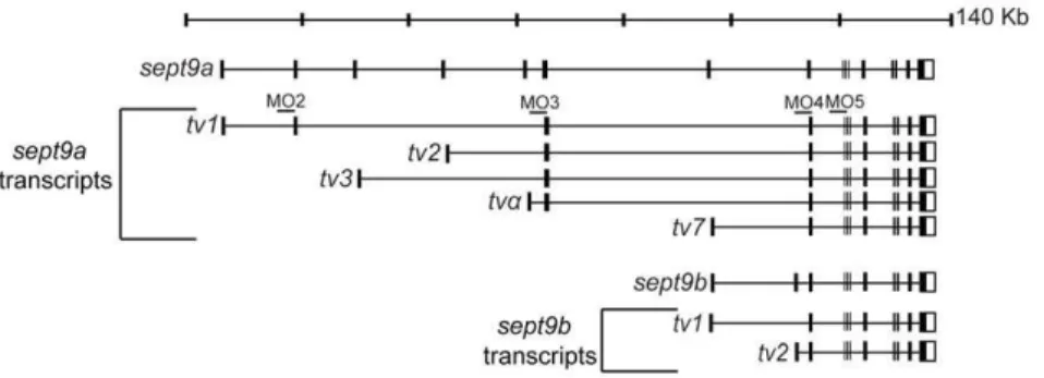
Through a combination of genetic sequence analysis and BLAST searches using known human SEPT9 transcripts, we identified multiple mRNA transcripts produced from two different zebrafishsept9genes,sept9aand sept9b.sept9ais located on chromosome 3, whereas sept9b is found on chromosome 6. sept9a produces transcripts homologous to the longest human SEPT9 isoforms 1, 2, and 3, the shortest human variant SEPT9_v7 (NCBI NM_001113496; named SEPT9_v5 in earlier literature [7]) and a unique transcript not identified in other vertebrates which we have denotedsept9a_tva. Similar to humans,
through the use of alternate 59 exons, sept9a _tv1, 2, 3 anda, generate predicted protein products with unique N-termini of 32, 18, 7, and 10 amino acids respectively (Fig. 1, and Fig. 2). sept9a_tv7 encodes a truncated version of the longer transcripts. sept9bappears to express two humanSEPT9_v7 homologues with alternate 59 UTRs, sept9b_tv1 and sept9b_tv2 (Fig. 1). The predicted protein sequences of zebrafishsept9a_tv1, 2, and 3 are 73–74% similar and 61–62% identical to their human homo-logues, respectively (Fig. 2). sept9a_tv7 and sept9b_tv1 and _tv2 primarily encode the GTP binding domain found in all transcripts, and are highly conserved. These transcripts are 92% similar and 87% identical to each other at the amino acid level and 87–81% similar, 78–80% identical to the human sequence. We did not identify transcripts homologous to human SEPT9transcript variants 4, 5, and 6 (NCBI NM_001113495, NM_001113492, and NM_001113494;SEPT9_v5andv6known as v4* and v4 in previous literature [7,15,18]). However, the start codon in human SEPT9_v5 and v6 is not conserved from mammals to zebrafish (Fig. 2) and to date, only a single EST of humanSEPT9_v4has been identified in a teratocarcinoma cell line, suggesting that these transcripts may not be expressed in zebrafish. It is possible that furthersept9transcripts are expressed in zebrafish yet were not identified in this analysis.
Figure 1. SEPT9 transcripts are conserved between zebrafish and mammals.Zebrafish possess twosept9genes,sept9aandsept9b, that encode multiple mRNA transcripts homologous to mammalian transcripts. Zebrafish also express a transcript not currently found in mammals, sept9a_tva. Sites of morpholino splice blockers are noted.
doi:10.1371/journal.pone.0010712.g001
Developmental expression of zebrafishsept9 genes To examine wheresept9message is expressed in the developing zebrafish embryo, we performed whole mountin situhybridization using a probe tosept9a_tv1. This probe is expected to recognize all sept9atranscripts, and possibly those ofsept9b. Probes designed to the individualsept9atranscripts were not synthesized because the unique regions of the longersept9aisoforms are not large enough to probe individually. Early maternal expression of sept9 was ubiquitous (Fig. 3A) and remained so through blastula stages (Fig. 3B). During gastrulation,sept9became more restricted to the axial mesoderm and endoderm (Fig. 3C, D).sept9was expressed primarily in the floor plate and ventral mesoderm during segmentation (Fig. 3E–H). At 24 hours post fertilization, sept9 was expressed in the intermediate cell mass, epidermis, branchial arches, and pectoral fin bud (Fig. 3I–K).
While we were unable to analyze the spatial expression of specific sept9 isoforms we could, using RT-PCR, determine the temporal expression pattern of the various sept9 transcripts. Zebrafish embryos at different developmental stages were collected for cDNA preparation and subjected to RT-PCR using transcript
specific primers (Fig. 3L).sept9a_tv2, 3, andaare expressed at the two cell stage consistent with the maternal sept9a expression observed in the in situs. Expression of sept9a_tv1, 7 and a, and
sept9b_tv1 commence at high stage, consistent with zygotic expression, which begins at this time. All five transcripts ofsept9a and both sept9b transcripts stabilize expression though the segmentation and pharyngula stages. The longest transcript, sept9a_tv1, has two phases of expression; one during the late blastula and early gastrula stages, and a second beginning during early somitogenesis.
Effect ofsept9a inhibition on zebrafish development Because a majority of sept9 transcripts, including the longest sept9a_tv1, derived from the sept9a locus, we decided to focus further studies on this set of isoforms. Therefore, we targeted all sept9atranscripts, orsept9a_tv1only, for depletion using antisense morpholinos directed to the splice acceptor site of the fifth (MO5) or second (MO2) exons ofsept9a_tv1, respectively (Fig. 1). Embryos were injected at the 1-cell stage with 1.25, 2.5 or 5 ng of morpholino. The observed phenotypes were dosage dependent
Figure 2. SEPT9 amino acid sequence is conserved between zebrafish and mammals.Amino-acid alignments of zebrafish Sept9a and Sept9b putative protein products show a high degree of conservation. An asterisk notes the starting methionine of human SEPT9_i5/6, which is not conserved in zebrafish. Arrowheads mark the starting methionines for human Sept9_v7, and zebrafish isoforms Sept9a_tv7 and Sept9b (both transcripts).
and were divided into three categories which we termed class I, class II and class III based on morphology (Fig. 4J). At 48 hpf, embryos that looked similar to wild-type but had epidermal defects and minor tail perturbations were categorized as class I, those that had a curved or shortened body axis/tail in addition to the defects in class I were categorized as class II and those that had a severely shortened tail/body axis were classified as class III. Both MO5 and MO2 produced classes I and II (Fig. 4A, B, D, E). However, the severe class III phenotype (data not shown) was not observed in MO2 embryos, even at 5 ng of morpholino.
Circulating blood cells could be observed in class I embryos, however, cells were often observed pooling in the intermediate cell mass (ICM) and tail region. Classes II and III showed an absence of blood circulation, and often lacked the presence of mature
hemoglobinized erythrocytes. All classes exhibited yolk extension as well as epidermal defects, most commonly seen in the tail and yolk regions. Epidermal aggregates and edema were frequently noted, often at the tip of the tail and in the ICM. Cardiac edema was regularly observed in embryos from all classes and worsened as development proceeded; class II and III embryos did not survive past 7 days.
The phenotypes resulting from inhibition ofsepta_tv1or allsept9a transcripts were not regularly observed in embryos injected with mismatch controls to either morpholino (MO2MM and MO5MM; Fig. 4C, F, J). To determine ifsept9a transcript levels were altered, RT-PCR was performed on 24 hpf MO5 and MO2 embryos injected with 2.5 ng morpholino (Fig. 4K). Levels of sept9a_tv1 and tv7 were undetectable in MO5 embryos, and
Figure 3. Expression ofsept9genes during zebrafish development.Detection ofsept9mRNA was carried out by whole-mount in situ hybridization using a probe targeted to allsept9isoforms on staged embryos from 256 cells to 24 hpf. Images in A–C are lateral views, animal pole to top; D and E are dorsal views, anterior to top; F and H are dorsal posterior views; G, I and J are lateral views, K is a dorsal view, anterior to left.A–C: sept9transcripts are ubiquitously expressed at early developmental states.D: At bud stage,sept9is expressed in endoderm and axis.E–H:sept9is expressed in the floor plate, ventral mesoderm and tail bud during segmentation.I–K: At 24 hpf,sept9is expressed throughout the epidermis, branchial arches, pectoral fin, and in the intermediate cell mass.L: Transcript specific primers were used to detectsept9aandsept9btranscripts in various stages of development by RT-PCR.sept9a_tv 2, 3, andaare expressed maternally. Amplification ofeIFaand total RNA without addition of reverse transcriptase were used as controls. a, axis; ep, epidermis; fp, floorplate; icm, intermediate cell mass; tb, tail bud; vm, ventral mesoderm. doi:10.1371/journal.pone.0010712.g003
sept9a_tv1 but not_tv7was decreased with MO2 when compared to wild-type. MO2 also did not affect an amplicon from exons 3–5, whereas MO5 greatly reduced the level of this product. Since these exons are shared with sept9a_tv2, 3, and a (Fig. 1), the residual product may be due to perduring maternal transcripts. Morpho-linos designed tosept9a_tv1exon 3 (inhibiting transcript variants 1– 3 anda) and exon 4 (inhibiting all transcripts) acceptor splice sites (Fig. 1) also produced the same classes of morphants observed in MO5 embryos (data not shown).
Co-injection of a low concentration (1 pg) ofsept9a_tv1mRNA with MO2 showed a partial rescue of the morphant phenotypes
providing further evidence that at least classes I and II are a result ofsept9a transcript inhibition (Fig. 4G). The phenotype of MO5 injected embryos could not be rescued by co-injection ofsept9a_tv1 mRNA (data not shown). It is possible thatsept9atranscripts have overlapping functions, and that over-expression of onlysept9a_tv1 cannot compensate for the loss of multiple transcripts. This also complicates interpretation of the class III phenotype, as it difficult to distinguish between a phenotype caused by morpholino artifact and one caused by knocking-down additionalsept9aisoforms that can not be rescued withsept9a_tv1. However, the observation that four differentsept9aMOs cause similar defects whereas mismatch
Figure 4. Charaterization ofsept9amorphant and overexpression embryos.Embryos were injected at the one-cell stage with morpholinos targeted to allsept9atranscripts (MO5),sept9a_tv1only (MO2), mismatch controls (MO5MM, MO2MM), orsept9a_tv1mRNA with and without MO2. Morphants shown were injected with 2.5 ng morpholino. At 48 hpf, the phenotypes were assessed by morphological criteria, according to severity.
A,D: Class I morphants had defects in epidermal integrity and yolk extension and minor curvature of the tail.B,E: Class II morphants had a curved body axis in addition to the defects observed in class I. Class III morphants had a severely shortened body axis (data not shown). Arrows indicate yolk extension defects. All classes exhibited defects in blood circulation.G: Coinjection of 1 pg ofsept9a_tv1mRNA with 5 ng of MO2 partially rescued the observed phenotypes.H,I: Embryos injected with as little as 4 pg ofsept9a_tv1mRNA often had phenotypes similar to those ofsept9amorphants including epidermal aggregates (arrow), blood pooling, and tail edema (bracket). (OE) indicates over expression.C,F: Control mismatch morpholinos did not present a phenotype.J: Graphical representation of MO classes at various concentrations. The number of embryos tested in each experiments is indicated by (n) on top of each column.K:sept9asplice morpholinos inhibitsept9atranscript splicing. RT-PCR analysis was performed on 24 hpf wild-type embryos, embryos injected with 2.5 ng MO5 or MO2 (pooled classes I and II), or 5 bp mismatch controls. MO5 embryos show a complete loss ofsept9a_tv1andsept9a_tv7. The presence of a low level ofsept9aexons 3–5 transcripts in MO5-injected embryos may be due to maternal mRNA. MO2 embryos show a decrease insept9a_tv1compared to wild-type while the other transcripts are not affected.
morpholinos result in no defect and that the morphant phenotype is rescued by co-injection ofsept9a_tv1mRNA, demonstrates that the observed morphant phenotypes are due to specific loss-of-function ofsept9and not toxicity.
Effect ofsept9a_tv1overexpression on zebrafish development
Recent studies in cultured cells have shown that humanSEPT9 appears to be highly regulated [12,13,14]. While attempting rescue ofsept9amorphant embryos, we found that increased levels ofsept9a_tv1 mRNA led to a number of embryonic developmental defects including alterations in convergence and extension, dorsalization, and cyclopia. However, the phenotypes did not clearly group into classes like the sept9a morphant embryos. Interestingly, many of the phenotypes were similar to those observed insept9amorphants including cardiac and tail fin edema, a curved tail and/or body axis, a loss of circulating blood cells with concentrated pools of cells in the tail region and ICM, and epidermal defects including regions of aggregated cells (Fig. 4H, I). Thus, some phenotypes were observed with both gain and loss of sept9a_tv1function, whereas other phenotypes were only found in gain-of-function experiments.
Knock-down and overexpression ofsept9acause an increase in cell death
Alterations in humanSEPT9have been shown to cause defects in cytokinesis, leading to changes in cell morphology and decreases in cellular growth [12,14]. To determine if the defects observed in the tails ofsept9a_tv1morphant and overexpression (OE) embryos included an increase in apoptotic cells, we used acridine orange (AO) to mark cell death. AO-positive cells were rarely observed in wild-type embryos, yet both sept9a MO2 and sept9a_tv1 OE embryos showed an increase in apoptotic cells in the tail indicating cell death (Fig. 5). These data suggest that both loss- and gain-of-function of sept9a in zebrafish lead to an increase in cell death possibly through defects in cell division.
Discussion
In this study we have shown that, like humans, zebrafish express multiplesept9transcripts. These transcripts are expressed through-out development in different tissues types including the ventral mesoderm and axis at early developmental stages, and the epidermis at later stages. We have demonstrated that inhibition and overexpression ofsept9atranscripts in zebrafish embryos lead to a myriad of phenotypes including edema, loss of blood
circulation, tail fin malformations, loss of epidermal integrity and increased cell death. Additionally, we have provided evidence that multiplesept9aMOs targeted to different splice sites yield similar phenotypes, and that overexpression ofsept9a_tv1causes develop-mental defects similar to those observed with the MOs. Thus, too much or too littlesept9afunction is deleterious for many embryonic cells, indicating that cells need to carefully regulatesept9a levels. The correct levels of sept9a, therefore, are needed to maintain tissue integrity and to allow normal cell division.
The fact that zebrafish posses twosept9orthologues (sept9aand sept9b) is not unusual, given the proposed genomic duplication event that occurred in teleost fish [19]. The two orthologues appear to have evolved such that only sept9a expresses longer isoforms possessing a proline-rich region, while both genes express shorter isoforms primarily consisting of a GTP-binding domain. The predicted polypeptides are highly similar to mammalian SEPT9 proteins, suggesting possible overlapping functions. While zebrafish do not appear to express homologues to humanSEPT9 transcripts 5 and 6, they do express two additional variant 7 transcripts from sept9b. It is possible that these transcripts are regulated in a manner similar toSEPT9_v5andv6[18]. Further studies are required to determine if sept9a and sept9b have overlapping functions in zebrafish.
RT-PCR analysis of human tissues has shown that a majority of SEPT9transcripts are expressed in almost every tissue type tested [6,7] and cultured cell lines express different combinations of SEPT9 proteins depending on the line [11,12,14,20]. However, whether different SEPT9 polypeptides interact with one another and the individual function of each transcript remains to be determined. We found that zebrafish express multiple sept9 transcripts from two different genes, and confirmed the role of sept9during zebrafish development. Moreover, we observed the same spectrum of phenotypes when we eliminated the largest sept9aisoform,sept9a_tv1, as when we eliminated allsept9aisoforms, providing the first evidence that the smaller isoforms cannot compensate for a lack of the largest isoform.
Recently, a number ofsept9a transcripts were identified in an analysis of hematopoietic genes isolated from zebrafish kidney marrow [21] and expression ofsept9bwas shown to be increased in embryos overexpressing etsrp, a transcription factor required for vasculogenesis and primitive myelopoiesis in zebrafish [22]. This studies support the hypothesis that sept9 genes play a role in hematopoiesis. However, the pericardial edema, loss of blood circulation and tail malformations observed in both sept9a morphant and OE embryos are also consistent with defects in osmoregulation observed when epidermal barrier function is lost [23] or if fish are exposed to toxins that impair homeostasis of the
Figure 5. Knockdown and overexpression (OE) ofsept9a_tv1results in an increase in apoptotic cells in the tail.Embryos at the one-cell stage were injected with 2.5 ng of MO2 or 4 pg ofsept9a_tv1mRNA and analyzed for acridine orange (AO) staining at 24 hpf.A–C: The tail fin of wild-type embryos is negative for AO indicating few apoptotic cells.D–I: Both class II MO2 andsept9a_v1OE embryos show an increase in AO staining indicating increased cell death.
doi:10.1371/journal.pone.0010712.g005
skin or kidney [24,25]. Zebrafish will be a good model system for future studies examining the roles of various sept9 isoforms in developmental processes such as hematopoiesis and epidermal development.
Acknowledgments
M. L. would like to thank Dr. Phillip Chance for his support and guidance, and Dr. Dan Doherty for intellectual contributions and thoughtful discussions.
Author Contributions
Conceived and designed the experiments: MLL DCW MCH DK. Performed the experiments: MLL DCW. Analyzed the data: MLL DCW DK. Contributed reagents/materials/analysis tools: MLL DCW DK. Wrote the paper: MLL DCW DK.
References
1. Hall PA, Russell SE (2004) The pathobiology of the septin gene family. J Pathol 204: 489–505.
2. Kinoshita M (2003) The septins. Genome Biol 4: 236.
3. McIlhatton MA, Burrows JF, Donaghy PG, Chanduloy S, Johnston PG, et al. (2001) Genomic organization, complex splicing pattern and expression of a human septin gene on chromosome 17q25.3. Oncogene 20: 5930–5939. 4. Osaka M, Rowley JD, Zeleznik-Le NJ (1999) MSF (MLL septin-like fusion), a
fusion partner gene of MLL, in a therapy-related acute myeloid leukemia with a t(11;17)(q23;q25). Proc Natl Acad Sci U S A 96: 6428–6433.
5. Russell SE, McIlhatton MA, Burrows JF, Donaghy PG, Chanduloy S, et al. (2000) Isolation and mapping of a human septin gene to a region on chromosome 17q, commonly deleted in sporadic epithelial ovarian tumors. Cancer Res 60: 4729–4734.
6. Burrows JF, Chanduloy S, McIlhatton MA, Nagar H, Yeates K, et al. (2003) Altered expression of the septin gene, SEPT9, in ovarian neoplasia. J Pathol 201: 581–588.
7. Scott M, Hyland PL, McGregor G, Hillan KJ, Russell SE, et al. (2005) Multimodality expression profiling shows SEPT9 to be overexpressed in a wide range of human tumours. Oncogene 24: 4688–4700.
8. Amir S, Wang R, Matzkin H, Simons JW, Mabjeesh NJ (2006) MSF-A interacts with hypoxia-inducible factor-1alpha and augments hypoxia-inducible factor transcriptional activation to affect tumorigenicity and angiogenesis. Cancer Res 66: 856–866.
9. Kuhlenbaumer G, Hannibal MC, Nelis E, Schirmacher A, Verpoorten N, et al. (2005) Mutations in SEPT9 cause hereditary neuralgic amyotrophy. Nat Genet 37: 1044–1046.
10. Landsverk ML, Ruzzo EK, Mefford HC, Buysse K, Buchan JG, et al. (2009) Duplication within the SEPT9 gene associated with a founder effect in North American families with hereditary neuralgic amyotrophy. Hum Mol Genet 18: 1200–1208.
11. Nagata K, Kawajiri A, Matsui S, Takagishi M, Shiromizu T, et al. (2003) Filament formation of MSF-A, a mammalian septin, in human mammary epithelial cells depends on interactions with microtubules. J Biol Chem 278: 18538–18543.
12. Surka MC, Tsang CW, Trimble WS (2002) The mammalian septin MSF localizes with microtubules and is required for completion of cytokinesis. Mol Biol Cell 13: 3532–3545.
13. Robertson C, Church SW, Nagar HA, Price J, Hall PA, et al. (2004) Properties of SEPT9 isoforms and the requirement for GTP binding. J Pathol 203: 519–527.
14. Gonzalez ME, Peterson EA, Privette LM, Loffreda-Wren JL, Kalikin LM, et al. (2007) High SEPT9_v1 expression in human breast cancer cells is associated with oncogenic phenotypes. Cancer Res 67: 8554–8564.
15. Chacko AD, Hyland PL, McDade SS, Hamilton PW, Russell SH, et al. (2005) SEPT9_v4 expression induces morphological change, increased motility and disturbed polarity. J Pathol 206: 458–465.
16. Westerfield M (1993) The zebrafish book: a guide for the laboratory use of zebrafish (Brachydanio rerio). Eugene: University of Oregon Press.
17. Webb AE, Driever W, Kimelman D (2008) psoriasis regulates epidermal development in zebrafish. Dev Dyn 237: 1153–1164.
18. McDade SS, Hall PA, Russell SE (2007) Translational control of SEPT9 isoforms is perturbed in disease. Hum Mol Genet 16: 742–752.
19. Meyer A, Schartl M (1999) Gene and genome duplications in vertebrates: the one-to-four (-to-eight in fish) rule and the evolution of novel gene functions. Curr Opin Cell Biol 11: 699–704.
20. Sudo K, Ito H, Iwamoto I, Morishita R, Asano T, et al. (2007) SEPT9 sequence alternations causing hereditary neuralgic amyotrophy are associated with altered interactions with SEPT4/SEPT11 and resistance to Rho/Rhotekin-signaling. Hum Mutat 28: 1005–1013.
21. Song HD, Sun XJ, Deng M, Zhang GW, Zhou Y, et al. (2004) Hematopoietic gene expression profile in zebrafish kidney marrow. Proc Natl Acad Sci U S A 101: 16240–16245.
22. Gomez GA, Veldman MB, Zhao Y, Burgess S, Lin S (2009) Discovery and characterization of novel vascular and hematopoietic genes downstream of etsrp in zebrafish. PLoS One 4: e4994.
23. Kiener TK, Selptsova-Friedrich I, Hunziker W (2008) Tjp3/zo-3 is critical for epidermal barrier function in zebrafish embryos. Dev Biol 316: 36–49. 24. Hentschel DM, Park KM, Cilenti L, Zervos AS, Drummond I, et al. (2005)
Acute renal failure in zebrafish: a novel system to study a complex disease. Am J Physiol Renal Physiol 288: F923–929.